

C57BL/6-Tnfrsf4tm1(TNFRSF4)Bcgen Tnfsf4tm1(TNFSF4)Bcgen/Bcgen • 120543
on this page

OX40L: Enhances effector T cell function, sustains their survival and suppresses apoptosis

OX40
OX40L

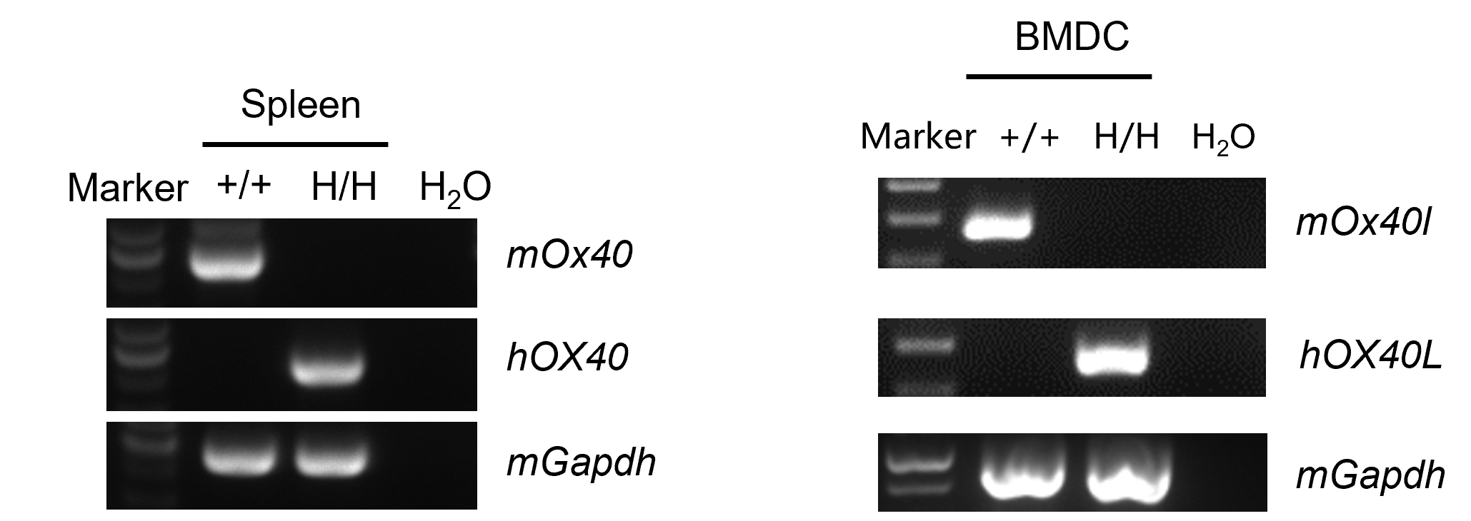
Strain specific analysis of OX40 and OX40L gene expression in wild-type C57BL/6JNifdc mice and homozygous B-hOX40/hOX40L mice by RT-PCR. Spleen and bone marrow cells were collected from wild-type C57BL/6JNifdc mice(+/+) and homozygous B-hOX40/hOX40L mice (H/H;H/H). DCs were induced from bone marrow cells. cDNA libraries were synthesized by reverse transcription, followed by PCR with mouse or human OX40/OX40L primers. Mouse OX40 mRNA was only detectable in spleen from wild-type mice Human OX40 mRNA was exclusively detectable in spleen from homozygous B-hOX40/hOX40L mice but not in wild-type mice. Mouse Ox40l mRNA was detectable in bone marrow-derived DC cells (BMDC) of wild-type mice. Human OX40L mRNA was detectable only in BMDC from B-hOX40/hOX40L mice but not in wild-type mice.
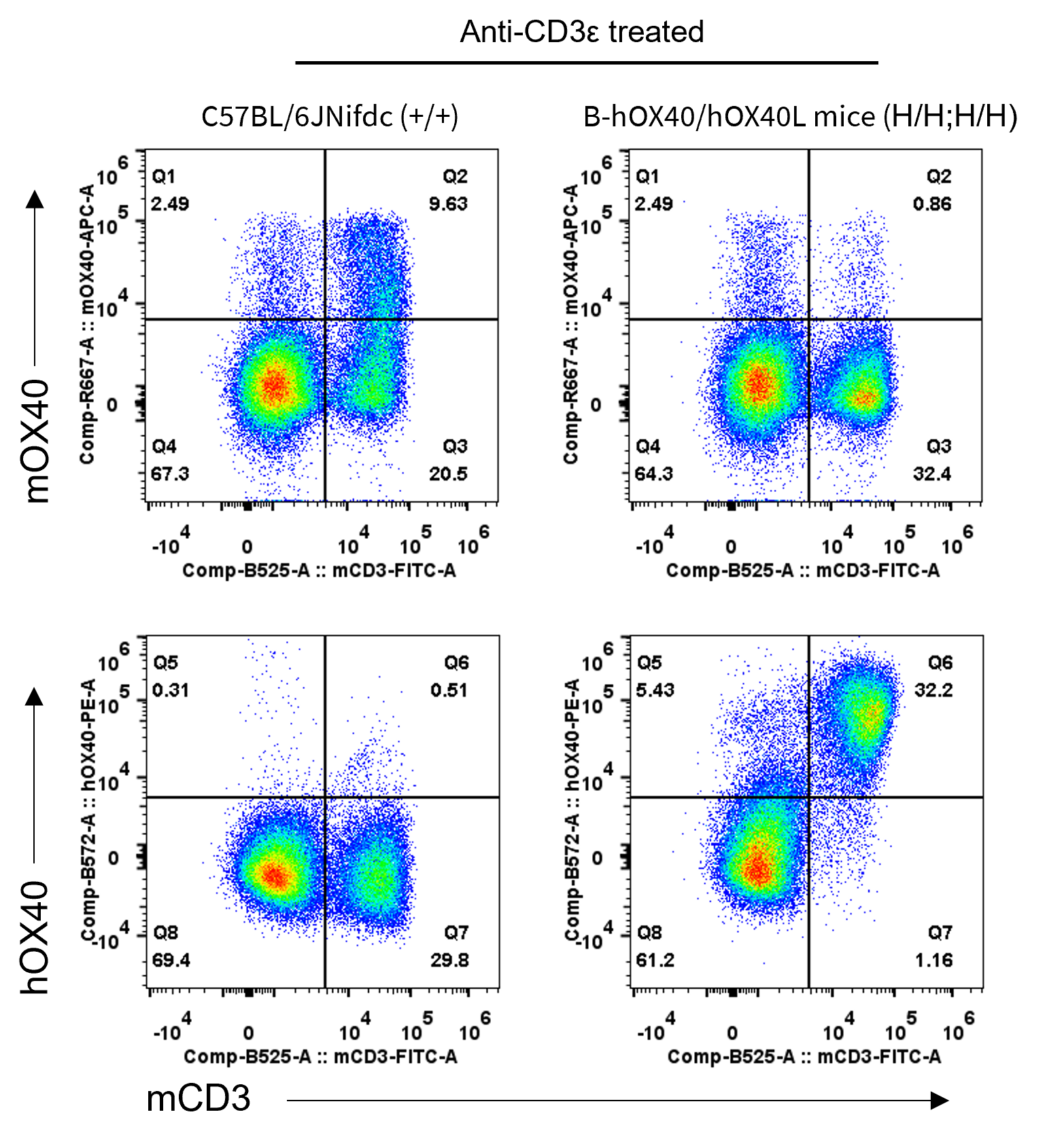
Strain specific OX40 expression analysis in homozygous B-hOX40/hOX40L mice by flow cytometry. Splenocytes were collected from wild-type C57BL/6JNifdc(+/+) and homozygous B-hOX40/hOX40L mice (H/H;H/H) stimulated with anti-CD3ε in vivo, and protein expression was analyzed with anti-mouse OX40 antibody (Biolegend, 119414) and anti-human OX40 antibody (Biolegend, 350004) by flow cytometry. Mouse OX40 was only detectable in T cells of wild-type C57BL/6JNifdc mice. Human OX40 was only detectable in T cells of homozygous B-hOX40/hOX40L mice, but not in wild-type C57BL/6JNifdc mice.
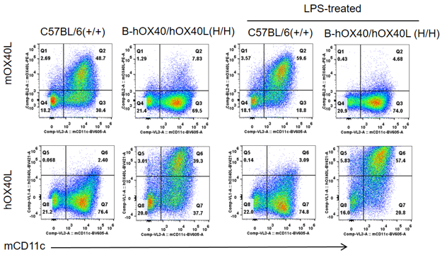
Strain specific OX40L expression analysis in homozygous B-hOX40/hOX40L mice by flow cytometry. Bone marrow cells were collected from wild-type C57BL/6JNifdc(+/+) and homozygous B-hOX40/hOX40L mice (H/H;H/H). DCs were induced from bone marrow cells and stimulated with LPS. Then DCs were analyzed by flow cytometry with anti-OX40L antibodies. Mouse OX40L was detectable in bone marrow-derived DC cells (BMDC) from wild-type C57BL/6JNifdc mice. Human OX40L was exclusively detectable in bone BMDC from homozygous B-hOX40/hOX40L mice (H/H;H/H) but not wild-type C57BL/6JNifdc mice.
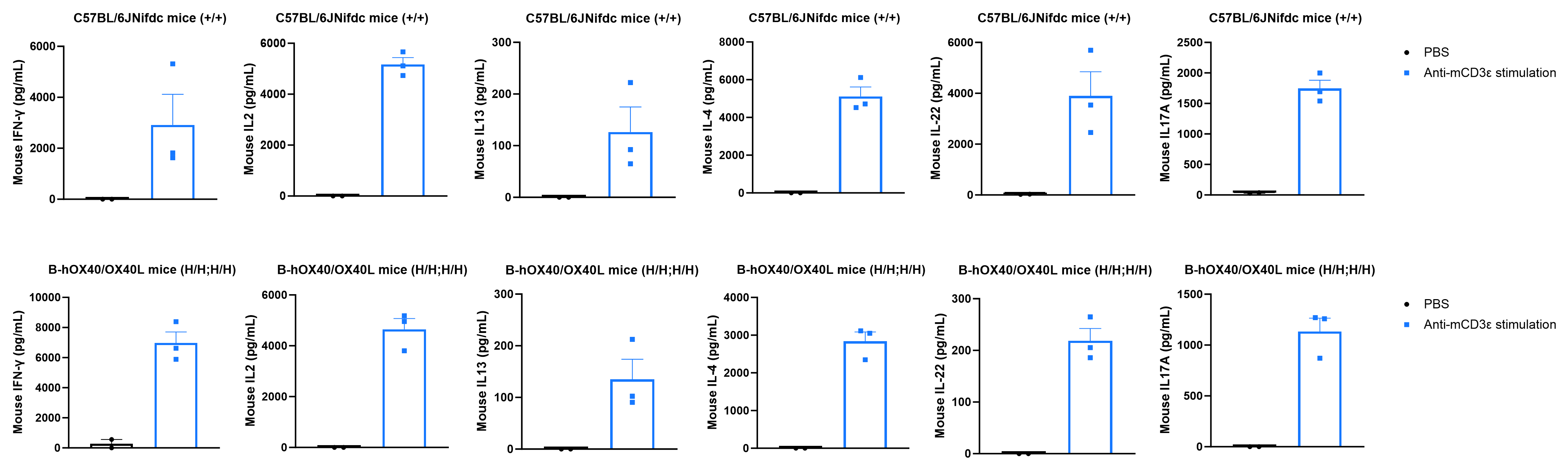
Strain specific cytokines expression analysis in wild-type C57BL/6JNifdc mice and homozygous humanized B-hOX40/OX40L mice by ELISA. Serum was collected from wild-type C57BL/6JNifdc mice (+/+) (female, n=3, 7-week-old) and homozygous B-hOX40/OX40L mice (H/H;H/H) (female, n=3, 7-week-old) stimulated with anti-mouse CD3ε antibody (37.5 μg/mL, 200 μL/mouse, i.p.) for 2 hrs in vivo. Expression level of mouse IL4, IL13, IL2, IL22, IL17A, and IFN-γ were analyzed by ELISA. After mCD3ε stimulation, a significant increase of mouse IL4, IL13, IL2, IL22, IL17A, and IFN-γ were detected in C57BL/6JNifdc mice (n=3) and homozygous B-hOX40/OX40L mice (n=3). Under the stimulation of mCD3ε, the downstream cytokines of the humanized mice can fully respond, which proves that the signaling pathway of humanized mice is complete. Values are expressed as mean ± SEM.
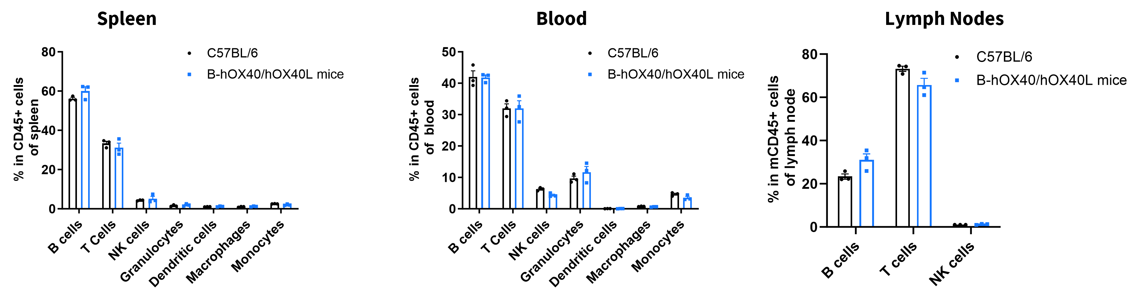
Analysis of leukocyte subpopulations by flow cytometry in immune organs and blood. Splenocytes, peripheral blood and lymph nodes were isolated from female C57BL/6 and B-hOX40/hOX40L mice (female, 6-week-old, n = 3). Single live cells were gated on the CD45⁺ population and analyzed by flow cytometry as indicated. Values are expressed as mean ± SEM.
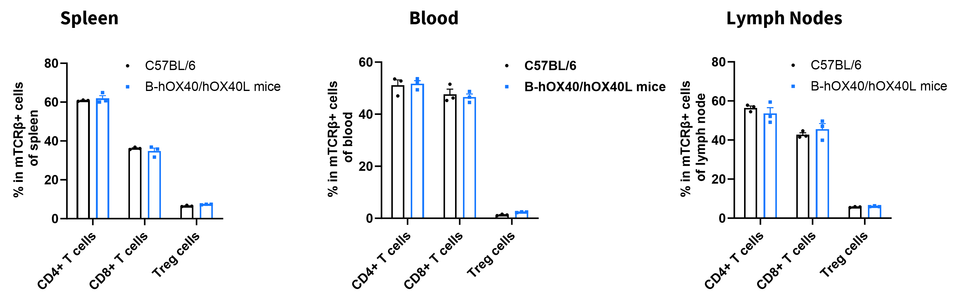
Analysis of T-cell subpopulations by flow cytometry in immune organs and blood. Splenocytes, peripheral blood, and lymph nodes were isolated from female C57BL/6 and B-hOX40/hOX40L mice (female, 6-week-old, n = 3). Single live cells were gated on the TCRβ⁺ T-cell population and analyzed by flow cytometry as indicated. Values are expressed as mean ± SEM.

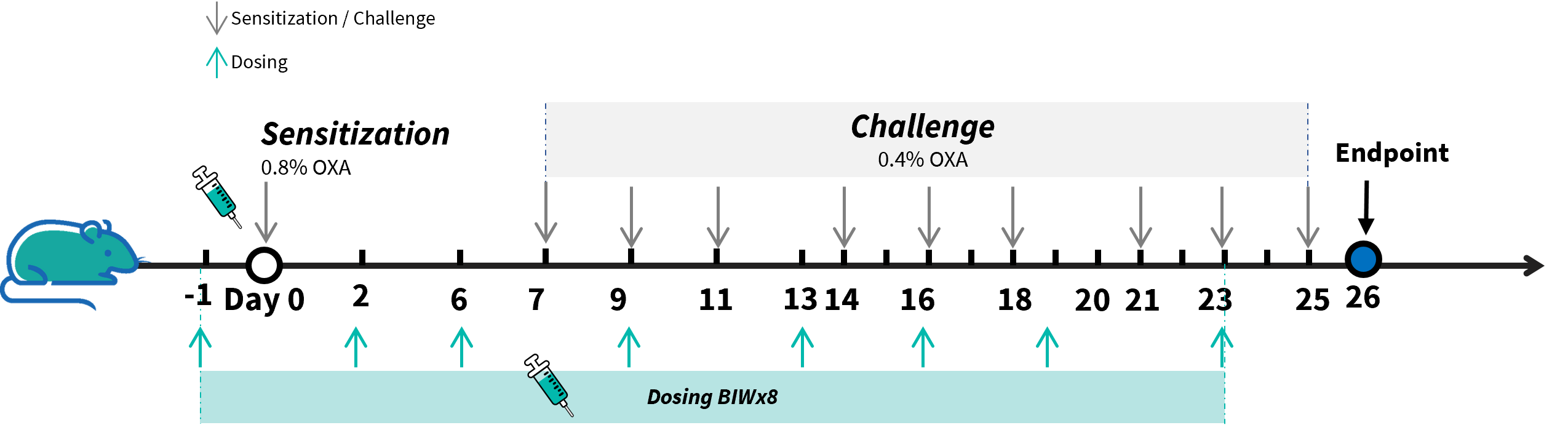
Experimental schedule for induction of AD model and in vivo efficacy of anti-human OX40L antibody. OXA was applied to dorsal and ear skin of mice on day 0, and then challenge to the same site of skin nine times from days 7 to 25. Anti-human OX40L antibody (provided by a client) was administered by intraperitoneal injection twice a week on days -1 to 23. Serum was collected at the endpoint on day 26. AD: atopic dermatitis; OXA: oxazolone.
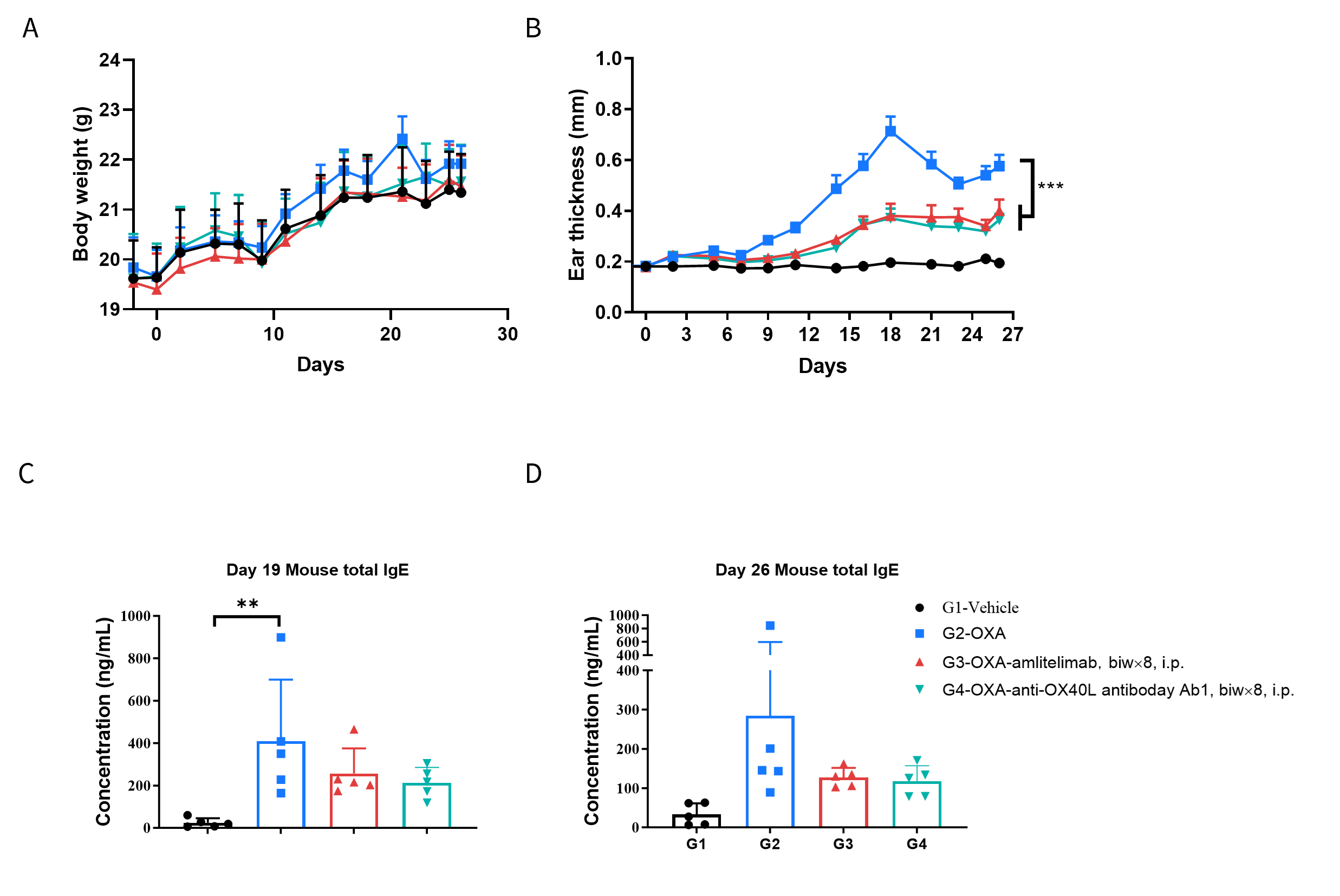
Efficacy of anti-human OX40L antibody in B-hOX40/hOX40L mice. Mice in each group were intraperitoneally injected with anti-hOX40L antibody (provided by a client, n=5). (A) Body weight changes during the treatment. (B) Statistical analysis of ear thickness in each group. (C&D) Total IgE levels in serum. The results showed that compared to the untreated group (G2), the group of mice treated with anti-OX40L antibody showed a significant reduction in ear thickness. Serum was collected at the study endpoint. IgE level was analyzed by ELISA. The results showed that the levels of total IgE in mice treated with anti-OX40L antibody was lower than that in untreated mice. Values are expressed as mean ± SEM. Significance was determined by two-way ANOVA test. *P < 0.05, **P < 0.01, ***P < 0.001.

Efficacy of anti-human OX40L antibody in B-hOX40/hOX40L mice. Ear tissues were collected at the study endpoint and analyzed with H&E. The results showed that compared to the untreated group (G2), the group of mice treated with anti-OX40L antibody (provided by a client) showed a significant reduction in epidermal thickness and pathological score of ear skin. Values are expressed as mean ± SEM. Significance was determined by two-way ANOVA test. *P < 0.05, **P < 0.01, ***P < 0.001, ****P < 0.0001. AD: Atopic dermatitis.
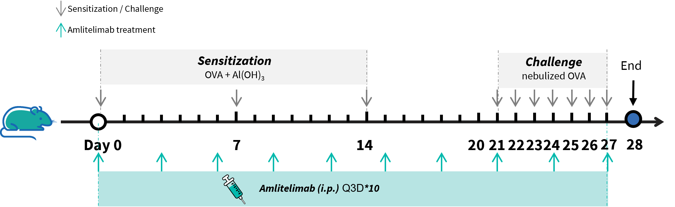
Experimental schedule for induction of asthma and in vivo efficacy of anti-human OX40L antibody. OVA + Al(OH)3 was injected intraperitoneally on days 0, 7, and 14; followed by daily nebulization with OVA for the challenge phase from days 21 to 27. Anti-human OX40L antibody amlitelimab analog (in-house) was administered by intraperitoneal injection every 3 days on days 0 to 27. Serum was collected at the endpoint on day 28. OVA: ovalbumin.

Analysis of immune cells in BALF (Bronchoalveolar fluid) by flow cytometry. B-hOX40/hOX40L mice (female, 7-week-old, n=6) were immunized with OVA to induce asthma. Anti-human OX40L antibody (amlitelimab analog, synthesized in-house) was intraperitoneally injected from day 0 to day 27. BALF was collected at the end of the experiment to detect inflammatory cell infiltration in lung tissue. The results showed that the number of CD45+ cells and eosinophils of BALF in the amlitelimab treated group (G3) decreased significantly compared with the OVA -induced untreated group (G2). Data indicated that anti-human OX40L antibodies could effectively reduce the number and proportion of eosinophils in B-hOX40/hOX40L mice induced with OVA. Values are expressed as mean ± SEM. Significance was determined by two-way ANOVA test. *P < 0.05, **P < 0.01, ***P < 0.001, ****P < 0.0001.
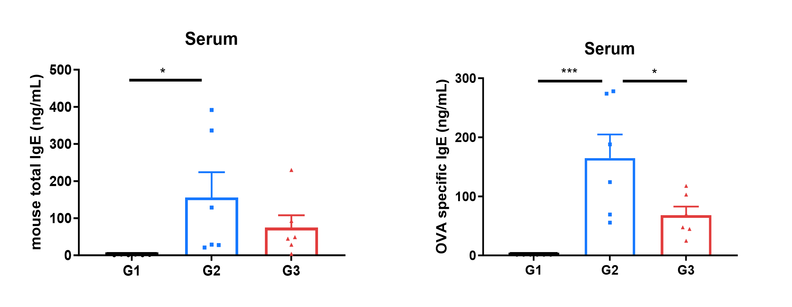
Mouse total IgE and OVA-specific IgE in serum were reduced in the mouse asthma model treated with anti-human OX40L antibody. Serum was collected at the study endpoint. IgE level was analyzed by ELISA. The results showed that the level of OVA specific IgE in mice treated with amlitelimab analog (in-house) was lower than that in untreated mice. Values are expressed as mean ± SEM. Significance was determined by unpaired t-test. *P < 0.05, **P < 0.01, ***P < 0.001.
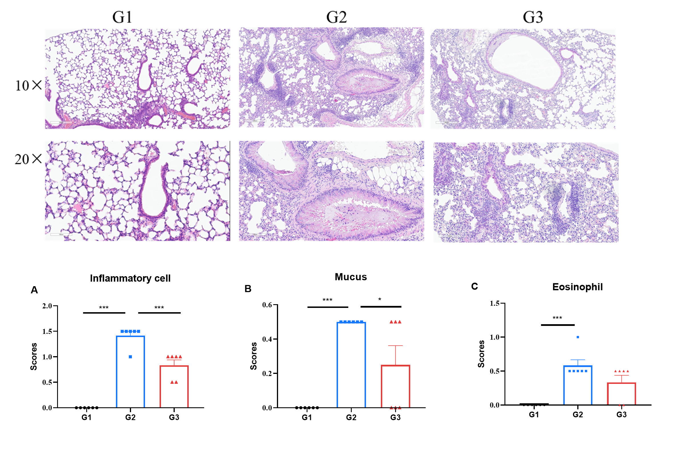
H&E staining of asthma-like model in B-hOX40/hOX40L mice. Lung tissues were collected at the study endpoint and analyzed with H&E staining. The results showed that compared to the untreated group (G2), the group of mice treated with amlitelimab analog (in-house) showed a significant reduction in inflammatory infiltration and mucus secretion in lung tissue, indicating that B-hOX40/hOX40L mice provide a powerful preclinical model for in vivo evaluation of anti-human OX40L antibodies. Values are expressed as mean ± SEM. Significance was determined by unpaired t-test. *P < 0.05, **P < 0.01, ***P < 0.001.
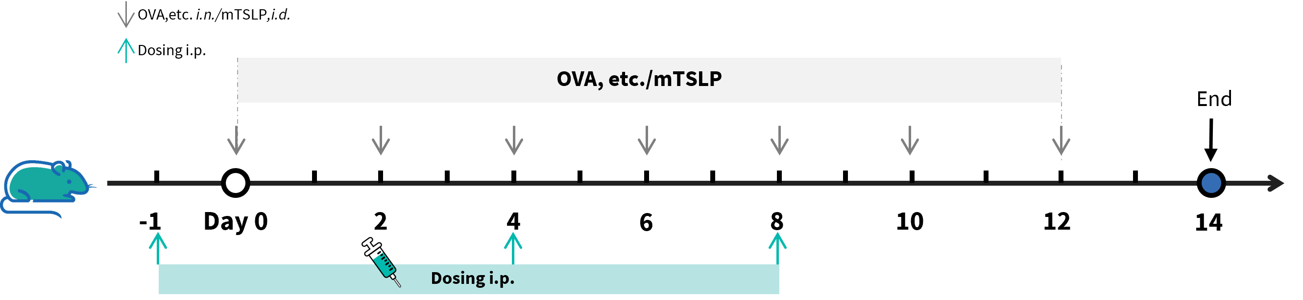
Experimental schedule for the induction of AD and asthma model and in vivo efficacy of anti-human OX40L antibody in B-hOX40/hOX40L mice. B-hOX40/hOX40L mice (male, 11-week-old, n=6) were immunized with OVA etc. inducer to induce asthma and injected with mTSLP to induced AD model. The anti-human OX40L antibody amlitelimab analog (in-house) was administered by intraperitoneal injection (n = 6).

Efficacy of anti-human OX40L antibody in B-hOX40/hOX40L mice. (A&B) Ear thickness and body weight changes during the treatment. (C) Total IgE levels in serum. The results showed that compared to the untreated group (G2), the group of mice treated with amlitelimab (in-house) showed a significant reduction in ear thickness. Serum was collected at the study endpoint. IgE level was analyzed by ELISA. The results showed that the levels of total IgE in mice treated with amlitelimab analog (in-house) was lower than that in untreated mice. Values are expressed as mean ± SEM. Significance was determined by two-way ANOVA test. *P < 0.05, **P < 0.01, ***P < 0.001.
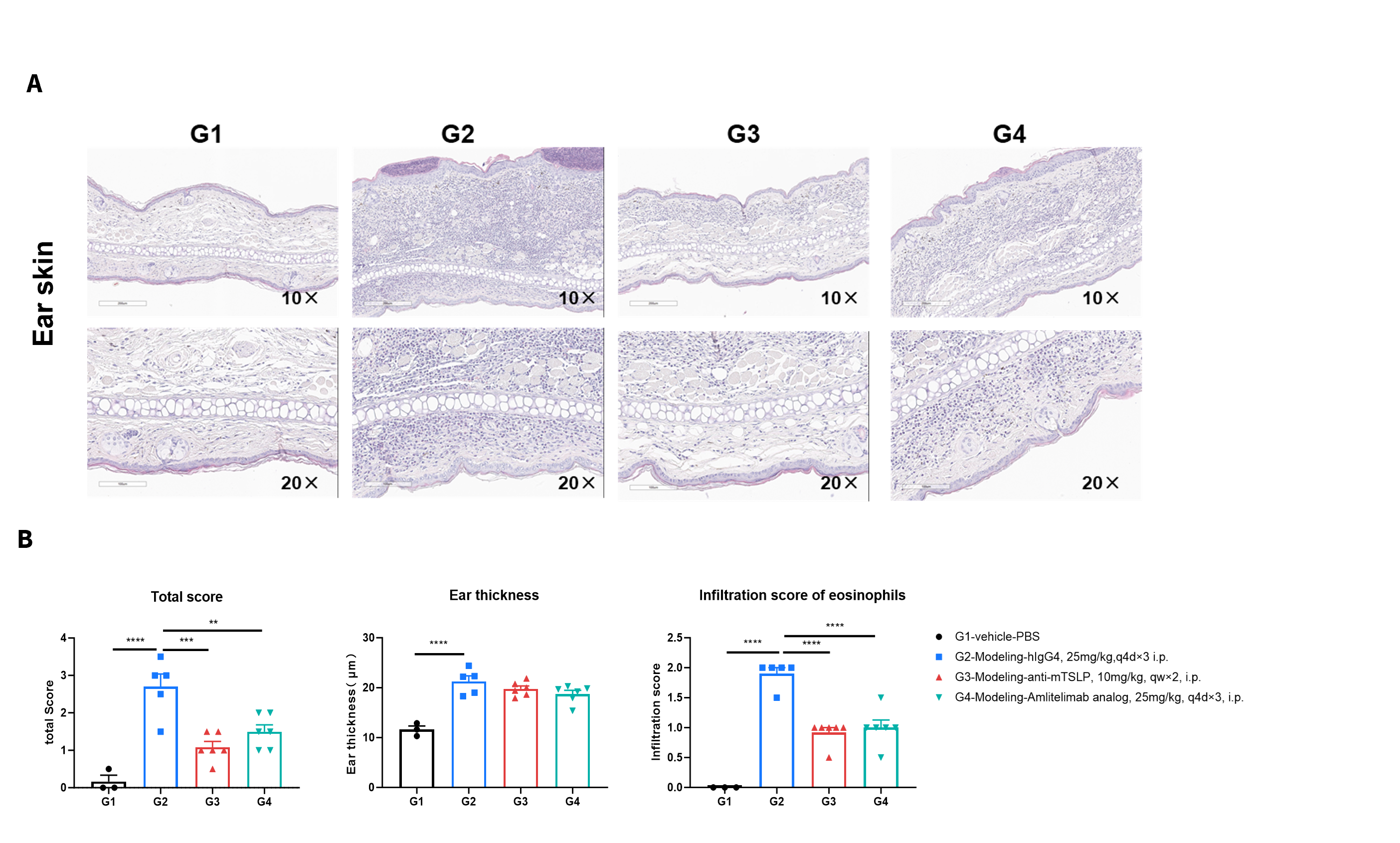
H&E staining of asthma-like model in B-hOX40/hOX40L mice. ear tissues were collected at the study endpoint and analyzed with H&E staining. The results showed that compared to the untreated group (G2), the group of mice treated with amlitelimab (in-house) showed a significant reduction in histopathology score and score of eosinophil infiltration. Values are expressed as mean ± SEM. Significance was determined by two-way ANOVA test. *P < 0.05, **P < 0.01, ***P < 0.001, ****P < 0.0001. AD: Atopic dermatitis.
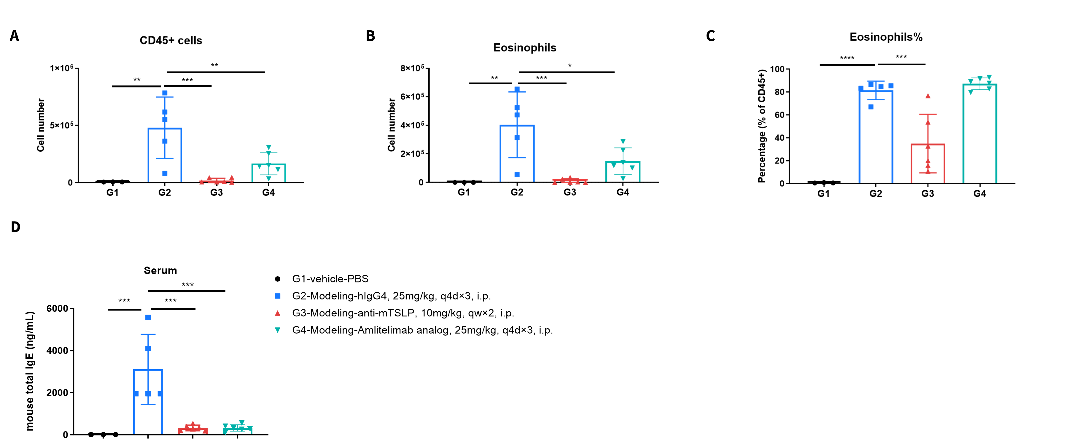
Analysis of immune cells in BALF (Bronchoalveolar fluid) by flow cytometry. B-hOX40/hOX40L mice (female, 7-week-old, n=6) were immunized with OVA to induce asthma. Anti-human OX40L antibody (amlitelimab analog, synthesized in-house) was intraperitoneally injected from day 0 to day 27. BALF was collected at the end of the experiment to detect inflammatory cell infiltration in lung tissue. The results showed that the number of CD45+ cells and eosinophils of BALF in the amlitelimab treated group (G3) decreased significantly compared with the OVA -induced untreated group (G2). Data indicated that anti-human OX40L antibodies could effectively reduce the number and proportion of eosinophils in B-hOX40/hOX40L mice induced with OVA. Values are expressed as mean ± SEM. Significance was determined by two-way ANOVA test. *P < 0.05, **P < 0.01, ***P < 0.001, ****P < 0.0001.
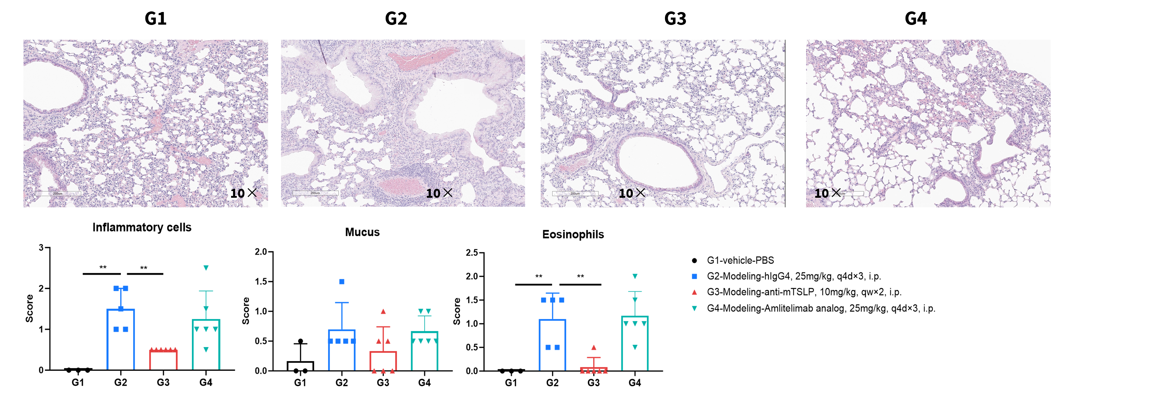
Analysis of immune cells in BALF (Bronchoalveolar fluid) by flow cytometry. B-hOX40/hOX40L mice (female, 7-week-old, n=6) were immunized with OVA to induce asthma. Anti-human OX40L antibody (amlitelimab analog, synthesized in-house) was intraperitoneally injected from day 0 to day 27. BALF was collected at the end of the experiment to detect inflammatory cell infiltration in lung tissue. The results showed that the number of CD45+ cells and eosinophils of BALF in the amlitelimab treated group (G3) decreased significantly compared with the OVA -induced untreated group (G2). Data indicated that anti-human OX40L antibodies could effectively reduce the number and proportion of eosinophils in B-hOX40/hOX40L mice induced with OVA. Values are expressed as mean ± SEM. Significance was determined by two-way ANOVA test. *P < 0.05, **P < 0.01, ***P < 0.001, ****P < 0.0001.
