Description
Overview of PD-1, VEGFA, and CTLA4 in Cancer Immunotherapy and Angiogenesis
- Signaling Pathway: PD-1: Upon binding to its ligands PD-L1 or PD-L2, PD-1 recruits the phosphatases SHP-1 and SHP-2 via its intracellular ITIM and ITSM motifs, suppressing TCR and CD28 signaling. This inhibits downstream pathways including PI3K/AKT, RAS/MAPK, and NF-κB, leading to reduced T-cell proliferation, cytokine production, and cytotoxic activity. VEGFA: VEGFA primarily binds to VEGFR-2 (KDR/FLK-1) on endothelial cells, triggering receptor dimerization and tyrosine kinase activation. This activates downstream signaling cascades including PI3K/AKT, MAPK/ERK, PLCγ/PKC, and SRC pathways, promoting endothelial cell proliferation, migration, survival, and neovascularization. CTLA4: CTLA4 competes with the co-stimulatory receptor CD28 for binding to CD80 (B7-1) and CD86 (B7-2) on antigen-presenting cells, exhibiting significantly higher binding affinity. CTLA4 engagement attenuates CD28-mediated co-stimulation, suppresses PI3K/AKT signaling, reduces IL-2 production, and limits T-cell activation and expansion.
- Therapeutic Inhibition: PD-1 blockade with monoclonal antibodies restores exhausted T-cell function and enhances antitumor immunity. CTLA4 blockade enhances T-cell priming and expansion by relieving inhibitory signaling during the early stages of immune activation. VEGFA inhibition suppresses tumor angiogenesis, normalizes tumor vasculature, and improves immune cell infiltration into the tumor microenvironment. Emerging therapeutic strategies combine these pathways to achieve synergistic antitumor effects.
Targeting strategy
Gene targeting strategy for B-hPD-1/hCTLA4 mice. The exon 2 of mouse Pdcd1 gene that encode the IgV domain was replaced by human PDCD1 exon 2 in B-hPD-1/hCTLA4 mice. The exon 2 of mouse Ctla4 gene that encodes the extracellular domain was replaced by human CTLA4 exon 2 in B-hPD-1/hCTLA4 mice.
mRNA Expression Analysis
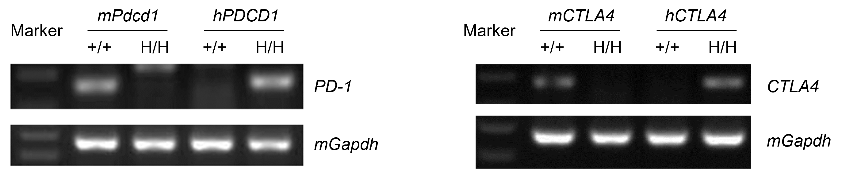
Strain specific analysis of PD-1 and CTLA4 gene expression in C57BL/6 and hPD-1/hCTLA4 mice by RT-PCR. Mouse Pdcd1 and Ctla4 mRNA were detectable only in spleen of wild-type mice (+/+). Human PDCD1 and CTLA4 mRNA were detectable only in spleen of B-hPD-1/hCTLA4 mice (H/H), but not in wild-type (+/+) mice.
PD-1 Protein Expression in Spleen
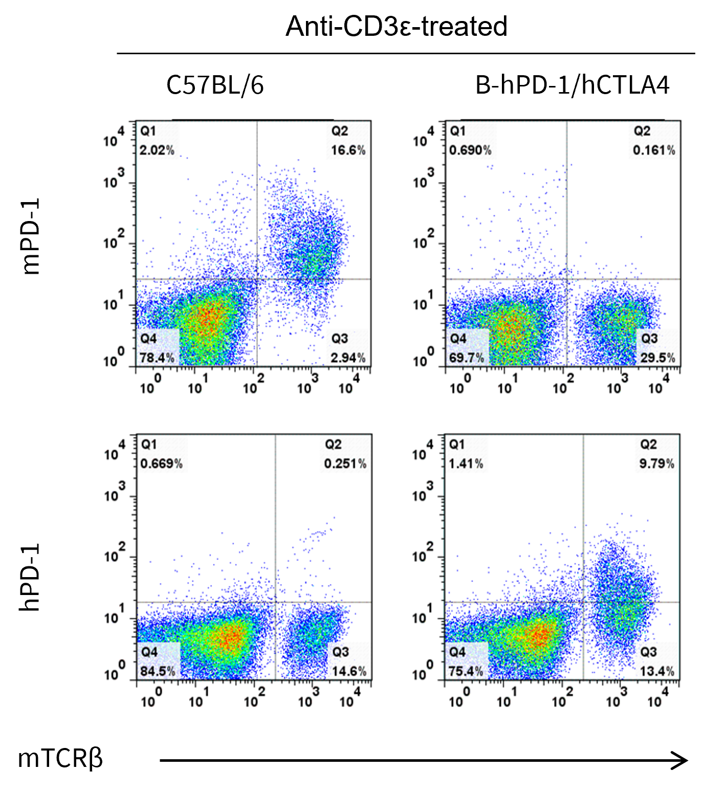
- Mouse PD-1 was detected on T cells populations in wild-type C57BL/6 mice, but not in B-hPD-1/hCTLA4 mice.
- Human PD-1 was detected on T cells populations in B-hPD-1/hCTLA4 mice, but not in wild-type C57BL/6 mice.
Mouse and human PD-1 expression analysis in splenocytes. Splenocytes were collected from wild-type C57BL/6 mice and homozygous B-hPD-1/hCTLA4 mice stimulated with anti-CD3ε in vivo. PD-1 expression on T cells was analyzed by flow cytometry using species-specific anti-PD-1 antibodies.
Combination Therapy of anti-hPD-1 antibody with anti-hCTLA4 antibody in the Subcutaneous MC38 Model in B-hPD-1/hCTLA4 Mice
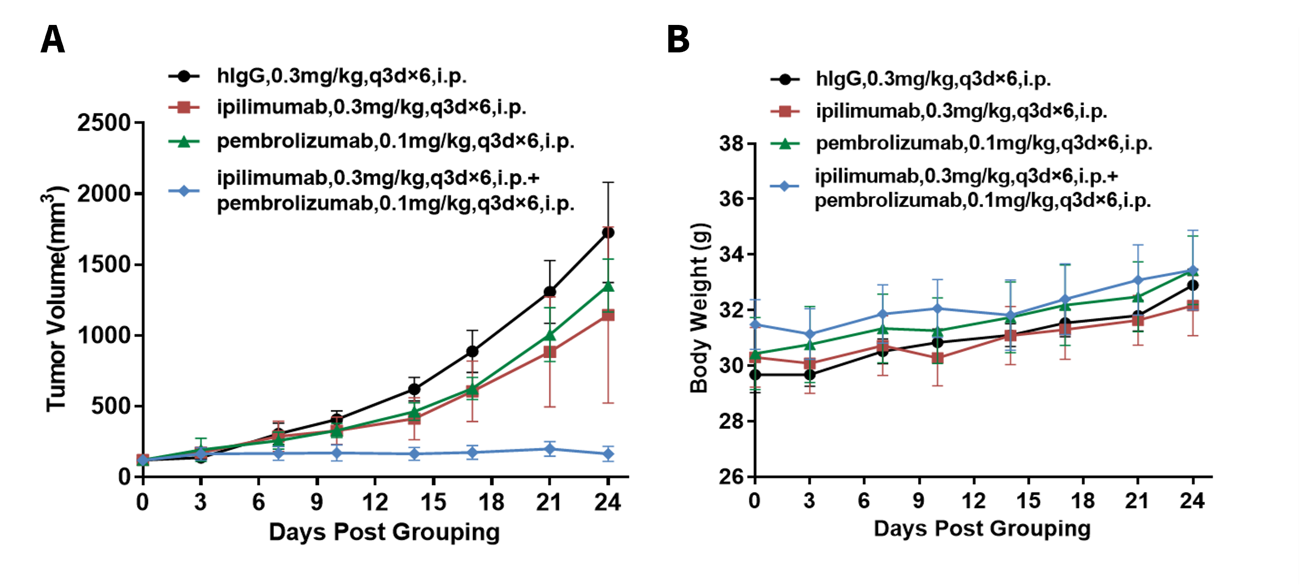
Antitumor activity of anti-human PD-1 antibody combined with anti-human CTLA4 antibody in B-hPD-1/hCTLA4 mice. Murine colon cancer MC38 cells were subcutaneously implanted into homozygous B-hPD-1/hCTLA4 mice (female, 6-7 week-old, n=5). Mice were grouped when tumor volume reached approximately 100 mm3, at which time they were treated with pembrolizumab and ipilimumab with doses and schedules indicated in panel. (A) Tumor growth curves. (B) Body weight changes during treatment. As shown in panel A, combination of pembrolizumab and ipilimumab shows more inhibitory effects than individual groups, demonstrating that the B-hPD-1/hCTLA4 mice provide a powerful preclinical model for in vivo evaluating combination therapy efficacy of hPD-1 antibodies and hCTLA4 antibodies. Values are expressed as mean ± SEM. (All antibodies were made in house)
Efficacy Evaluation of Anti-PD-1/VEGFA/CTLA4 TsAbs in the Subcutaneous B-hVEGFA MC38 Model in B-hPD-1/hCTLA4 Mice
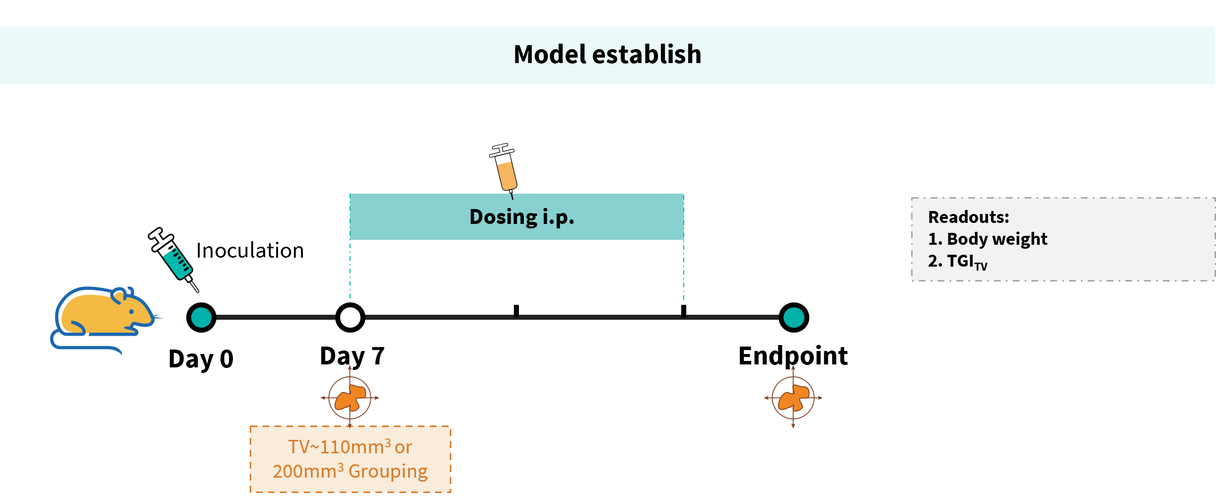
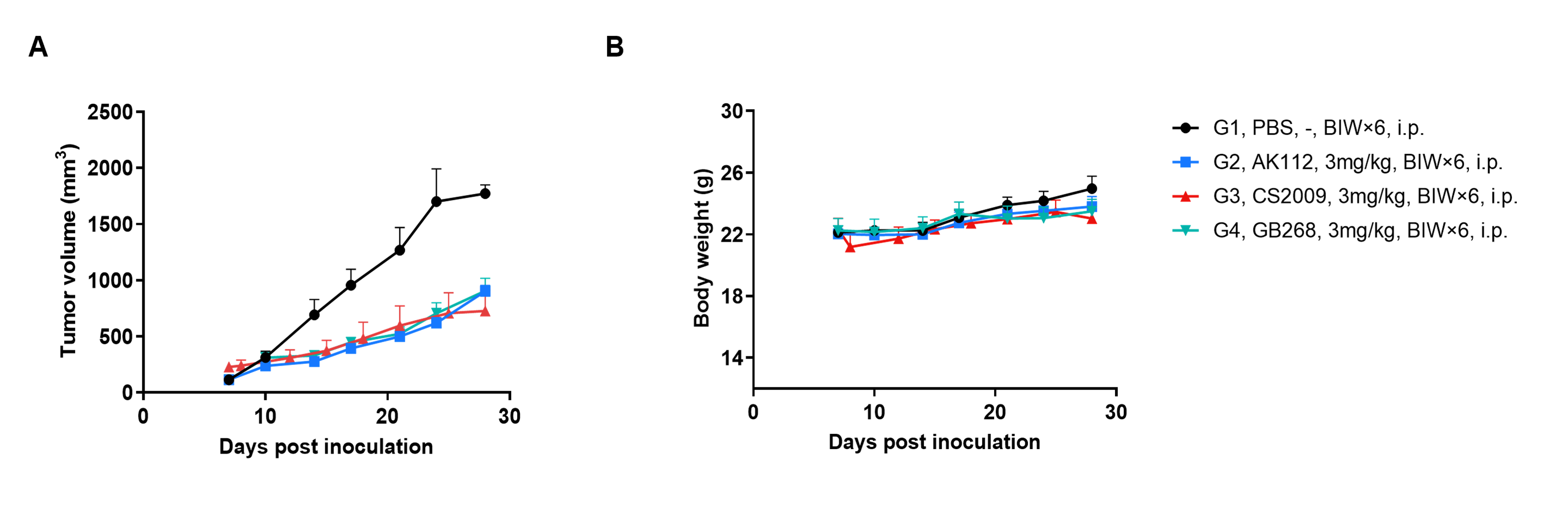
Establishment of a B-hVEGFA MC38 mouse model and in vivo efficacy evaluation of an anti-human PD-1/VEGFA/CTLA4 trispecific antibody. B-hVEGFA MC38 mouse colon carcinoma cells were subcutaneously implanted into homozygous B-hPD-1/hCTLA4 mice (female, 10 weeks old, n=4). When the mean tumor volume reached approximately 110 mm³ (AK112 and GB268 group) or 220 mm³ (CS2009 group), mice were randomized and treated intraperitoneally with either the anti-PD-1/VEGFA bispecific antibody (AK112) and anti-PD-1/VEGFA/CTLA4 trispecific antibody (CS2009 or GB268). The all antibodies were provided by the client, and the experiment was conducted in collaboration with the client.
Efficacy Evaluation of Anti-PD-1/VEGFA/CTLA4 Tri-specific Abs in the Subcutaneous B-hVEGFA MC38 Model in B-hPD-1/hCTLA4 Mice
Efficacy of an anti-human PD-1/VEGFA/CTLA4 trispecific antibody in B-hPD-1/hCTLA4 mice. (A) Tumor growth curves. (B) Body weight changes. As shown in panel A, anti-PD-1/VEGFA bispecific antibody (AK112) and anti-PD-1/VEGFA/CTLA4 trispecific antibody (CS2009 or GB268) that shown in the panel were efficacious in controlling tumor growth in B-hPD-1/hCTLA4 mice, demonstrating that the B-hPD-1/hCTLA4 mice provide a powerful preclinical model for in vivo evaluation of anti-PD-1/VEGFA/CTLA4 Tri-specific antibodies. The antibodies were provided by the client, and the experiment was conducted in collaboration with the client. Values are expressed as mean ± SEM.
* When publishing results obtained using this animal model, please acknowledge the source as follows: The animal model [B-hPD-1/hCTLA4 mice] (Cat# 120519) was purchased from Biocytogen.