mRNA Expression by RT-PCR
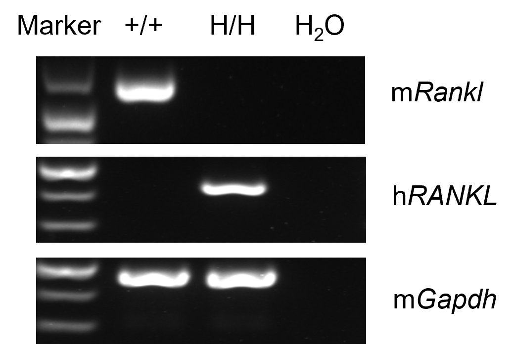
- Mouse Rankl mRNA was detectable only in wild-type C57BL/6JNIfdc mice.
- Human RANKL mRNA was detectable only in homozygous B-hRANKL mice but not in wild-type mice.
Strain specific analysis of RANKL mRNA expression in wild-type C57BL/6 mice and homozygous B-hRANKL mice by RT-PCR. Thymus RNA was isolated from wild-type C57BL/6 mice (+/+) and homozygous B-hRANKL mice (H/H), then cDNA libraries were synthesized by reverse transcription, followed by PCR with mouse or human RANKL primers. Mouse Rankl mRNA was detectable only in wild-type C57BL/6JNIfdc mice. Human RANKL mRNA was detectable only in homozygous B-hRANKL mice but not in wild-type mice.
RANKL Protein Expression Analysis
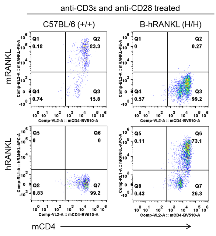
- Mouse RANKL was detectable only in wild-type C57BL/6 mice.
- Human RANKL was exclusively detectable in homozygous B-hRANKL mice but not in wild-type mice.
Strain specific analysis of RANKL expression in wild-type C57BL/6 and homozygous B-hRANKL mice by flow cytometry. CD4+ T cells were isolated from the spleen of wild-type C57BL/6 mice (+/+) and homozygous B-hRANKL mice (H/H), and then were stimulated with anti-mCD3ε (2 μg/mL) and anti-mCD28 antibodies (5 μg/mL) in vitro. After two days culture, RANKL expression was analyzed by flow cytometry with species-specific anti-RANKL antibody (anti-mouse RANKL antibody, Biolegend, 510005; anti-human RANKL antibody, Biolegend, 347507). Mouse RANKL was detectable only in wild-type C57BL/6 mice. Human RANKL was exclusively detectable in homozygous B-hRANKL mice but not in wild-type mice.
Analysis of Leukocyte Subpopulations
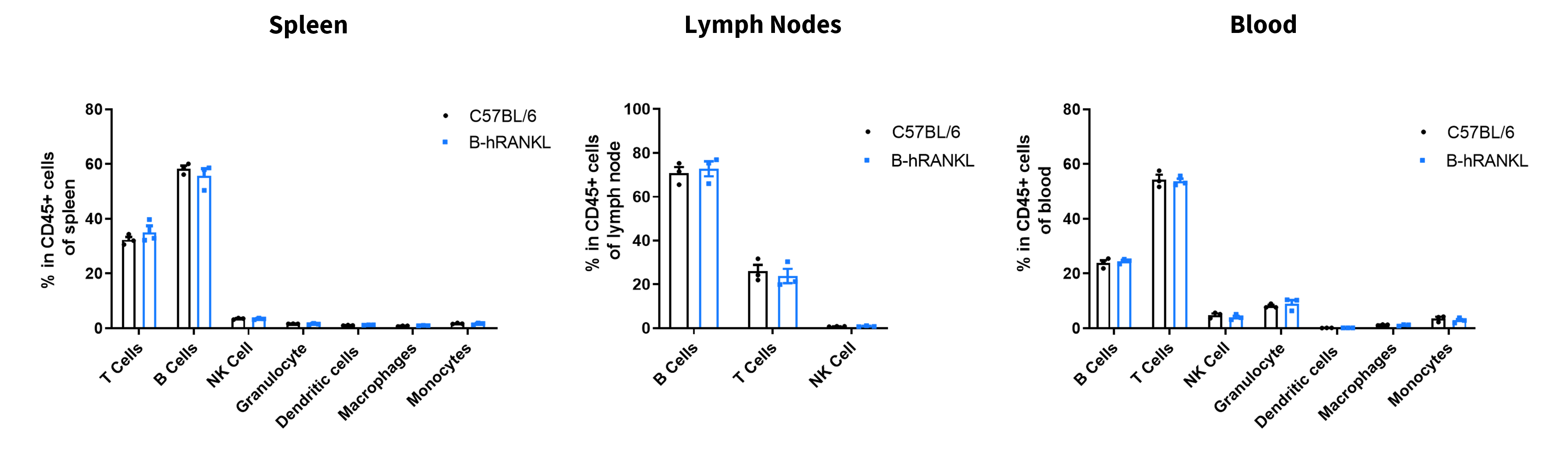
- The frequencies of B cells, T cells, NK cells, granulocytes, DCs, macrophages and monocytes in homozygous B-hRANKL mice were similar to those in C57BL/6 mice, demonstrating that humanization of RNAKL does not change the frequency or distribution of these cell types in spleen, lymph node and blood.
Analysis of leukocyte subpopulation by flow cytometry in immune organs. Spleen, lymph nodes and blood were isolated from wild-type C57BL/6 mice and homozygous B-hRANKL mice (female, 6-week-old, n=3). Single live cells were gated on the CD45+ population and analyzed by flow cytometry as indicated. Values are expressed as mean ± SEM.
Analysis of T Cell Subpopulations
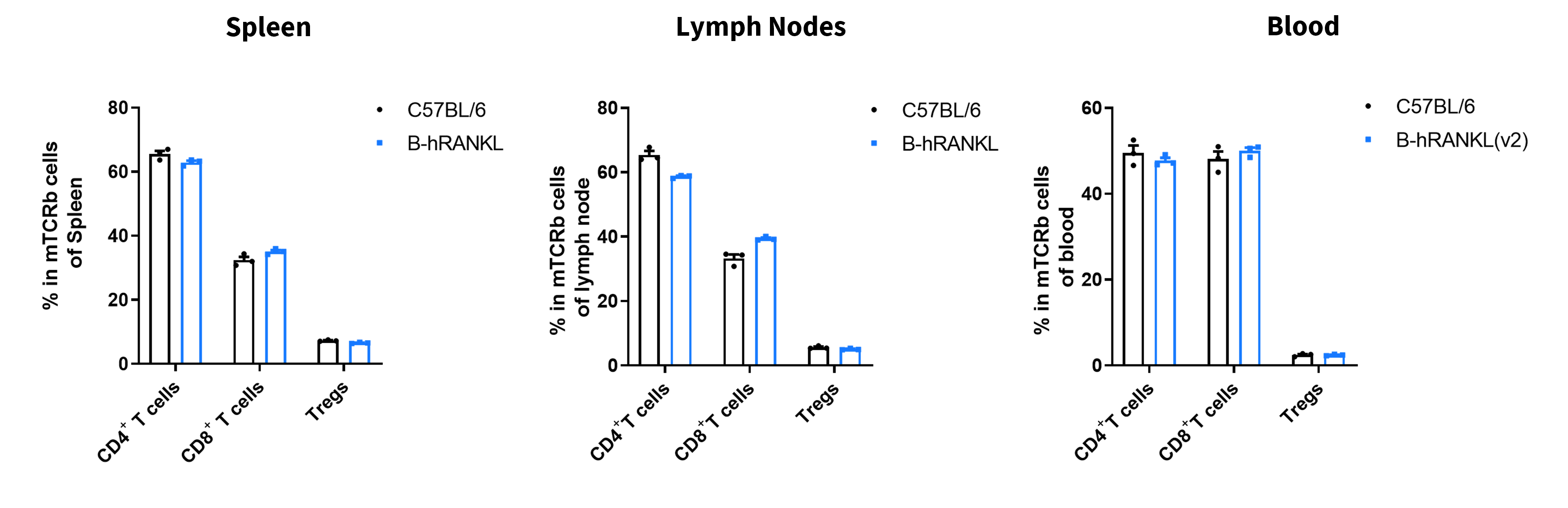
- The frequencies of CD4+ T, CD8+ T and Tregs in homozygous B-hRANKL mice were comparable to those in C57BL/6 mice, demonstrating that introduction of human RANKL in place of its mouse counterpart does not change the overall development, differentiation of distribution of these T cell sub types in spleen, lymph nodes and blood.
Analysis of T cell subpopulation by flow cytometry in immune organs. Spleen, lymph nodes and blood were isolated from wild-type C57BL/6 and homozygous B-hRANKL mice(female, 6-week-old, n=3). Single live cells were gated on the TCRb+ T cell population and analyzed by flow cytometry as indicated. Values are expressed as mean ± SEM.
Antibody Binding Assay
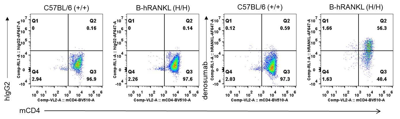
- Anti-hRANKL antibody denosumab (in house) can bind CD4+ T cells from B-hRANKL mice, but can not bind the cells from wild-type C57BL/6 mice.
Antibody binding analysis in homozygous B-hRANKL mice. CD4+ T cells were isolated from the spleen of wild-type C57BL/6 mice (+/+) and homozygous B-hRANKL mice (H/H), and then were stimulated with anti-mCD3ε (2 μg/mL) and anti-mCD28 antibodies (5 μg/mL) in vitro. After two days culture, single live cells were gated for CD4+ population and used for further analysis. Anti-hRANKL antibody denosumab (in house) can bind CD4+ T cells from B-hRANKL mice, but can not bind the cells from wild-type C57BL/6 mice.
mTRAcP-5b Expression Analysis
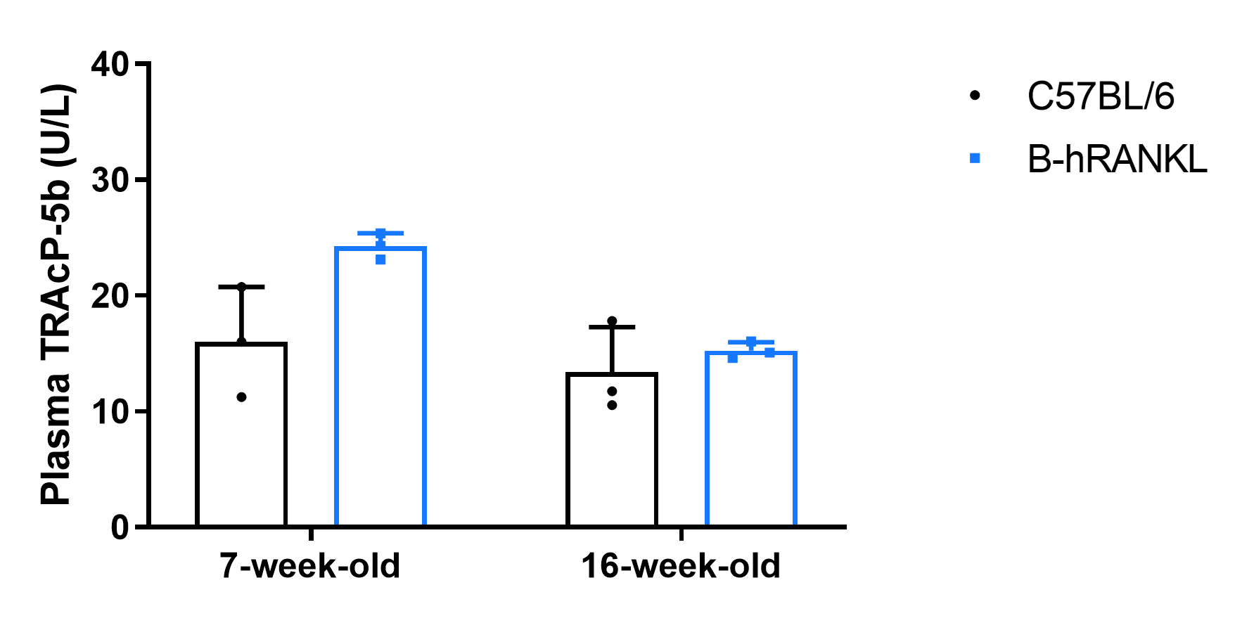
- Mouse TRAcP-5b concentration in plasma were comparable between homozygous B-hRANKL mice and wild-type mice.
Mouse TRAcP-5b concentration assay in homozygous B-hRANKL mice by ELISA. Plasma was collected from wild-type C57BL/6 mice (+/+) and homozygous B-hRANKL mice (H/H) (n=3 per group, 7-week-old and 16-week-old), and then were analyzed by ELISA with mouse TRAcP-5b ELISA kit (IDS, SB-TR103). At both age time point, concentrations of mouse TRAcP-5b were comparable between homozygous B-hRANKL mice and wild-type mice. Values are expressed as mean ± SEM.
In vivo efficacy for anti-human RNAKL antibody with mouse osteoporosis model
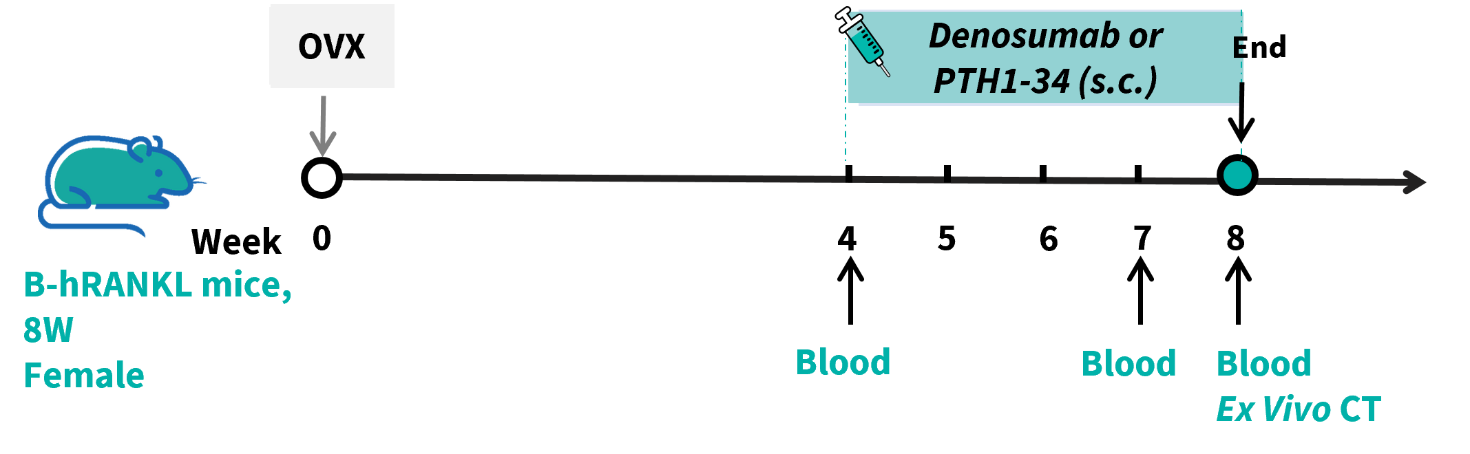
Ovariectomy induced osteoporosis in B-hRANKL mice. Mice were first randomly separated to receive ovariectomy or sham surgery. The ovariectomized mice were then randomly divided into 3 treatment groups (OVX, Denosumab and PTH1-34) at 4 weeks after surgery (n = 6). Mice were sacrificed at 4 weeks after treatment. Serum samples were collected at weeks 4, 7 and 8 to detect CTX-1 and osteocalcin. After sacrifice, proximal tibias were harvested for ex vivo micro-CT analysis of bone mineral density.
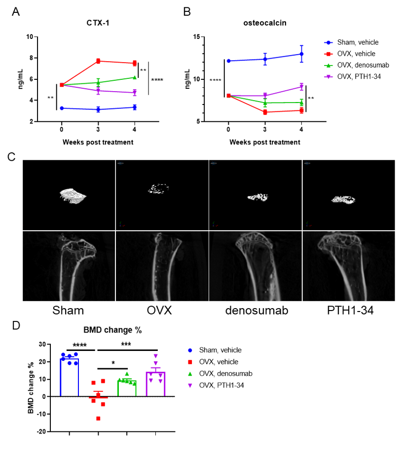
Treatment with in-house anti-human RANKL antibody denosumab exerted therapeutic effects on osteoporosis in ovariectomized B-hRANKL mice. The concentration of serum bone resorption marker CTX-1 (A) and serum bone formation marker OC (B). (C) Representative micro-CT images of proximal tibias. (D) The BMD change of the proximal tibia. Compared with the sham-operated group, the OVX vehicle group exhibited elevated serum CTX-1levels, accompanied by significant reductions in serum OC concentration and BMD, confirming the successful establishment of an osteoporosis model in B-hRANKL mice. Relative to the untreated OVX group, intervention with either denosumab or PTH1-34 significantly lowered circulating CTX-1 levels, whereas serum OC levels and tibial BMD percentage change were both substantially restored. These results indicated that the anti-human RANKL antibody could effectively ameliorates osteoporosis in B-hRANKL mice. The B-hRANKL mice serve as a powerful preclinical mouse model for in vivo evaluating efficacy of anti-human RANKL antibodies. Values are expressed as mean ± SEM.
Note: Data are shared upon the client's approval.
* When publishing results obtained using this animal model, please acknowledge the source as follows: The animal model [B-hRANKL mice] (Cat# 111072) was purchased from Biocytogen.