

C57BL/6-Tfrctm1(TFRC)Bcgen Slc3a2tm1(SLC3A2)Bcgen/Bcgen • 113596

Background:
Verification:
Key Advantages
Applications

TFR1 humanization (B-hTFR1 mice):
CD98HC humanization (B-hCD98HC mice):

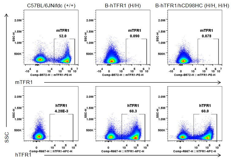
Strain-specific TFR1 expression was analyzed by flow cytometry in bone marrow erythrocytes from wild-type C57BL/6JNifdc mice (+/+), homozygous TFR1 humanized mice (H/H), and homozygous TFR1/CD98HC humanized mice (H/H, H/H). Bone marrow cells were analyzed with anti-mouse TFR1 antibody (BioLegend, 113808) and anti-human TFR1 antibody (BioLegend, 334108). Mouse TFR1 was detected only in erythrocytes of wild-type mice, whereas humanized TFR1 was exclusively detected in erythrocytes of homozygous TFR1 humanized mice and TFR1/CD98HC humanized mice, but not in wild-type mice.
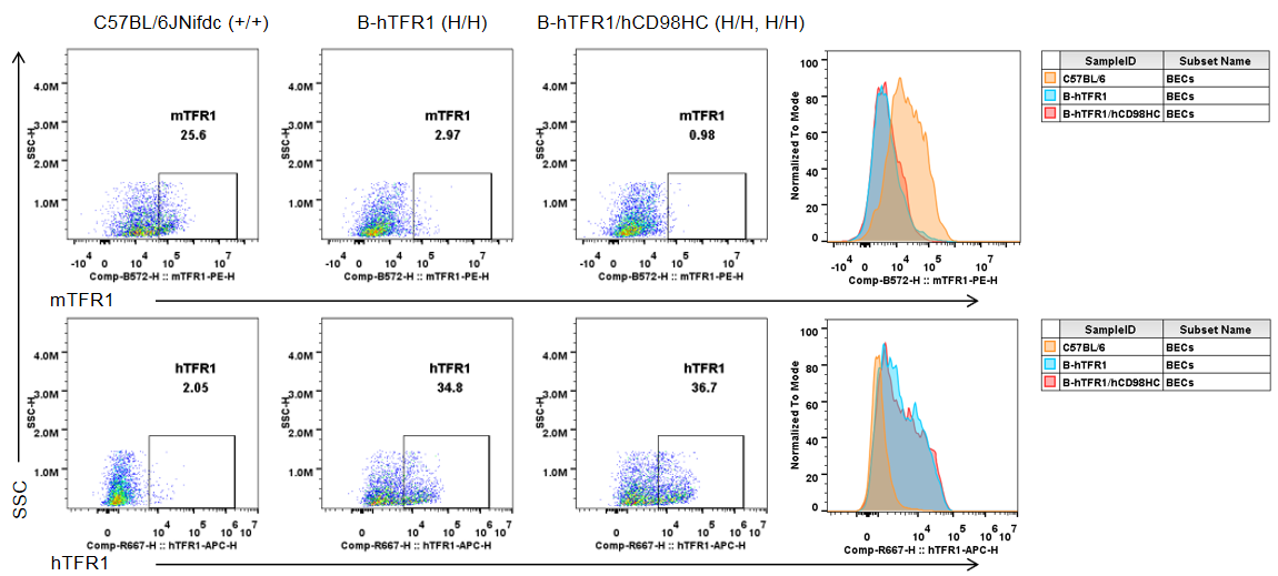
Brain cells were collected from wild-type C57BL/6JNifdc mice (+/+), homozygous TFR1 humanized mice (H/H), and homozygous TFR1/CD98HC humanized mice (H/H, H/H). Flow cytometry was performed using anti-mouse TFR1 antibody (BioLegend, 113808) and anti-human TFR1 antibody (BioLegend, 334108). Mouse TFR1 was detectable only in brain endothelial cells of wild-type mice, whereas humanized TFR1 was exclusively detected in brain endothelial cells of homozygous TFR1 humanized mice and TFR1/CD98HC humanized mice.
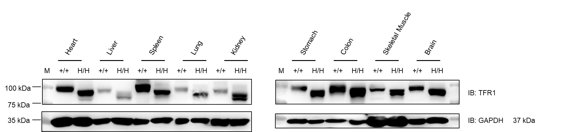
Western blot analysis was performed on tissue lysates from heart, liver, spleen, lung, kidney, stomach, colon, muscle, and brain of wild-type C57BL/6JNifdc mice (+/+) and homozygous TFR1 humanized mice (H/H). Samples were probed with anti-TFR1 antibody (Abcam, ab214039), which is cross-reactive between mouse and human TFR1. TFR1 protein was detectable in all examined tissues from both genotypes. GAPDH was used as a loading control. M: marker. Total protein loaded per lane: 40 μg.

Protein expression of TFR1 in the spinal cord, cerebral cortex, hippocampus, and cerebellum was analyzed by western blot in wild-type C57BL/6JNifdc mice (+/+) and homozygous TFR1/CD98HC humanized mice (H/H). Tissue lysates were probed with anti-transferrin receptor antibody (Abcam, ab214039), which is cross-reactive between species. TFR1 was detectable in CNS tissues from both wild-type and humanized mice. (A) Male. (B) Female. Total protein loaded per lane: 40 μg.
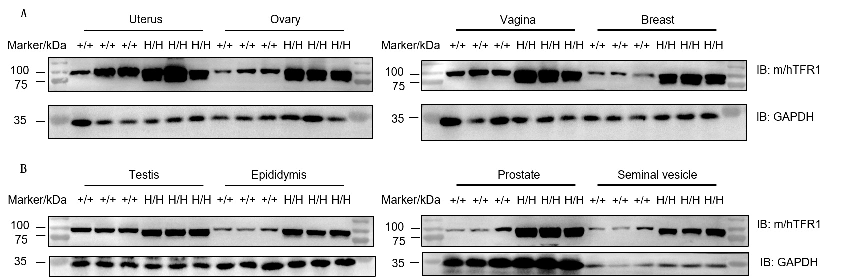
Western blot analysis of uterus, ovary, vagina, breast, testis, epididymis, prostate, and seminal vesicle was performed in wild-type C57BL/6JNifdc mice (+/+) and homozygous TFR1/CD98HC humanized mice (H/H) using anti-transferrin receptor antibody (Abcam, ab214039). TFR1 protein was detectable in all examined reproductive organs from both genotypes due to antibody cross-reactivity. Total protein loaded per lane: 30 μg.
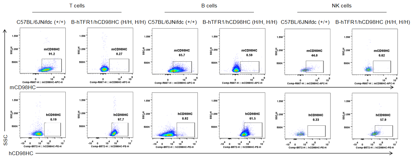
Strain specific CD98HC expression analysis in wild-type C57BL/6JNifdc and homozygous B-hTFR1/hCD98HC mice by flow cytometry. Blood cells were collected from wild-type C57BL/6JNifdc (+/+) and homozygous B-hTFR1/hCD98HC mice (H/H, H/H) and analyzed by flow cytometry with anti-mouse CD98HC antibody (Biolegend, 128211) and anti-human CD98HC antibody (231161-CD98BBBB-h1.L produced in-house).
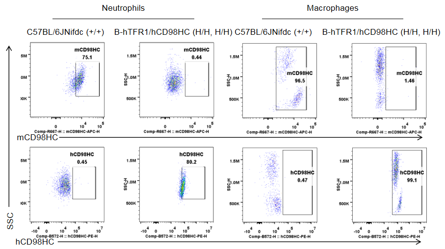
Strain specific CD98HC expression analysis in wild-type C57BL/6JNifdc and homozygous B-hTFR1/hCD98HC mice by flow cytometry. Blood cells were collected from wild-type C57BL/6JNifdc (+/+) and homozygous B-hTFR1/hCD98HC mice (H/H, H/H) and analyzed by flow cytometry with anti-mouse CD98HC antibody (Biolegend, 128211) and anti-human CD98HC antibody (231161-CD98BBBB-h1.L produced in-house).
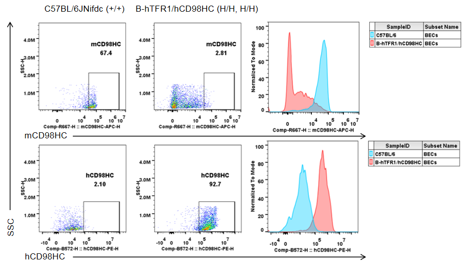
Strain specific CD98HC expression in wild-type C57BL/6JNifdc and homozygous B-hTFR1/hCD98HC mice by flow cytometry. Brain cells were collected from wild-type C57BL/6JNifdc (+/+) and homozygous B-hTFR1/hCD98HC mice (H/H, H/H) and analyzed by flow cytometry with anti-mouse CD98HC antibody (Biolegend, 128211) and anti-human CD98HC antibody (231161-CD98BBBB-h1.L produced in-house).
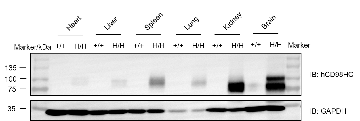
Western blot analysis of hCD98HC protein expression in homozygous B-hCD98HC mice.
Various tissue lysates were collected from wild-type C57BL/6 and homozygous B-hCD98HC mice (H/H), and then analyzed by western blot with species-specific anti-human CD98 antibody (Abcam, ab307587). 50 μg total proteins were loaded for western blotting analysis. M, marker.
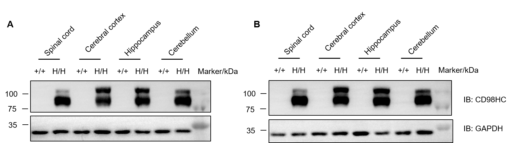
Protein expression analysis of CD98HC in homozygous B-hTFR1/hCD98HC mice. Various tissue lysates were collected from wild-type C57BL/6JNifdc mice (+/+) and homozygous B-hTFR1/hCD98HC mice (H/H), and then analyzed by western blot with anti-CD98 antibody (abcam, ab307587). 40 μg total protein was loaded for western blotting analysis. (A) Male. (B) Female.
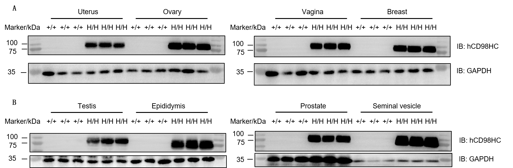
Protein expression analysis of CD98HC in homozygous B-hTFR1/hCD98HC mice. Various tissue lysates were collected from wild-type C57BL/6JNifdc mice (+/+) and homozygous B-hTFR1/hCD98HC mice (H/H), and then analyzed by western blot with anti-CD98 antibody (Abcam, ab307587). 30 μg total protein was loaded for western blotting analysis.
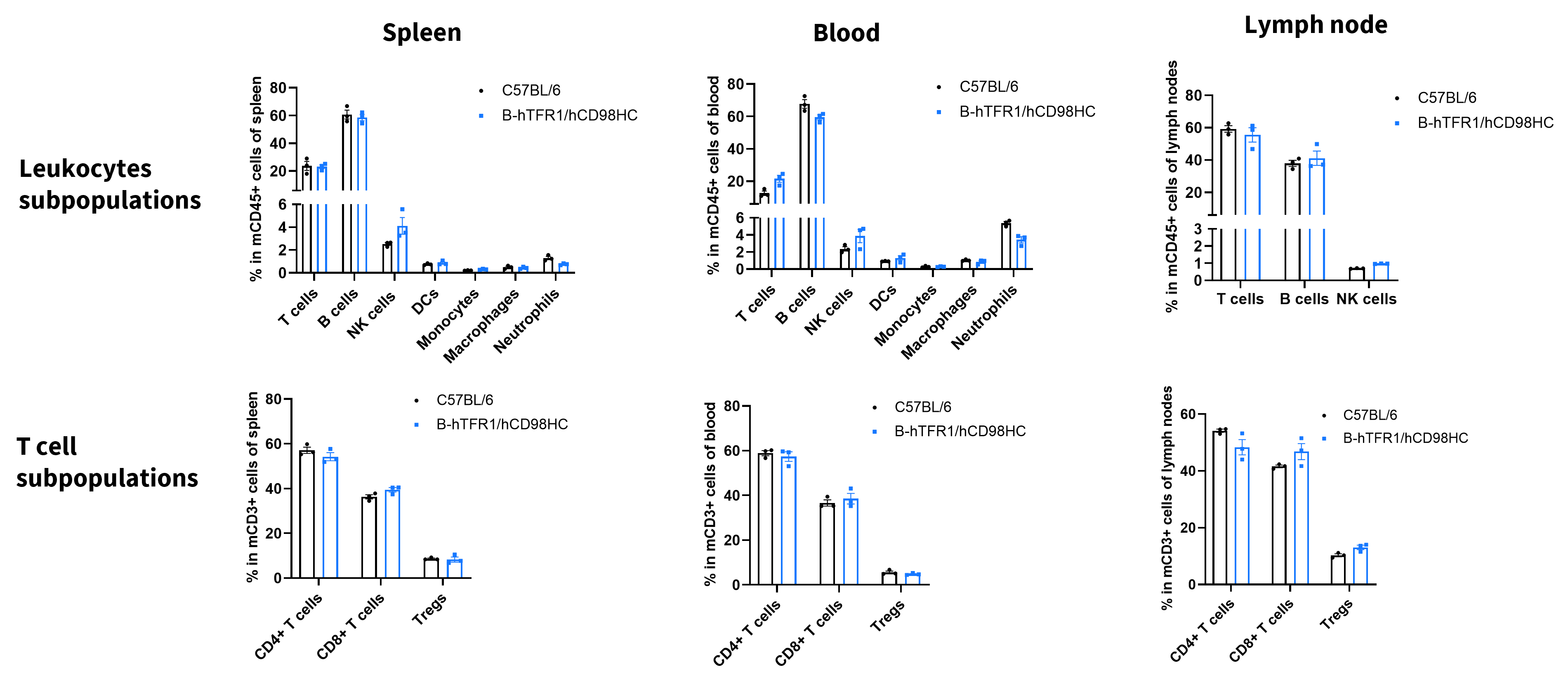
Values are expressed as mean ± SD.

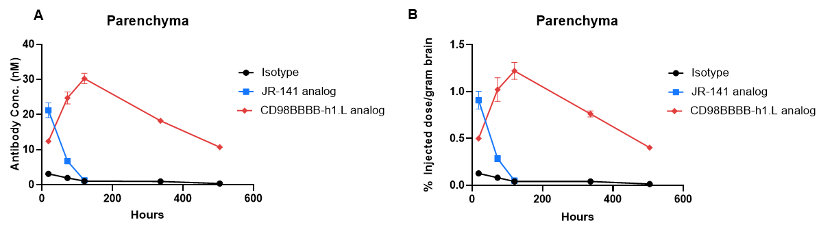
In vivo PK evaluation and comparison of anti-human TFR1 and anti-CD98HC antibody. B-hTFR1/hCD98HC mice (n=2, female, 8-week-old) were injected with control IgG (10 mpk) anti-human TFR1 antibody (TFR1 Ab, JR-141 analog, monovalent, produced in house, 12.56 mpk) and anti-human CD98HC antibody (CD98HC Ab, CD98BBBB-h1.L analog, monovalent, produced in house, 13.3 mpk) via tail vein. Brain were taken for in vivo PK evaluation after dosing 18 h and 3, 5, 14, 21 days. Brain concentrations (A) and % of injection/gram brain (B) were quantified. As shown in panel, anti-human TFR1 antibody exhibited higher brain exposure in 24 h after dose, while anti-CD98HC antibody exhibited higher brain exposure in 72 h after dose. The results confirmed that B-hTFR1/hCD98HC mice enables uptake of an intravenously administered anti-human TFR1 antibody or anti-human CD98HC antibody, and this mice can be used for the comparison of penetration efficacy of shuttle molecules targeting TFR1 or CD98HC. Graphs represent mean ± SEM.
