

C57BL/6-Tnfsf15tm2(TNFSF15)Bcgen Itga4tm1(ITGA4)Bcgen Itgb7tm1(ITGB7)Bcgen/Bcgen • 113037
on this page

TL1A: A key inflammation cytokine in chronic intestinal inflammation and fibrosis-related diseases
α4β7: A central pathway for directing lymphocyte migration to the gut and other mucosal sites during inflammation

TL1A
ITGA4
ITGB7

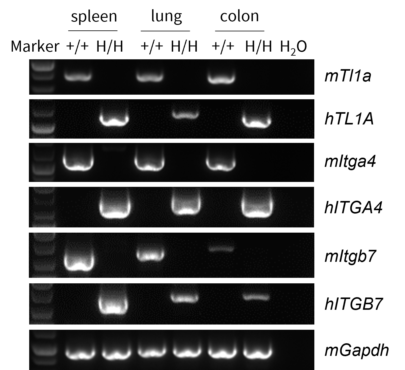
Species specific analysis of TL1A, ITGA4 and ITGB7 gene expression in wild-type C57BL/6JNifdc mice and homozygous B-hTL1A/hα4β7 mice by RT-PCR. Spleen, Colon and lung were collected from wild-type C57BL/6JNifdc mice (+/+) and homozygous B-hTL1A/hα4β7 mice (H/H;H/H;H/H). Mouse Tl1a, Itga4 and Itgb7 mRNA were detectable only in wild-type C57BL/6JNifdc mice. Human TL1A, ITGA4, and ITGB7 mRNA were detectable only in homozygous B-hTL1A/hα4β7 mice, but not in wild-type C57BL/6JNifdc mice.
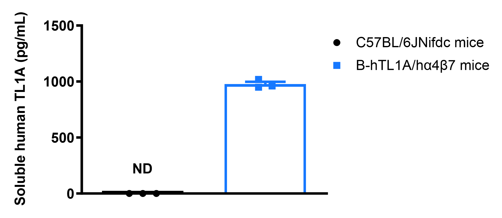
Soluble TL1A expression analysis in B-hTL1A/hα4β7 mice by ELISA. Bone marrow derived dendritic cells (BMDC) were produced by culturing the bone marrow from wild-type C57BL/6JNifdc mice (+/+) and homozygous B-hTL1A/hα4β7 mice (H/H;H/H;H/H), which were stimulated with LPS in vitro. After stimulation, the supernatants were collected and the level of soluble TL1A was analyzed by ELISA. Soluble human TL1A was exclusively detectable in homozygous B-hTL1A/hα4β7 mice, but not in wild-type C57BL/6JNifdc mice. Values are expressed as mean ± SEM. ND: not detectable.
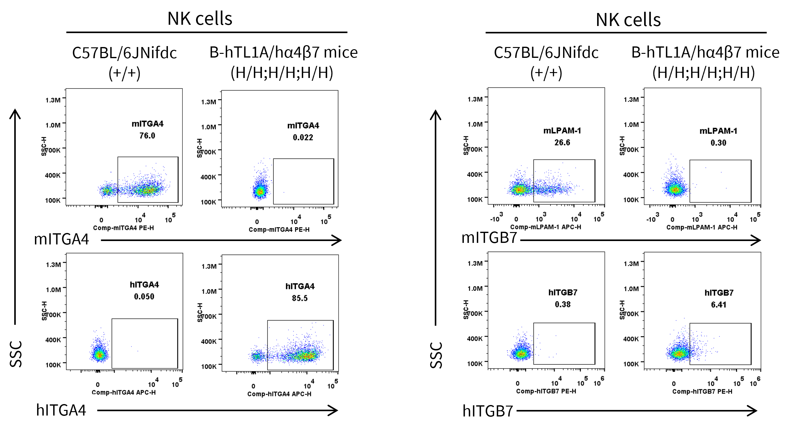
Strain specific ITGA4 and ITGB7 expression analysis in wild-type C57BL/6JNifdc mice and homozygous humanized B-hTL1A/hα4β7 mice by flow cytometry. Splenocytes were collected from wild-type C57BL/6JNifdc mice (+/+) and homozygous B-hTL1A/hα4β7 mice (H/H;H/H;H/H). Protein expression was analyzed with anti-mouse ITGA4 antibody (Biolegend, 103705), anti-mouse ITGB7 antibody (Biolegend, 120607), anti-human ITGA4 antibody (Biolegend, 304307) anti-human ITGB7 antibody (Invitrogen, MA5-23541) by flow cytometry. Mouse ITGA4 and ITGB7 were only detectable in wild-type C57BL/6JNifdc mice. Human ITGA4 and ITGB7 were exclusively detectable in homozygous B-hTL1A/hα4β7 mice, but not in wild-type C57BL/6JNifdc mice.
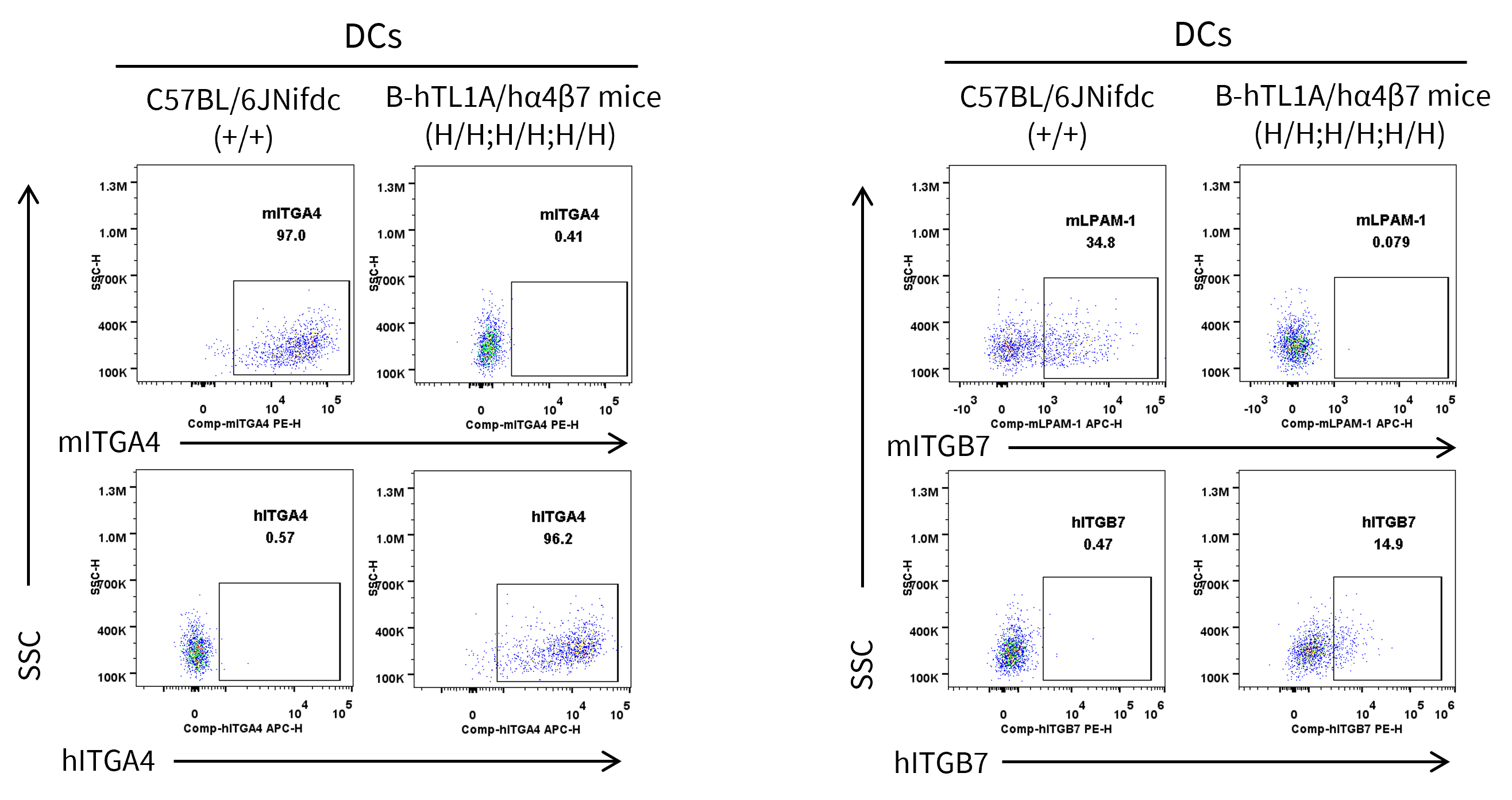
Strain specific ITGA4 and ITGB7 expression analysis in wild-type C57BL/6JNifdc mice and homozygous humanized B-hTL1A/hα4β7 mice by flow cytometry. Splenocytes were collected from wild-type C57BL/6JNifdc mice (+/+) and homozygous B-hTL1A/hα4β7 mice (H/H;H/H;H/H). Protein expression was analyzed with anti-mouse ITGA4 antibody (Biolegend, 103705), anti-mouse ITGB7 antibody (Biolegend, 120607), anti-human ITGA4 antibody (Biolegend, 304307) anti-human ITGB7 antibody (Invitrogen, MA5-23541) by flow cytometry. Mouse ITGA4 and ITGB7 were only detectable in wild-type C57BL/6JNifdc mice. Human ITGA4 and ITGB7 were exclusively detectable in homozygous B-hTL1A/hα4β7 mice, but not in wild-type C57BL/6JNifdc mice.
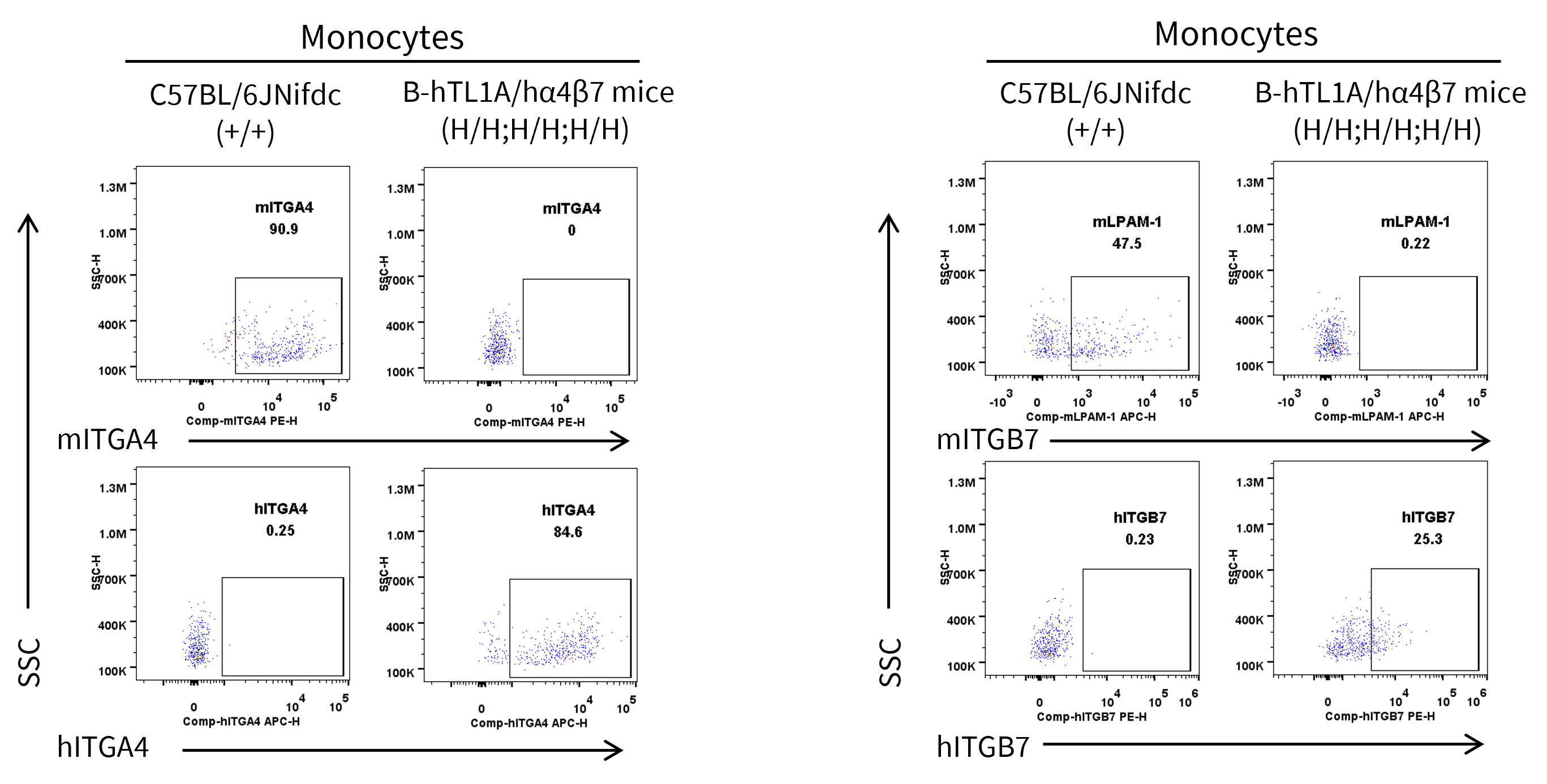
Strain specific ITGA4 and ITGB7 expression analysis in wild-type C57BL/6JNifdc mice and homozygous humanized B-hTL1A/hα4β7 mice by flow cytometry. Splenocytes were collected from wild-type C57BL/6JNifdc mice (+/+) and homozygous B-hTL1A/hα4β7 mice (H/H;H/H;H/H). Protein expression was analyzed with anti-mouse ITGA4 antibody (Biolegend, 103705), anti-mouse ITGB7 antibody (Biolegend, 120607), anti-human ITGA4 antibody (Biolegend, 304307) anti-human ITGB7 antibody (Invitrogen, MA5-23541) by flow cytometry. Mouse ITGA4 and ITGB7 were only detectable in wild-type C57BL/6JNifdc mice. Human ITGA4 and ITGB7 were exclusively detectable in homozygous B-hTL1A/hα4β7 mice, but not in wild-type C57BL/6JNifdc mice.
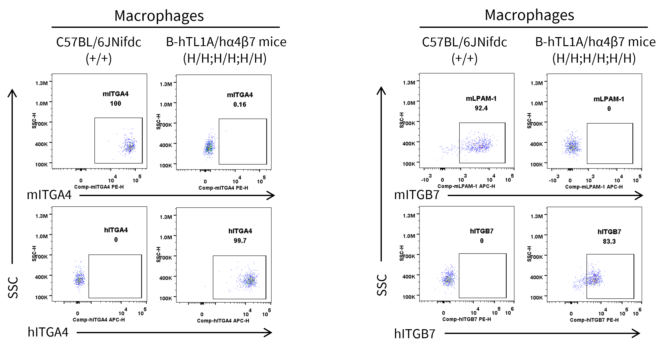
Strain specific ITGA4 and ITGB7 expression analysis in wild-type C57BL/6JNifdc mice and homozygous humanized B-hTL1A/hα4β7 mice by flow cytometry. Splenocytes were collected from wild-type C57BL/6JNifdc mice (+/+) and homozygous B-hTL1A/hα4β7 mice (H/H;H/H;H/H). Protein expression was analyzed with anti-mouse ITGA4 antibody (Biolegend, 103705), anti-mouse ITGB7 antibody (Biolegend, 120607), anti-human ITGA4 antibody (Biolegend, 304307) anti-human ITGB7 antibody (Invitrogen, MA5-23541) by flow cytometry. Mouse ITGA4 and ITGB7 were only detectable in wild-type C57BL/6JNifdc mice. Human ITGA4 and ITGB7 were exclusively detectable in homozygous B-hTL1A/hα4β7 mice, but not in wild-type C57BL/6JNifdc mice.

Analysis of leukocyte subpopulations by flow cytometry in immune organs and blood. Splenocytes, peripheral blood, and lymph nodes were isolated from female C57BL/6JNifdc and B-hTL1A/hα4β7 mice (female, 8-week-old, n = 3). Single live cells were gated on the CD45⁺ population and analyzed by flow cytometry as indicated. Values are expressed as mean ± SEM.

Analysis of T-cell subpopulations by flow cytometry in immune organs and blood. Splenocytes, peripheral blood, and lymph nodes were isolated from female C57BL/6JNifdc and B-hTL1A/hα4β7 mice (female, 8-week-old, n = 3). Single live cells were gated on the CD3⁺ T-cell population and analyzed by flow cytometry as indicated. Values are expressed as mean ± SEM.
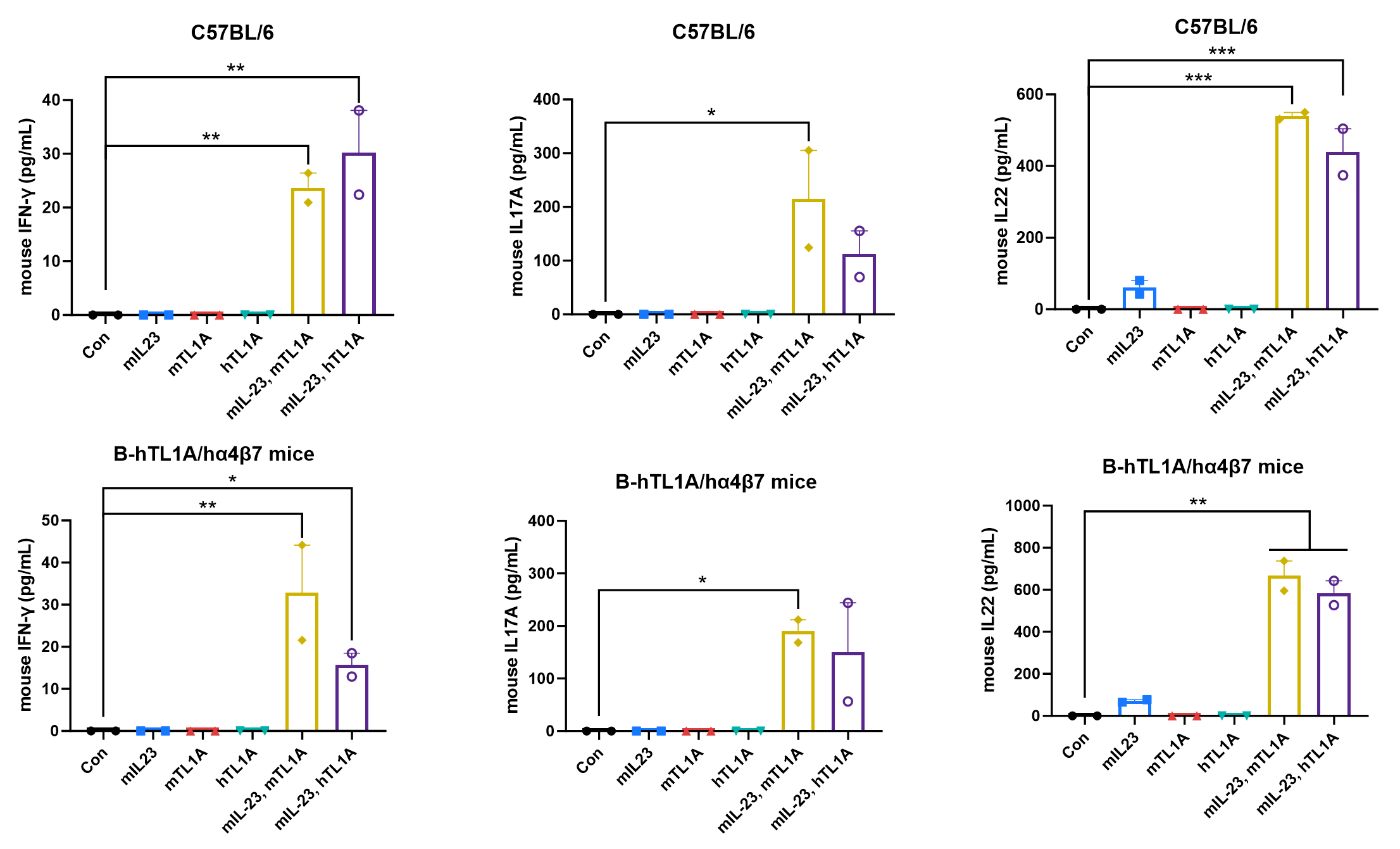
Ex vivo functional analysis in B-hTL1A/hα4β7 mice. Splenocytes were collected from wild-type C57BL/6 mice (+/+) and homozygous B-hTL1A/hα4β7 mice (H/H;H/H;H/H), then the production of mouse IFN-γ, mouse IL17A, and mouse IL22 in supernatants were assessed by ELISA after 72 h of incubation with mIL23 (10 ng/mL), mTL1A (300 ng/mL), and hTL1A (300 ng/mL) in vitro.
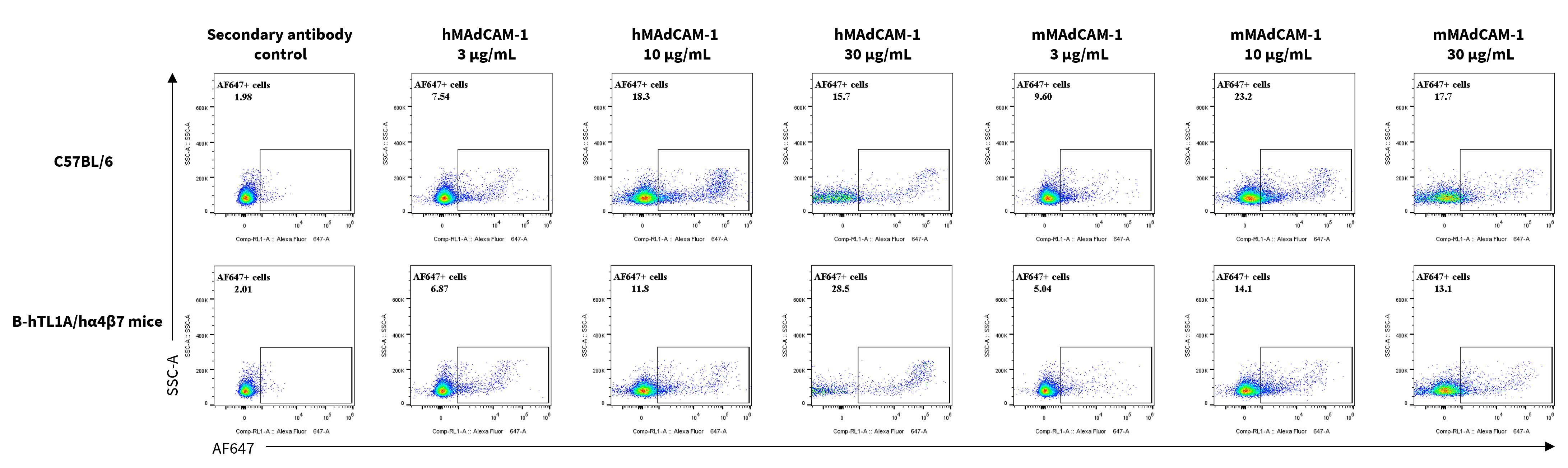
Assessment of MAdCAM-1 binding in C57BL/6 and B-hTL1A/hα4β7 mice. Splenocytes were collected from homozygous B-hTL1A/hα4β7 mice (H/H;H/H;H/H), and were incubated with human MAdCAM-1 protein (3, 10, 30 μg/mL) or mouse MAdCAM-1 protein (3, 10, 30 μg/mL) in vitro for 1 h. The binding of MAdCAM-1 to α4β7 were detected by flow cytometry.

Growth curve of wild-type C57BL/6JNifdc and B-hTL1A/hα4β7 mice. Eight-week-old mice were grouped by sex (10 males and 10 females). Body weight was measured on the same day of every week, until 13 weeks. The minimum and maximum body weights shown in the table were calculated from the mean ± SD. The growth curve of the B-hTL1A/hα4β7 mice was similar to the growth curve of C57BL/6JNifdc mice.
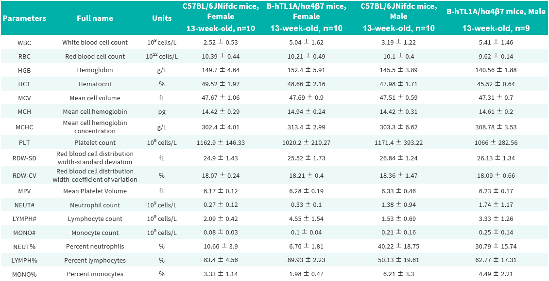
Complete blood count (CBC) of B-hTL1A/hα4β7 mice. Values are expressed as mean ± SD.
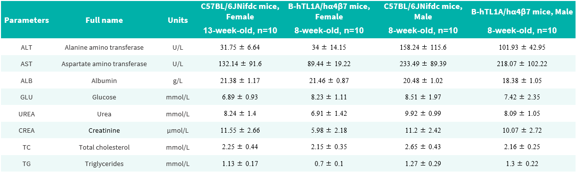
Blood biochemical parameters of B-hTL1A/hα4β7 mice are shown. Values are expressed as mean ± SD.
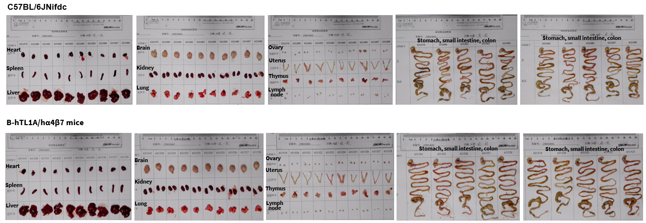
Organs of female C57BL/6JNifdc (13-week-old, n=10) and B-hTL1A/hα4β7 mice (13-week-old, n=10).
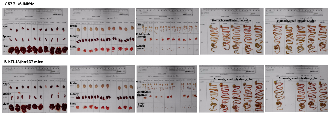
Organs of male C57BL/6JNifdc (13-week-old, n=10) and B-hTL1A/hα4β7 mice (13-week-old, n=9).
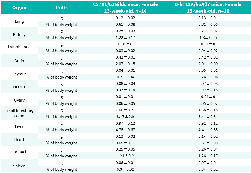
Average weight of the main organs of female C57BL/6 and B-hTL1A/hα4β7 mice.
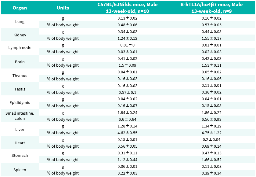
Average weight of the main organs of male C57BL/6 and B-hTL1A/hα4β7 mice.
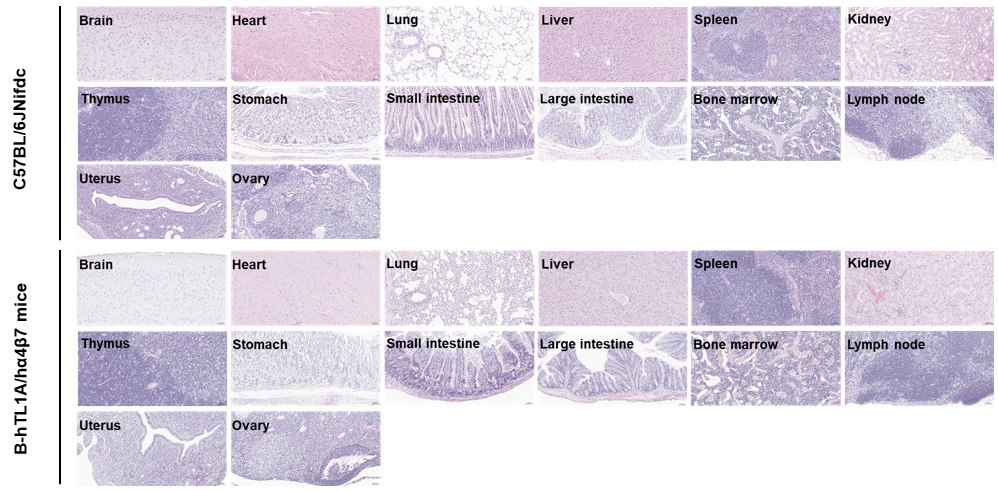
Histopathological analysis of organs in female C57BL/6JNifdc and B-hTL1A/hα4β7 mice. Major organs from C57BL/6JNifdc and B-hTL1A/hα4β7 mice were collected at 13 weeks of age and analyzed by H&E staining (female, n = 3).
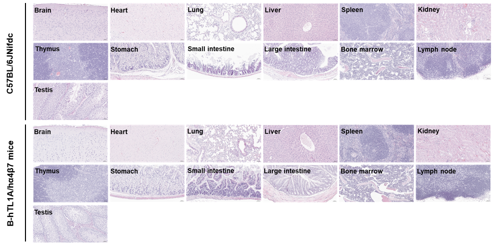
Histopathological analysis of organs in male C57BL/6JNifdc and B-hTL1A/hα4β7 mice. Major organs from C57BL/6JNifdc and B-hTL1A/hα4β7 mice were collected at 13 weeks of age and analyzed by H&E staining (female, n = 3).

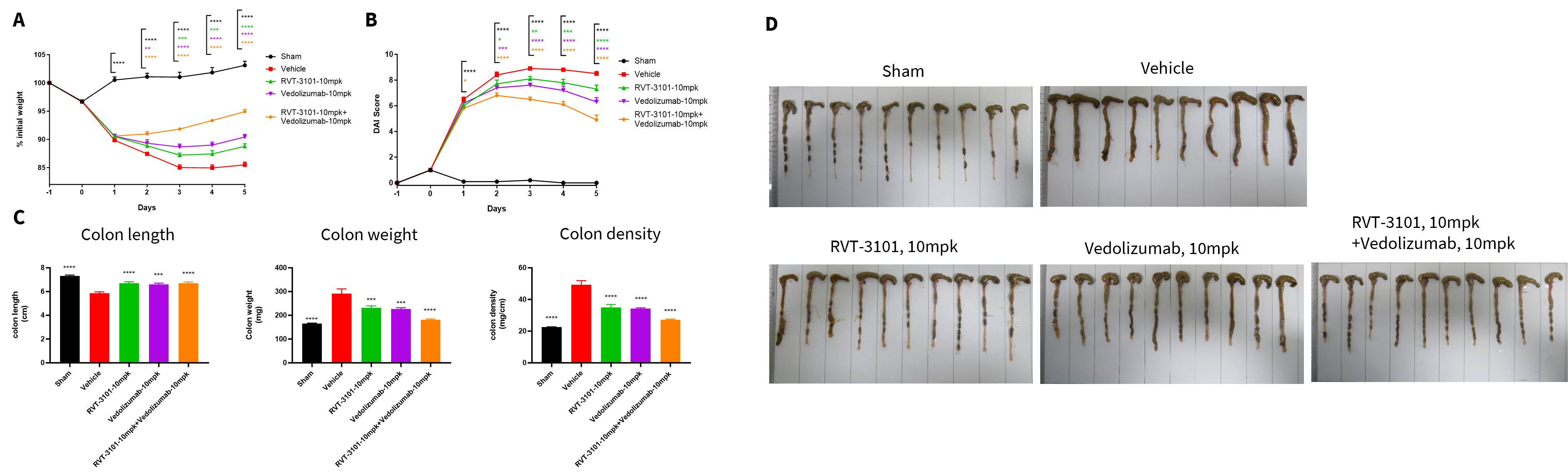
The therapeutic efficacy of anti-TL1A and anti-human α4β7 antibodies on the TNBS-induced acute colitis model in B-hTL1A/hα4β7 mice. TNBS solution was instilled into the colon lumen of B-hTL1A/hα4β7 mice (female, 8-10 weeks-old, n=8). The control group (Sham) received intrarectal injections of 50% ethanol. The treatment groups received anti-TL1A antibody RVT-3101 (10 mpk, provided by WuXi AppTec), anti-human α4β7 antibody Vedolizumab (10 mpk, provided by WuXi AppTec) alone or in combination. (A) Body weight change. (B) DAI score. (C) Colon Index. (D) Colon photo. An acute colitis disease model induced by TNBS was established in B-hTL1A/hα4β7 mice, and administration of anti-TL1A antibody RVT-3101 and anti-human α4β7 antibody Vedolizumab effectively improved TNBS-induced acute colitis, and their combination provided better efficacy. Values are expressed as mean ± SEM. *p<0.05, **p<0.01, ***p<0.001, ****p<0.0001, versus Vehicle, ANOVA.
Note: This experiment was conducted by WuXi AppTec using B-hTL1A/hα4β7 mice.
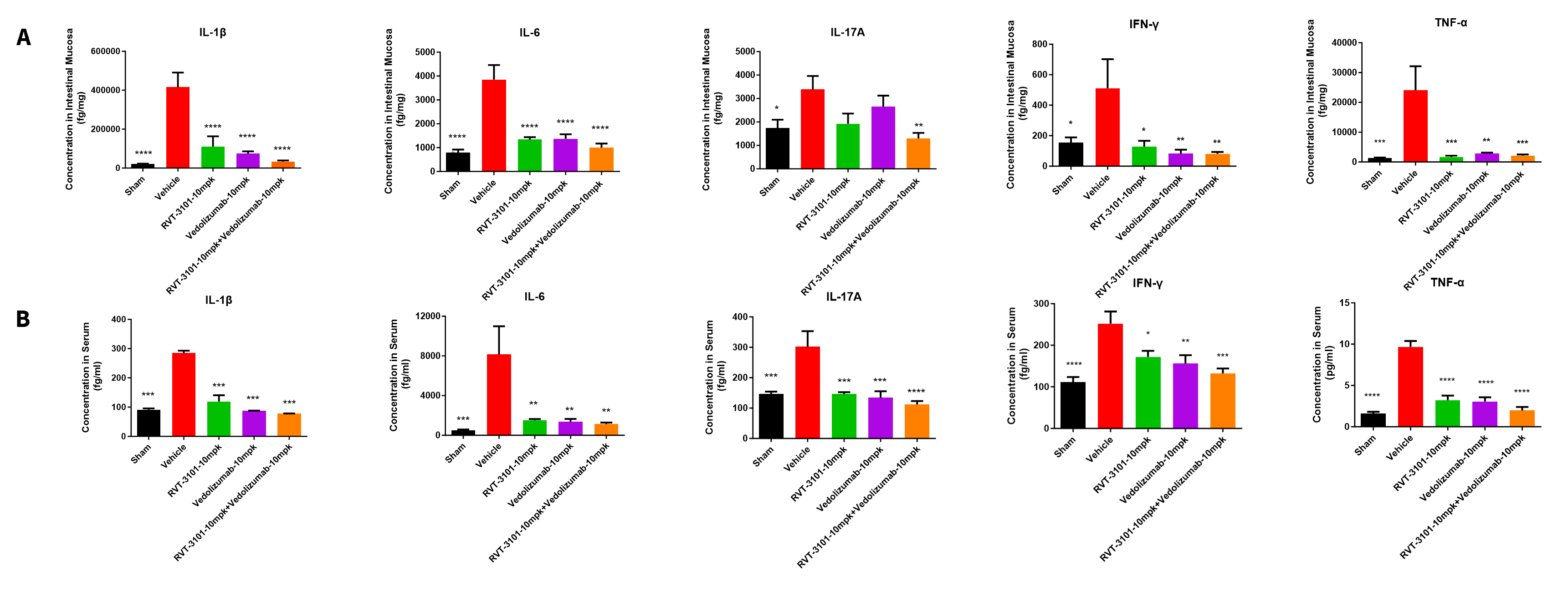
Analysis of inflammatory cytokines on the TNBS-induced acute colitis model in B-hTL1A/hα4β7 mice. TNBS solution was instilled into the colon lumen of B-hTL1A/hα4β7 mice (female, 8-10 weeks-old, n=8). The control group (Sham) received intrarectal injections of 50% ethanol. The treatment groups received anti-TL1A antibody RVT-3101 (10 mpk, provided by WuXi AppTec), anti-human α4β7 antibody Vedolizumab (10 mpk, provided by WuXi AppTec) alone or in combination. (A) The concentrations of IL-1β, IL-6, IL-17A, IFN-γ, TNF-α in intestinal mucosa. (B) The concentrations of IL-1β, IL-6, IL-17A, IFN-γ, TNF-α in serum. Administration of anti-TL1A antibody RVT-3101 and anti-human α4β7 antibody Vedolizumab markedly reduced the production of inflammatory cytokines in TNBS-induced acute colitis mice, and their combination provided better efficacy. The results indicate that B-hTL1A/hα4β7 mice are a powerful tool for evaluating in vivo efficacy of the combination of anti-TL1A antibody and anti-human α4β7 antibody. Values are expressed as mean ± SEM. *p<0.05, **p<0.01, ***p<0.001, ****p<0.0001, versus Vehicle, ANOVA.
Note: This experiment was conducted by WuXi AppTec using B-hTL1A/hα4β7 mice.
