

C57BL/6-Cd3etm1(CD3E)Bcgen Cd3dtm1(CD3D)Bcgen Cd3gtm1(CD3G)Bcgen Cd8atm1(CD8A)Bcgen Cd8btm1(CD8B)Bcgen/Bcg • 114717
on this page


CD3EDG
CD8

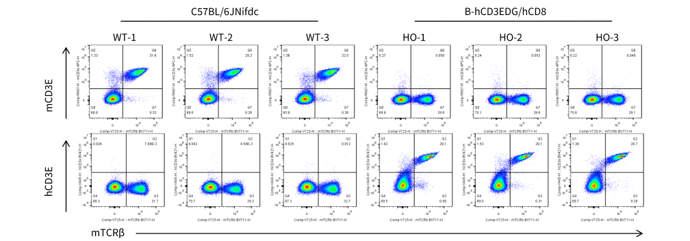
Strain specific CD3E expression analysis in homozygous B-hCD3EDG/hCD8 mice by flow cytometry. Splenocytes were collected from wild-type C57BL/6JNifdc mice (WT) and homozygous B-hCD3EDG/hCD8 mice (HO), and analyzed by flow cytometry with species-specific anti-mouse CD3ε antibody (Biolegend, 100312) and anti-human CD3ε antibody (BD Pharmingen, 562426).
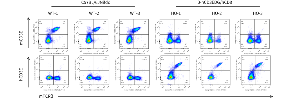
Strain specific CD3E expression analysis in homozygous B-hCD3EDG/hCD8 mice by flow cytometry. Blood was collected from wild-type C57BL/6JNifdc mice (WT) and homozygous B-hCD3EDG/hCD8 mice (HO), and analyzed by flow cytometry with species-specific anti-mouse CD3ε antibody (Biolegend, 100312) and anti-human CD3ε antibody (BD Pharmingen, 562426).
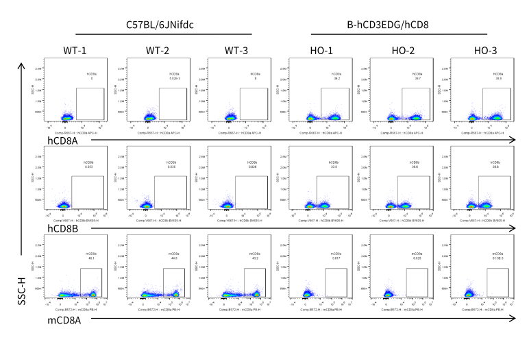
Strain specific CD8 expression analysis in homozygous B-hCD3EDG/hCD8 mice by flow cytometry. Splenocytes were collected from wild-type C57BL/6JNifdc mice (WT) and homozygous B-hCD3EDG/hCD8 mice (HO), and then analyzed by flow cytometry with species-specific anti-CD8 antibodies (anti-human CD8A, Biolegend, 300908; anti-mouse CD8A, Biolegend, 100708; anti-human CD8B, BD, 742392).
Strain specific CD8 expression analysis in homozygous B-hCD3EDG/hCD8 mice by flow cytometry. Splenocytes were collected from wild-type C57BL/6JNifdc mice (WT) and homozygous B-hCD3EDG/hCD8 mice (HO), and then analyzed by flow cytometry with species-specific anti-CD8 antibodies (anti-human CD8A, Biolegend, 300908; anti-mouse CD8A, Biolegend, 100708; anti-human CD8B, BD, 742392).
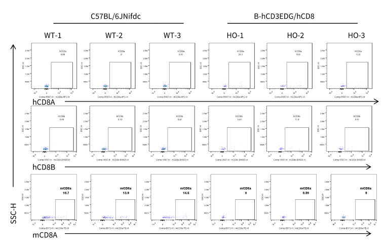
Strain specific CD8 expression analysis in homozygous B-hCD3EDG/hCD8 mice by flow cytometry. Splenocytes were collected from wild-type C57BL/6JNifdc mice (WT) and homozygous B-hCD3EDG/hCD8 mice (HO), and then analyzed by flow cytometry with species-specific anti-CD8 antibodies (anti-human CD8A, Biolegend, 300908; anti-mouse CD8A, Biolegend, 100708; anti-human CD8B, BD, 742392).
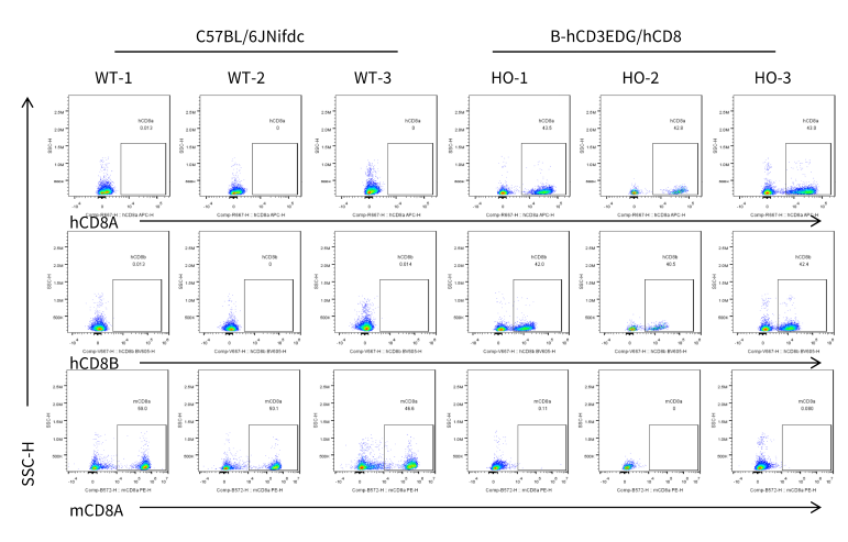
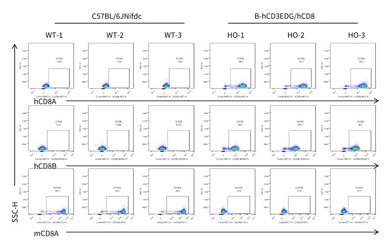
Strain specific CD8 expression analysis in homozygous B-hCD3EDG/hCD8 mice by flow cytometry. Blood cells were collected from wild-type C57BL/6JNifdc mice (WT) and homozygous B-hCD3EDG/hCD8 mice (HO), and then analyzed by flow cytometry with species-specific anti-CD8 antibodies (anti-human CD8A, Biolegend, 300908; anti-mouse CD8A, Biolegend, 100708; anti-human CD8B, BD, 742392).
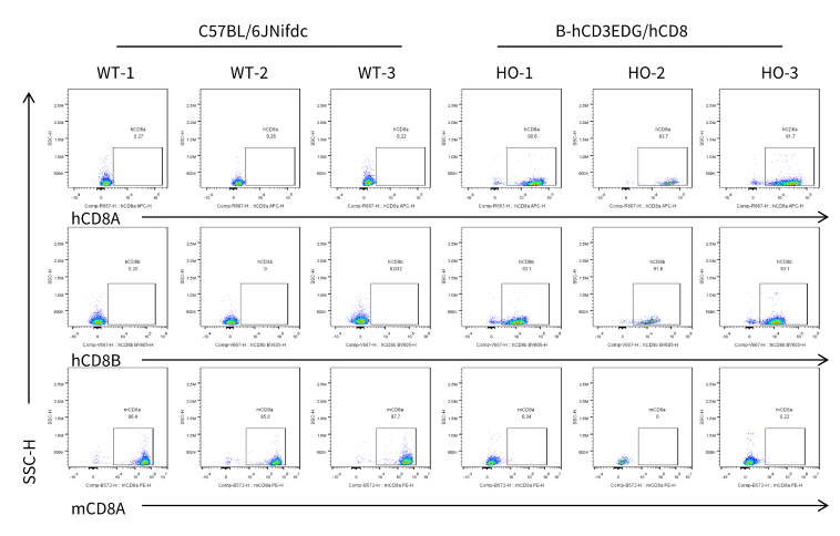
Strain specific CD8 expression analysis in homozygous B-hCD3EDG/hCD8 mice by flow cytometry. Blood cells were collected from wild-type C57BL/6JNifdc mice (WT) and homozygous B-hCD3EDG/hCD8 mice (HO), and then analyzed by flow cytometry with species-specific anti-CD8 antibodies (anti-human CD8A, Biolegend, 300908; anti-mouse CD8A, Biolegend, 100708; anti-human CD8B, BD, 742392).
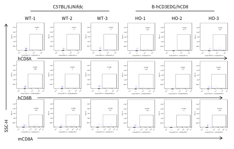
Strain specific CD8 expression analysis in homozygous B-hCD3EDG/hCD8 mice by flow cytometry. Blood cells were collected from wild-type C57BL/6JNifdc mice (WT) and homozygous B-hCD3EDG/hCD8 mice (HO), and then analyzed by flow cytometry with species-specific anti-CD8 antibodies (anti-human CD8A, Biolegend, 300908; anti-mouse CD8A, Biolegend, 100708; anti-human CD8B, BD, 742392).
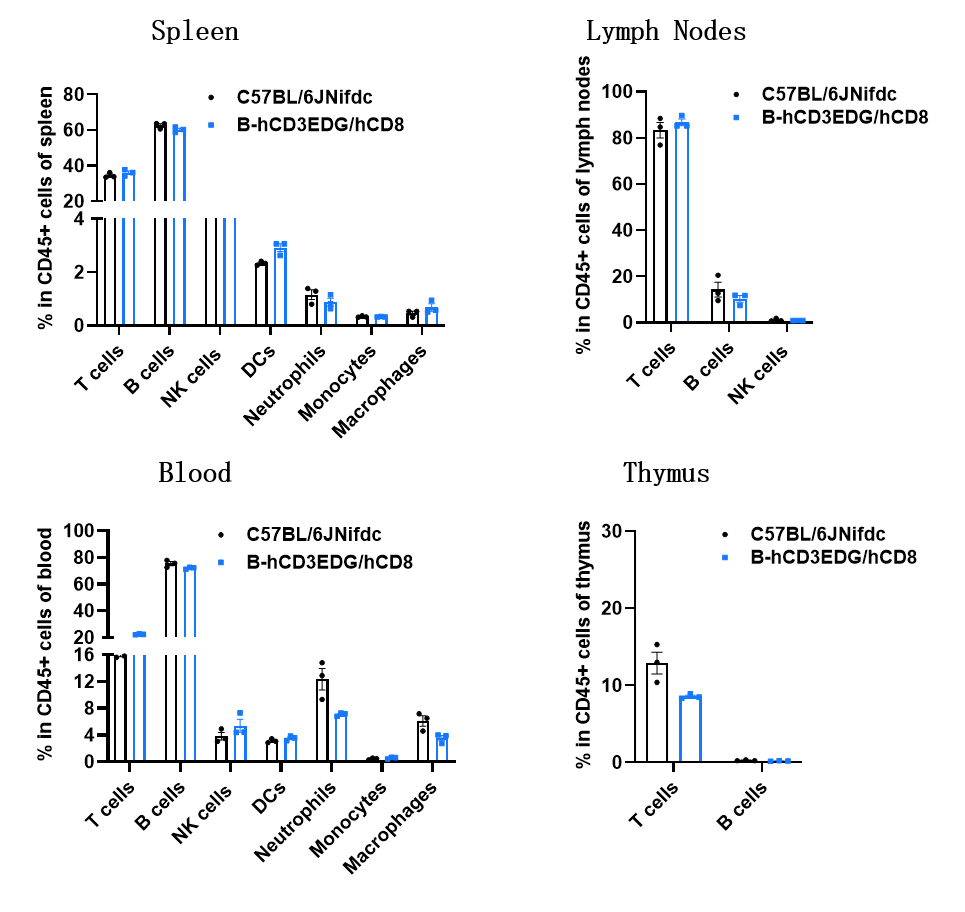
Analysis of leukocyte subpopulations by flow cytometry in immune organs and blood. Splenocytes, peripheral blood, lymph nodes, and thymus were isolated from C57BL/6JNifdc and B-hCD3EDG/hCD8 mice (male, 9-week-old, n = 3). Single live cells were gated on the CD45⁺ population and analyzed by flow cytometry as indicated. Values are expressed as mean ± SEM.
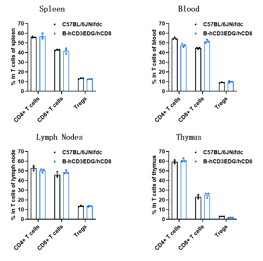
Analysis of T-cell subpopulations by flow cytometry in immune organs and blood. Splenocytes, peripheral blood, lymph nodes, and thymus were isolated from C57BL/6JNifdc and B-hCD3EDG/hCD8 mice (male, 9-week-old, n = 3). Single live cells were gated on the TCRβ⁺ T-cell population and analyzed by flow cytometry as indicated. Values are expressed as mean ± SEM.
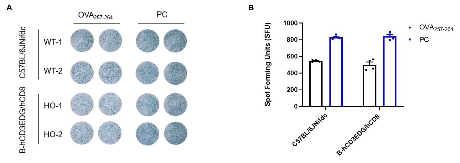
Detection of OVA-induced immune responses in B-hCD3EDG/hCD8 mice by IFN-γ ELISpot assay. Male wild-type C57BL/6JNifdc mice and B-hCD3EDG/hCD8 mice at the age of 9–10 weeks were immunized with intraperitoneal injection of 0.3 mg of OVA protein (Simga, A5503-25MG) and 30 μg poly (I:C) (InvivoGen, tlrl-pic). Mice were immunized with OVA two times at 1-week interval. One week after the last immunization, mice were sacrificed. The splenocytes were extracted, stimulated with OVA peptide257–264, or Cell Activation Cocktail (without Brefeldin A), (BioLegend, 42330) as positive control (PC), and then measured for IFN-γ secretion. (A) Representative results showing stimulation of splenocytes harvested from immunized mice with OVA peptide257–264, or positive control in duplicates. (B) Summary of results. These data indicate that B-hCD3EDG/hCD8 mice have normal T cell immunogenic function.
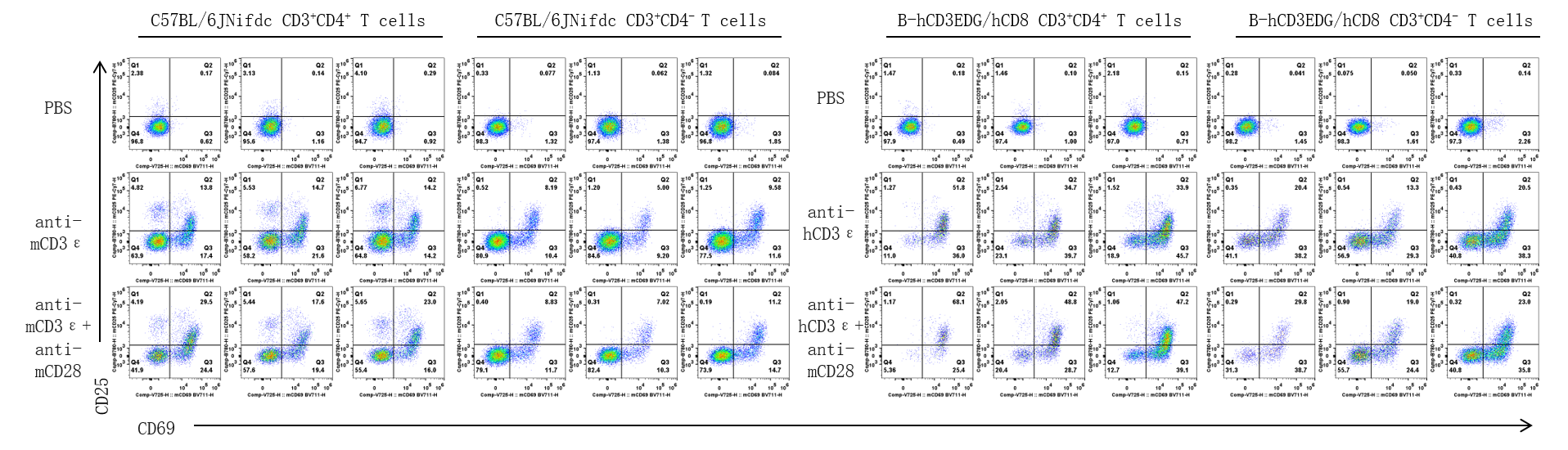
CD25 and CD69 expression analysis in wild-type C57BL/6JNifdc mice and homozygous humanized B-hCD3EDG/hCD8 mice by flow cytometry. T cells were isolated from splenocytes of C57BL/6JNifdc and B-hCD3EDG/hCD8 mice (male, 9-week-old, n=3), and were incubated in the presence of anti-mouse CD3ε antibody (2 ug/ml, BioXcell, BE0001-2), or anti-human CD3ε antibody (2 ug/ml, BioXcell, BE0231) with or without anti-mCD28 antibody (5 ug/ml, BioXcell, BE0015-1) for 24h. T cell proliferation was tested by flow cytometry. T cell activation in B-hCD3EDG/hCD8 mice was significantly up-regulated by anti-hCD3ε antibody and anti-mCD28 antibody.
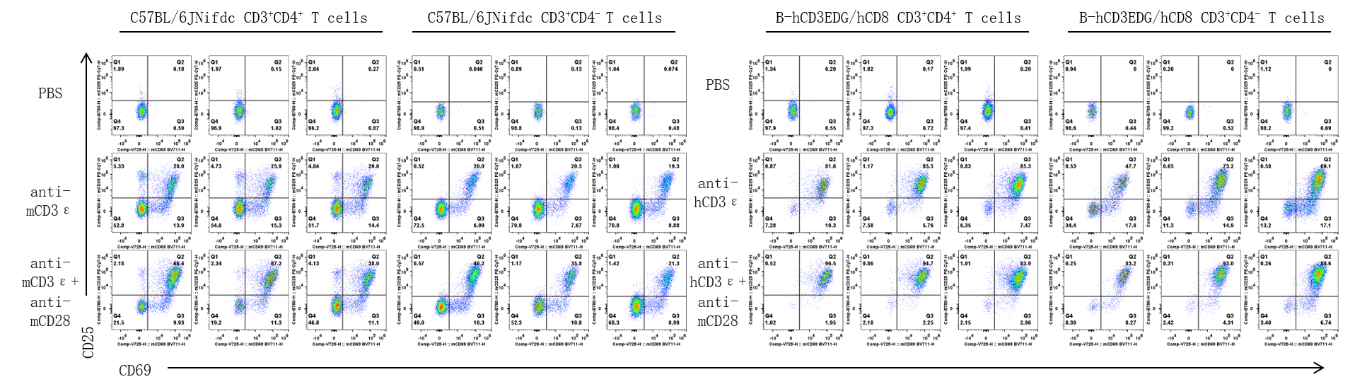
CD25 and CD69 expression analysis in wild-type C57BL/6JNifdc mice and homozygous humanized B-hCD3EDG/hCD8 mice by flow cytometry. T cells were isolated from splenocytes of C57BL/6JNifdc and B-hCD3EDG/hCD8 mice (male, 9-week-old, n=3), and were incubated in the presence of anti-mouse CD3ε antibody (2 ug/ml, BioXcell, BE0001-2), or anti-human CD3ε antibody (2 ug/ml, BioXcell, BE0231) with or without anti-mCD28 antibody (5 ug/ml, BioXcell, BE0015-1) for 48h. T cell proliferation was tested by flow cytometry. T cell activation in B-hCD3EDG/hCD8 mice was significantly up-regulated by anti-hCD3ε antibody and anti-mCD28 antibody.
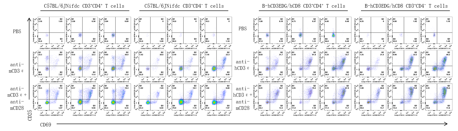
CD25 and CD69 expression analysis in wild-type C57BL/6JNifdc mice and homozygous humanized B-hCD3EDG/hCD8 mice by flow cytometry. T cells were isolated from splenocytes of C57BL/6JNifdc and B-hCD3EDG/hCD8 mice (male, 9 week-old, n=3), and were incubated in the presence of anti-mouse CD3ε antibody (2 ug/ml, BioXcell, BE0001-2), or anti-human CD3ε antibody (2 ug/ml, BioXcell, BE0231) with or without anti-mCD28 antibody (5 ug/ml, BioXcell, BE0015-1) for 72h. T cell proliferation was tested by flow cytometry. T cell activation in B-hCD3EDG/hCD8 mice was significantly up-regulated by anti-hCD3ε antibody and anti-mCD28 antibody.
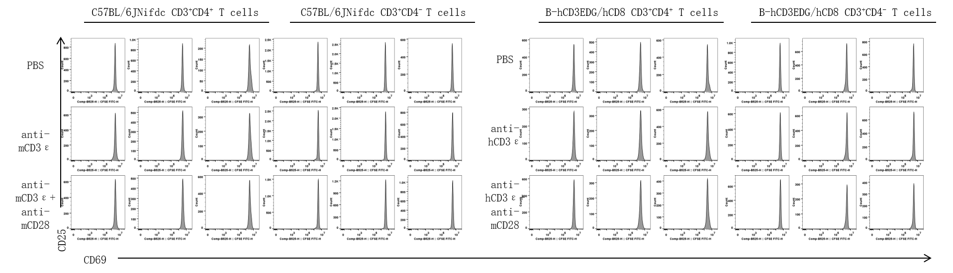
The proliferation of CD4+ T cells and CD8+ T cells was analyzed by flow cytometry. T cells were isolated from splenocytes of C57BL/6JNifdc and B-hCD3EDG/hCD8 mice (female, 13 week-old, n=3), and were incubated in the presence of anti-mouse CD3ε antibody (2 ug/ml, BioXcell, BE0001-2), or anti-human CD3ε antibody (2 ug/ml, BioXcell, BE0231) with or without anti-mCD28 antibody (5 ug/ml, BioXcell, BE0015-1) for 24h. T cell proliferation was tested by flow cytometry.
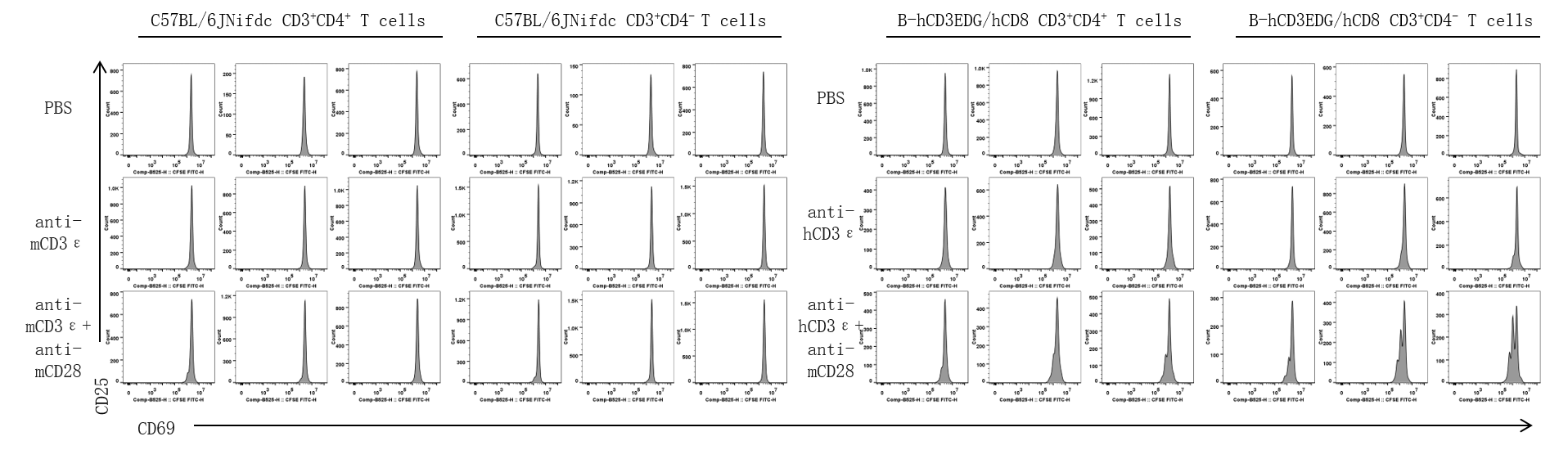
The proliferation of CD4+ T cells and CD8+ T cells was analyzed by flow cytometry. T cells were isolated from splenocytes of C57BL/6JNifdc and B-hCD3EDG/hCD8 mice (female, 13 week-old, n=3), and were incubated in the presence of anti-mouse CD3ε antibody (2 ug/ml, BioXcell, BE0001-2), or anti-human CD3ε antibody (2 ug/ml, BioXcell, BE0231) with or without anti-mCD28 antibody (5 ug/ml, BioXcell, BE0015-1) for 48h. T cell proliferation was tested by flow cytometry.
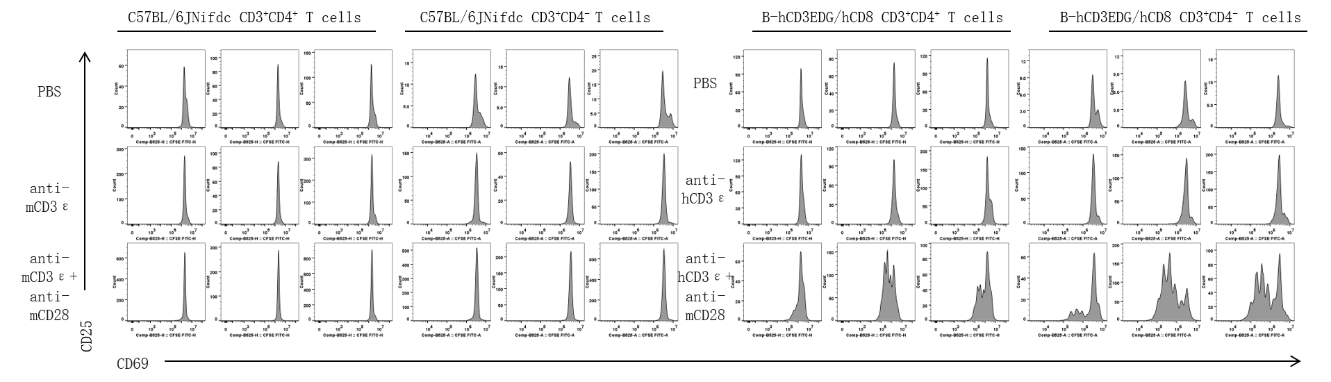
The proliferation of CD4+ T cells and CD8+ T cells was analyzed by flow cytometry. T cells were isolated from splenocytes of C57BL/6JNifdc and B-hCD3EDG/hCD8 mice (female, 13 week-old, n=3), and were incubated in the presence of anti-mouse CD3ε antibody (2 ug/ml, BioXcell, BE0001-2), or anti-human CD3ε antibody (2 ug/ml, BioXcell, BE0231) with or without anti-mCD28 antibody (5 ug/ml, BioXcell, BE0015-1) for 72h. T cell proliferation was tested by flow cytometry.
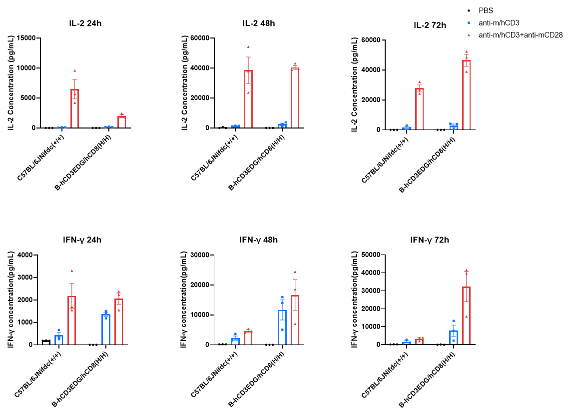
In vitro cytokine production (IFN-γ and IL-2) in B-hCD3EDG/hCD8 mice. T cells (2×10⁵) were isolated from the splenocytes of C57BL/6JNifdc and B-hCD3EDG/hCD8 mice (male, 9-week-old, n=3), incubated in the presence of anti-mCD3ε antibody (2 ug/ml,BioXcell,BE0001-2) or anti-hCD3ε antibody (2 ug/ml,BioXcell,BE0231) with or without anti-mCD28 antibody (5 ug/ml, BioXcell, BE0015-1) for 24h, 48h and 72h. IFN-γ and IL-2 productions were then tested using ELISA method.
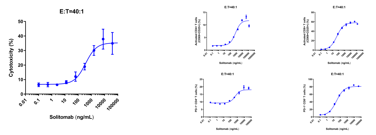
In vitro cytotoxicity evaluation of a CD3/EPCAM bispecific antibody against human EPCAM-expressing MC38 cells. B-hEPCAM MC38 cells were labeled with a violet cell tracker fluorescent dye and seeded into 96-well plates. On the following day, splenocytes isolated from B-hCD3EDG/hCD8 mice were added and cocultured with the target cells in the presence of various concentrations of the CD3/EPCAM bispecific antibody (Solitomab analog, commercial). Cytotoxicity and the proportion of CD4+ T / CD8+ T cell activation was assessed after 48 hours of incubation. Effector-to-target (E:T) cell ratios of 40:1 were used.

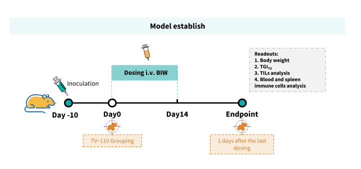
Establishment of a B-hEPCAM MC38 model and in vivo efficacy study of an anti-human CD3/EPCAM bispecific antibody. B-hEPCAM MC38 colon cancer cells were implanted subcutaneously into homozygous B-hCD3EDG/hCD8 mice (male, 7-week-old, n=5). When the average tumor volume reached approximately 110 mm³, mice were randomized and subsequently administered the anti-human CD3/EPCAM bispecific antibody (Solitomab analog, commercial) via intravenous injection.
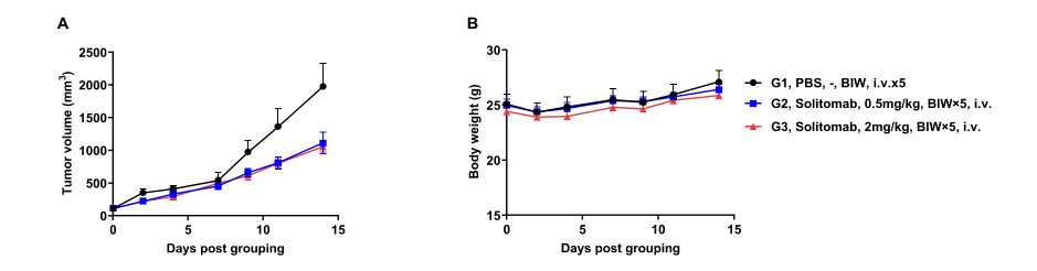
Antitumor activity of anti-human CD3/EPCAM bispecific antibody in B-hCD3EDG/hCD8 mice. (A) Tumor growth curves. (B) Body weight changes during treatment. As shown in panel A, CD3/EPCAM bispecific antibody (Solitomab analog, commercial) was efficacious in controlling tumor growth in B-hCD3EDG/hCD8 mice, demonstrating that the B-hCD3EDG/hCD8 mice provide a powerful preclinical model for in vivo evaluation of anti-human CD3-based TCE bi/multispecific antibodies. Values are expressed as mean ± SEM.
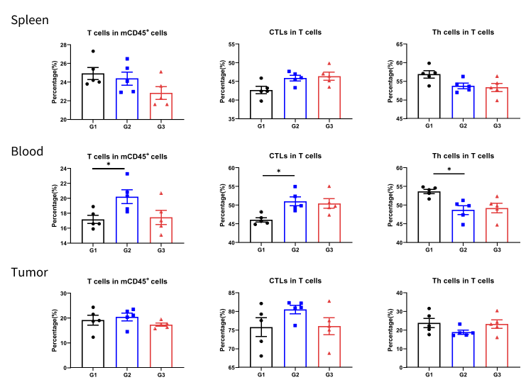
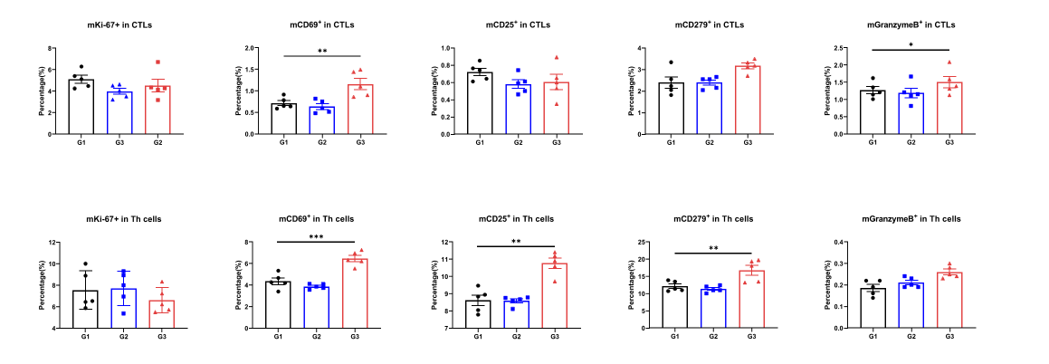
The proportions of mCD69+ CTL cells and mGranzyme B+ CTL cells in the spleen were increased in CD3/EPCAM BsAb treatment group.
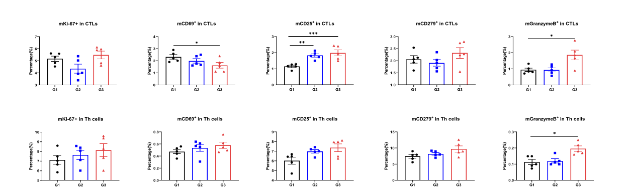
The proportions of mCD25+ CTL cells and mGranzyme B+ CTL cells in the blood were increased in CD3/EPCAM BsAb treatment group.
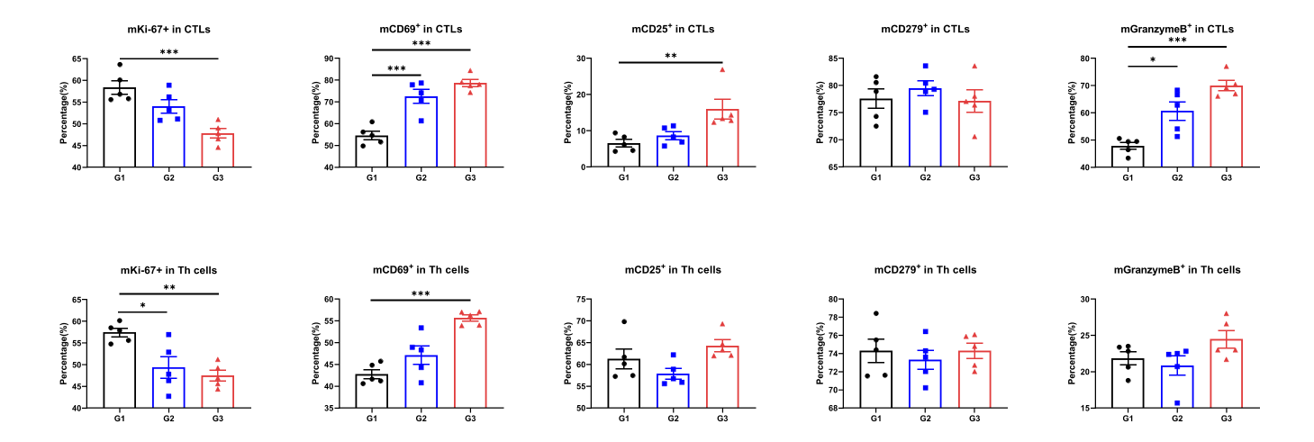
The proportions of mCD69+ CTL cells, mCD25+ CTL cells and mGranzyme B+ CTL cells in the tumor were increased, and the proportions of mKi67+ CTL cells and mKi67+ Th cells were decreased in CD3/EPCAM BsAb treatment group.
