
C57BL/6-Pdcd1tm3(PDCD1)Bcgen Cd274tm1(CD274)Bcgen Vegfatm1(VEGFA)Bcgen/Bcgen • 114019
| Product name | B-hPD-1 plus/hPD-L1/hVEGFA mice |
|---|---|
| Catalog number | 114019 |
| Strain name | C57BL/6-Pdcd1tm3(PDCD1)Bcgen Cd274tm1(CD274)Bcgen Vegfatm1(VEGFA)Bcgen/Bcgen |
| Strain background | C57BL/6 |
| NCBI gene ID | 5133,29126,7422 (Human) |
| Aliases | PD1; PD-1; CD279; SLEB2; hPD-1; hPD-l; hSLE1; ADMIO4; AIMTBS; B7-H; B7H1; PDL1; PD-L1; ADMIO5; hPD-L1; PDCD1L1; PDCD1LG1; VPF; VEGF; MVCD1; L-VEGF |
Gene targeting strategy for B-hPD-1 plus/hPD-L1/hVEGFA mice.
A chimeric CDS that encodes human PDCD1 extracellular domain, mouse Pdcd1 transmembrane and cytoplasmic domain, followed by WPRE-pA is inserted right after mouse Pdcd1 ATG to replace the exon 1 of mouse Pdcd1 gene. The chimeric PDCD1 protein expression will be driven by endogenous mouse Pdcd1 promoter, while mouse Pdcd1 gene transcription and translation will be disrupted.
The exon 3 of mouse Cd274 gene that encodes the IgV domain was replaced by human CD274 exon 3 in B-hPD-1 plus/hPD-L1/hVEGFA mice.
The exons 1-8 of mouse Vegfa gene that encode the full-length protein were replaced by human VEGFA exons 1-8 in B-hPD-1 plus/hPD-L1/hVEGFA mice.
Note: The B-hPD-1 plus/hPD-L1/hVEGFA three knock-in model, was developed by breeding the B-hPD-1 plus mice, the B-hPD-L1 mice and the B-hVEGFA mice.
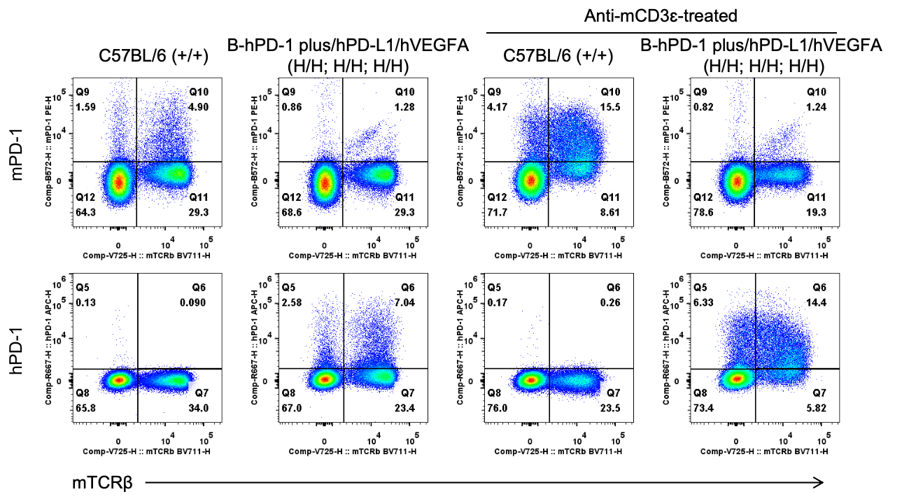
Strain specific PD-1 expression analysis in wild-type C57BL/6 and homozygous B-hPD-1 plus/hPD-L1/hVEGFA mice by flow cytometry. Splenocytes were collected from wild-type C57BL/6 mice (+/+) and homozygous B-hPD-1 plus/hPD-L1/hVEGFA mice (H/H) after stimulated with anti-mouse CD3ε antibody (7.5 μg, i.p.) in vivo for 24 hrs (female, 6-week-old, n=1) or not. Protein expression was analyzed with anti-mouse PD-1 antibody (Biolegend, 109104) and anti-human PD-1 antibody (Biolegend, 329908) by flow cytometry. Mouse PD-1 were detectable in wild-type C57BL/6 mice. Human PD-1 were exclusively detectable in homozygous B-hPD-1 plus/hPD-L1/hVEGFA mice but not in wild-type mice.
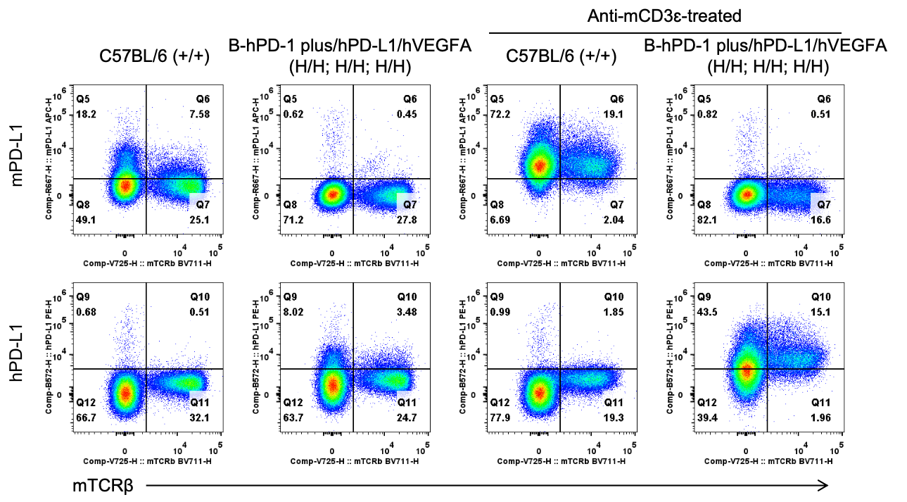
Strain specific PD-L1 expression analysis in wild-type C57BL/6 and homozygous B-hPD-1 plus/hPD-L1/hVEGFA mice by flow cytometry. Splenocytes were collected from wild-type C57BL/6 mice (+/+) and homozygous B-hPD-1 plus/hPD-L1/hVEGFA mice (H/H) after stimulated with anti-mouse CD3ε antibody (7.5 μg, i.p.) in vivo for 24 hrs (female, 6-week-old, n=1) or not. Protein expression was analyzed with anti-mouse PD-L1 antibody (Biolegend, 124312) and anti-human PD-L1 antibody (Biolegend, 329706) by flow cytometry. Mouse PD-L1 were detectable in wild-type C57BL/6 mice. Human PD-L1 were exclusively detectable in homozygous B-hPD-1 plus/hPD-L1/hVEGFA mice but not in wild-type mice.

Strain specific VEGFA expression analysis in wild-type C57BL/6 mice and homozygous B-hPD-1 plus/hPD-L1/hVEGFA mice by ELISA. Lung homogenates were collected from wild-type C57BL/6 mice (+/+) and homozygous B-hPD-1/hPD-L1 plus/hVEGFA mice (H/H; H/H; H/H). Expression level of mouse and human VEGFA were analyzed by ELISA (anti-mouse VEGFA antibody: R&D, MMV00; anti-human VEGFA antibody: R&D, DVE00). Mouse VEGFA was detectable in wild-type mice. Human VEGFA was exclusively detectable in homozygous B-hPD-1 plus/hPD-L1/hVEGFA mice but not in wild-type mice.
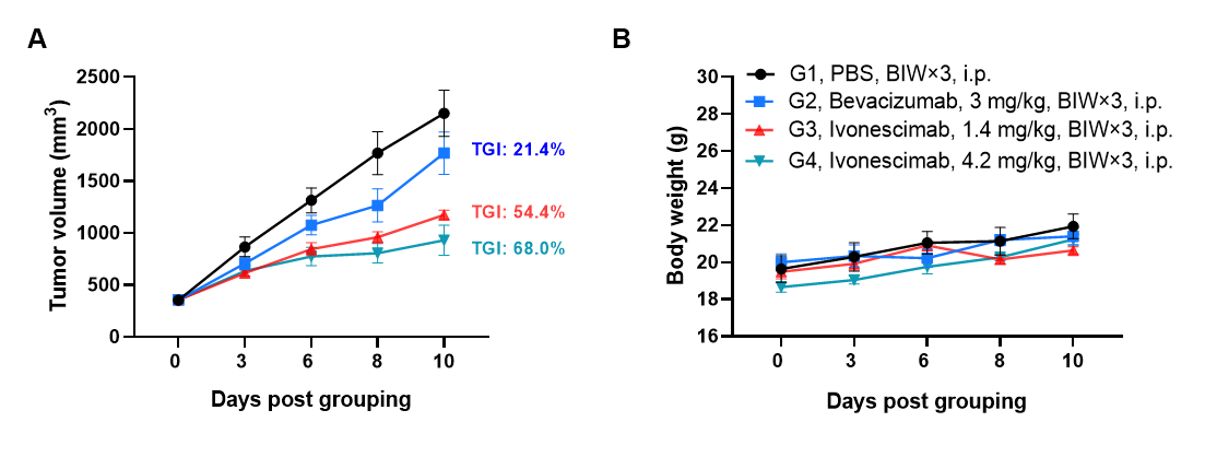
Antitumor activity of Ivonescimab (also known as AK112) in B-hPD-1 plus/hPD-L1/hVEGFA mice. Murine colon cancer B-hVEGFA/hPD-L1 plus MC38 cells were subcutaneously implanted into homozygous B-hPD-1 plus/hPD-L1/hVEGFA mice (Female, 9-week-old, n=6). Mice were grouped when tumor volume reached approximately 300 mm3, at which time they were intraperitoneally injected with Ivonescimab indicated in panel. As shown in panel A, Ivonescimab was efficacious in controlling tumor growth in B-hPD-1 plus/hPD-L1/hVEGFA mice. Values are expressed as mean ± SEM.
The overage of this tumor model is 61%.