Description
FOLR1: A GPI-anchored folate receptor and tumor-associated antigen
- Protein and function: FOLR1 encodes folate receptor alpha (FRα), a GPI-anchored glycoprotein that binds folate and mediates its uptake. The mature receptor has no transmembrane domain or cytoplasmic tail.
- Protein Expression: FRα occurs on selected normal epithelia, including kidney proximal tubules, lung and choroid plexus. Many ovarian cancers and subsets of lung and breast cancers overexpress FRα.
- Signaling Pathway: Folate uptake supports one-carbon metabolism and nucleotide synthesis. In experimental cancer models, FRα can engage gp130/JAK/STAT3 signaling. These effects depend on cellular context.
- Therapeutic strategies: ADCs deliver cytotoxic payloads after internalization. Antibodies can recruit Fc-dependent immune effectors. T-cell engagers and CAR-T cells redirect T-cell killing. Folate conjugates can deliver drugs or radionuclides.
Targeting strategy
FOLR1
- A human genomic segment containing FOLR1 exons 3–6 was introduced at the endogenous mouse Folr1 locus. The intended human precursor includes the N-terminal signal peptide, mature extracellular receptor and C-terminal GPI-anchoring signal peptide, which is removed during GPI attachment.
- The promoter and 5’UTR region of the mouse gene are retained. The design is intended to replace endogenous mouse FOLR1 protein expression.
- The design used human NM_016724.3 and mouse NM_001252552.2. The schematic represents the intended construct. RT-PCR clone sequencing identified abnormal splicing involving the mouse 5′ UTR and human exon 3 (see the FAQ).
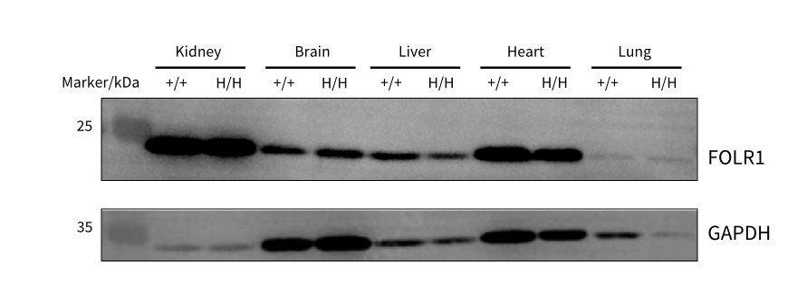
FOLR1 Immunoblot Analysis in Tissues
FOLR1 immunoblotting in normal tissues. Kidney, brain, liver, heart and lung lysates from wild-type B-NDG mice (+/+) and homozygous B-NDG hFOLR1 mice (H/H) were analyzed with Abcam ab230469 (40 μg protein per lane), with GAPDH as a loading control. Immunoreactive bands occurred in both genotypes, with weak lung signals. Mouse cross-reactivity or nonspecific binding cannot be excluded (Male, 8 weeks old, n = 1).
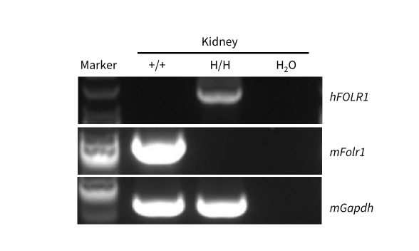
FOLR1 mRNA Expression in Kidney
FOLR1 transcript analysis in kidney by RT-PCR and clone sequencing. Kidney RNA from wild-type B-NDG mice (+/+) and homozygous B-NDG hFOLR1 mice (H/H) was reverse-transcribed to cDNA and amplified using mouse Folr1 or human FOLR1 primers. Of 15 sequenced RT-PCR clones, one matched the expected transcript and 14 showed abnormal splicing involving human exon 3 and the mouse 5′ UTR. PCR and clone selection may bias this ratio, which does not quantify transcript abundance in tissue.
* When publishing results obtained using this animal model, please acknowledge the source as follows: The animal model [B-NDG hFOLR1 mice] (Cat# 113372) was purchased from Biocytogen.