Results
Orthotopic Prostate Cancer Model: B-Tg(Luc) LNCap Clone FGC in B-NDG Mice
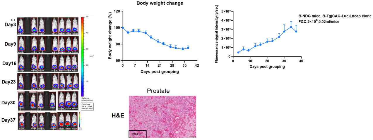
Establishment of orthotopic prostate cancer model.
The mouse orthotopic prostate cancer was generated by implanting B-Tg(Luc) LNCap clone FGC cells in the dorsal prostate lobes of B-NDG mice (M, n=7), , and the body weight and tumor volume of the mice were recorded weekly. The results showed that the tumor volume increased and body weight of mice gradually decreased during this study. This indicates that the cell line was successfully constructed as an orthotopic tumor model. H&E staining showed that Necrosis and hemorrhage were seen in prostate tumors, and inflammatory cell infiltration can be seen in seminal vesicles.
| Study ID: |
Groups |
Animal number |
Treatment |
Dosage (mg/kg) |
Lot No. |
Dosing Volume |
Dosing Route |
Treatment Schedule |
Treatment Times |
| 24P071536 |
G1 |
6 |
PBS |
- |
20240305 |
10μL/g |
i.v. |
QW |
2 |
| G2 |
6 |
DS-8201 |
3mpk |
83888105 |
10μL/g |
i.v. |
QW |
2 |
| G3 |
6 |
DS-8201 |
6mpk |
83888105 |
10μL/g |
i.v. |
QW |
2 |
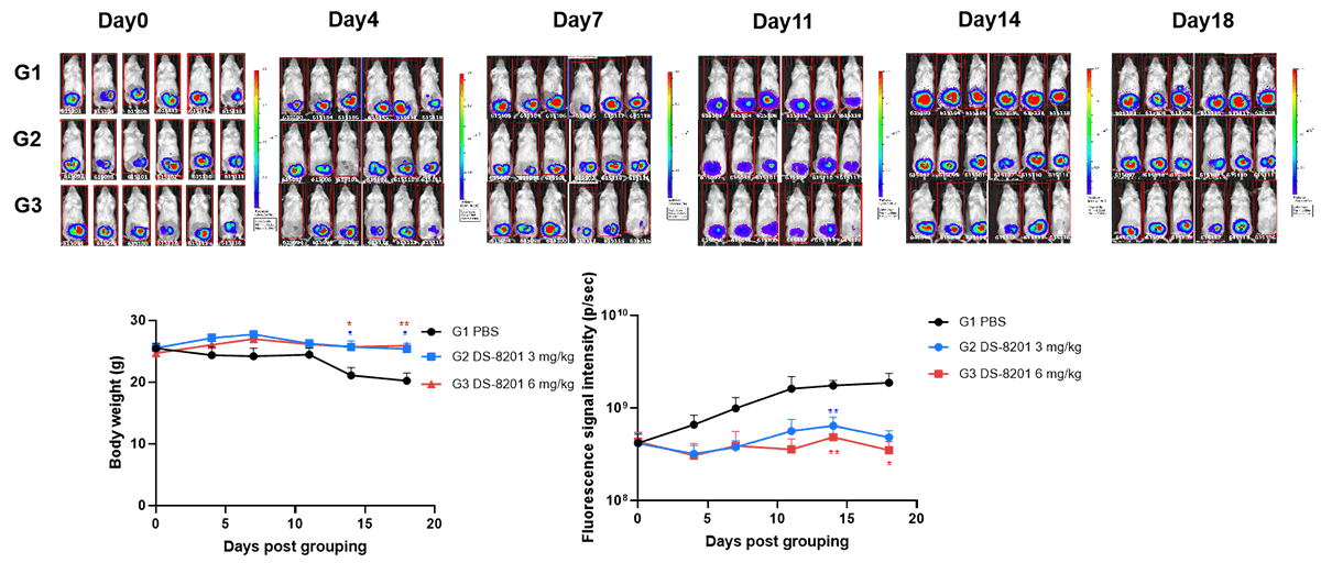
Evaluation of the efficacy of orthotopic prostate cancer model. The B-NDG (M, n=6) were inoculated with a volume of 20 μL of cell suspension (2×10⁶), mice were randomly divided into PBS and treatment groups on day 3 after tumor inoculation based on imaging values. The body weight and tumor fluorescence signal of the mice were recorded weekly. DS-8201 (3 and 6 mg/kg) significantly inhibited the growth of B-Tg(Luc) LNCap clone FGC orthotopic prostate cancer, demonstrating that this model can be well used for preclinical evaluation. Mice were dissected at the end of the experiment. Values are expressed as mean ± SEM. *p<0.05, **p<0.01.
Orthotopic Prostate Cancer Model: B-Tg(Luc) PC-3 in B-NDG Mice
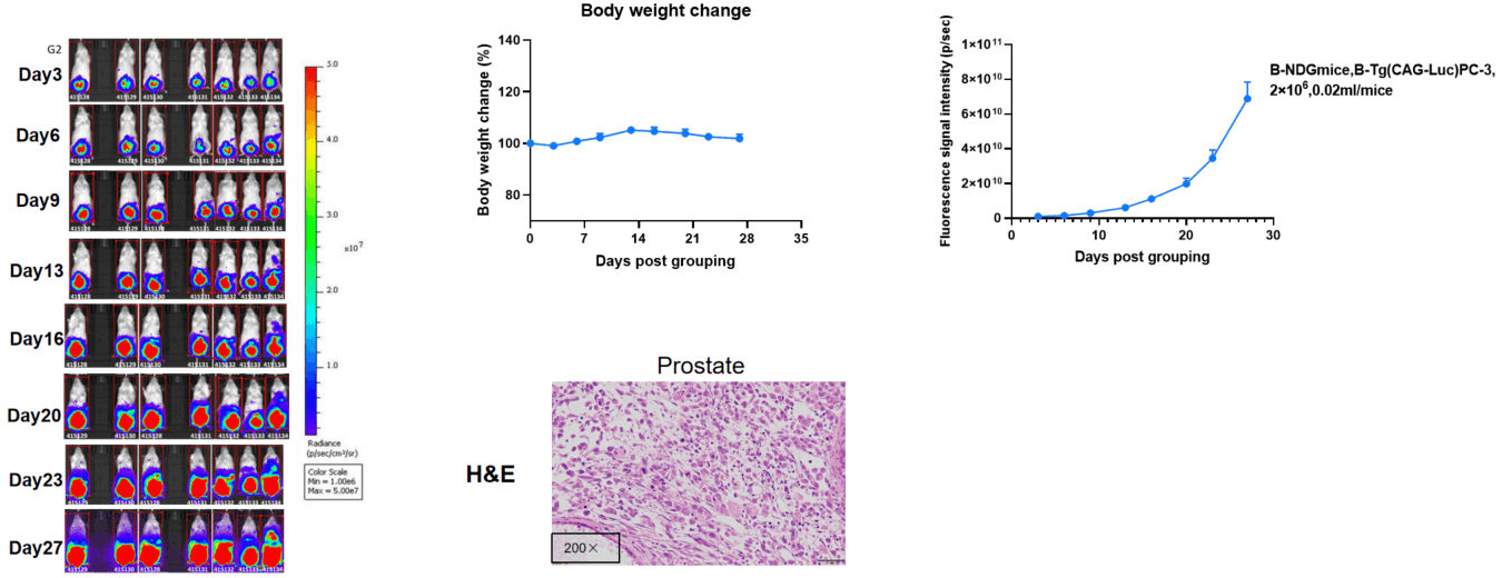
Establishment of orthotopic prostate cancer model.
The mouse orthotopic prostate cancer was generated by implanting B-Tg(Luc) PC-3 cells in the dorsal prostate lobes of B-NDG mice (M, n=7), and the body weight and tumor volume of the mice were recorded weekly. The results showed that the tumor volume increased and body weight of mice slightly change during this study. This indicated that the cell line was successfully constructed as an orthotopic tumor model. H&E staining showed that tumor cells were seen in prostate.