Description
CTLA4 (cytotoxic T-lymphocyte-associated protein 4) is an immune checkpoint receptor expressed on activated T cells and regulatory T cells, where it negatively regulates T-cell responses and helps maintain immune homeostasis. Because CTLA4 is a clinically validated immuno-oncology target, a humanized mouse model with species-specific target expression is valuable for evaluating anti-human CTLA4 therapeutics.
In CTLA4 humanized mice (B-hCTLA4 mice), exon 2 of the mouse Ctla4 gene, which encodes the extracellular domain, was replaced by human CTLA4 exon 2. This targeting strategy preserves the mouse genomic context while enabling expression of the human extracellular domain in place of the murine counterpart. Mouse Ctla4 mRNA and protein were detectable only in wild-type mice, whereas human CTLA4 mRNA and protein were exclusively detectable in CTLA4 humanized mice (B-hCTLA4 mice).
Key Advantages
- Species-specific human CTLA4 expression: CTLA4 humanized mice (B-hCTLA4 mice) enable selective expression of human CTLA4 in a murine background for anti-human CTLA4 drug development.
- Preserved immune homeostasis: Major leukocyte and T-cell subpopulations in spleen, blood, and lymph nodes remain comparable to wild-type controls after CTLA4 humanization.
- Suitable for in vivo immuno-oncology studies: CTLA4 humanized mice (B-hCTLA4 mice) support antitumor efficacy evaluation of anti-human CTLA4 antibodies in syngeneic tumor models.
Validation
- mRNA expression by RT-PCR: Human CTLA4 mRNA was detected in homozygous CTLA4 humanized mice (B-hCTLA4 mice), while mouse Ctla4 mRNA was detected in wild-type controls.
- Protein expression by flow cytometry: Human CTLA4 protein was detected in anti-CD3ε-stimulated splenic T cells from CTLA4 humanized mice (B-hCTLA4 mice), whereas mouse CTLA4 protein was detected in wild-type mice.
- Immune profiling by flow cytometry: Leukocyte and T-cell subpopulations in spleen, blood, and lymph nodes were assessed and found to be comparable between CTLA4 humanized mice (B-hCTLA4 mice) and wild-type controls.
- Functional efficacy in tumor model: Anti-human CTLA4 antibodies showed antitumor activity in MC38 tumor-bearing CTLA4 humanized mice (B-hCTLA4 mice).
Applications
In vivo efficacy, pharmacodynamic, and safety evaluation of anti-human CTLA4 antibodies and related immuno-oncology therapeutics in syngeneic tumor models.
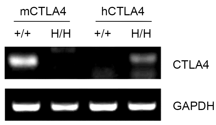
mRNA Expression Analysis of CTLA4 Humanized Mice (B-hCTLA4) by RT-PCR
Strain-specific analysis of CTLA4 mRNA expression of CTLA4 humanized mice (B-hCTLA4) by RT-PCR. Splenocytes were collected from wild-type (+/+) and homozygous CTLA4 humanized mice (B-hCTLA4, H/H). Mouse Ctla4 mRNA was detectable only in splenocytes of wild-type (+/+) mice, whereas human CTLA4 mRNA was detectable only in H/H mice, but not in +/+ mice.
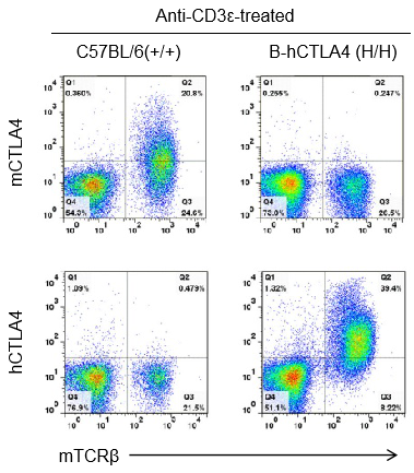
Protein Expression Analysis in T Cells of CTLA4 Humanized Mice (B-hCTLA4) by Flow Cytometry
Strain-specific CTLA4 protein expression analysis in T cells of CTLA4 humanized mice (B-hCTLA4) by flow cytometry. Splenocytes were collected from wild-type C57BL/6 (+/+) and homozygous CTLA4 humanized mice (B-hCTLA4, H/H) following anti-CD3ε treatment in vivo, and analyzed by flow cytometry using species-specific anti-CTLA4 antibodies. Mouse CTLA4 was detectable in wild-type mice, whereas human CTLA4 was exclusively detectable in homozygous CTLA4 humanized mice (B-hCTLA4), but not in wild-type mice.
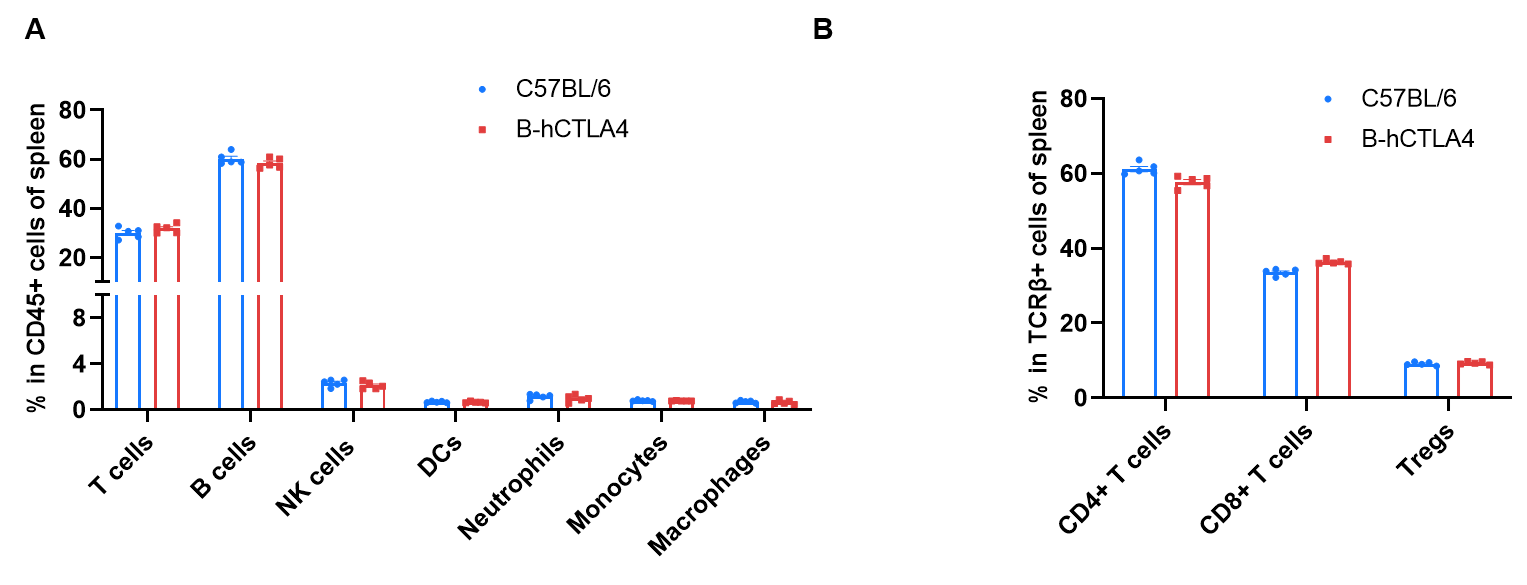
Frequency of Leukocyte Subpopulations in Spleen of CTLA4 Humanized Mice (B-hCTLA4) by Flow Cytometry
Frequency of leukocyte subpopulations in spleen of CTLA4 humanized mice (B-hCTLA4) by flow cytometry. Splenocytes were isolated from wild-type C57BL/6 mice (female, n=5, 9-week-old) and homozygous CTLA4 humanized mice (B-hCTLA4, female, n=5, 9-week-old). (A) Flow cytometry analysis of splenocytes was performed to assess the frequency of leukocyte subpopulations. (B) Frequency of T cell subpopulations. Percentages of T cells, B cells, NK cells, dendritic cells, neutrophils, monocytes, macrophages, CD4+ T cells, CD8+ T cells, and Tregs in CTLA4 humanized mice (B-hCTLA4) were similar to those in C57BL/6 mice. Values are expressed as mean ± SEM. Significance was determined by two-way ANOVA test (P < 0.05, P < 0.01, P < 0.001).
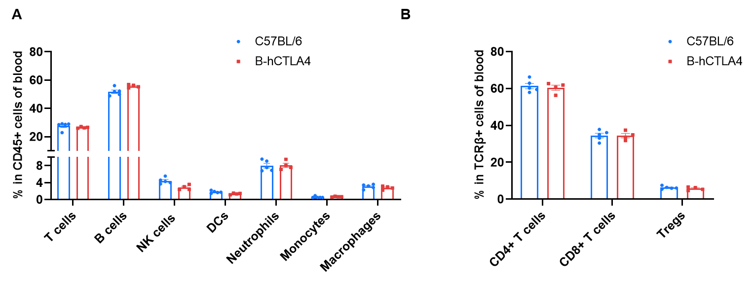
Frequency of Leukocyte Subpopulations in Blood of CTLA4 Humanized Mice (B-hCTLA4) by Flow Cytometry
Frequency of leukocyte subpopulations in blood of CTLA4 humanized mice (B-hCTLA4) by flow cytometry. Blood cells were isolated from wild-type C57BL/6 mice (female, n=5, 9-week-old) and homozygous CTLA4 humanized mice (B-hCTLA4, female, n=5, 9-week-old). (A) Flow cytometry analysis of blood cells was performed to assess the frequency of leukocyte subpopulations. (B) Frequency of T cell subpopulations. Percentages of T cells, B cells, NK cells, dendritic cells, neutrophils, monocytes, macrophages, CD4+ T cells, CD8+ T cells, and Tregs in CTLA4 humanized mice (B-hCTLA4) were similar to those in C57BL/6 mice. Values are expressed as mean ± SEM. Significance was determined by two-way ANOVA test (P < 0.05, P < 0.01, P < 0.001).
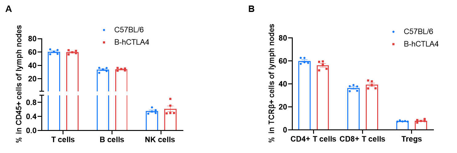
Frequency of Leukocyte Subpopulations in Lymph Nodes of CTLA4 Humanized Mice (B-hCTLA4) by Flow Cytometry
Frequency of leukocyte subpopulations in lymph nodes of CTLA4 humanized mice (B-hCTLA4) by flow cytometry. Lymph node cells were isolated from wild-type C57BL/6 mice (female, n=5, 9-week-old) and homozygous CTLA4 humanized mice (B-hCTLA4, female, n=5, 9-week-old). (A) Flow cytometry analysis of lymph node cells was performed to assess the frequency of leukocyte subpopulations. (B) Frequency of T cell subpopulations. Percentages of T cells, B cells, NK cells, CD4+ T cells, CD8+ T cells, and Tregs in CTLA4 humanized mice (B-hCTLA4) were similar to those in C57BL/6 mice. Values are expressed as mean ± SEM. Significance was determined by two-way ANOVA test (P < 0.05, P < 0.01, P < 0.001).
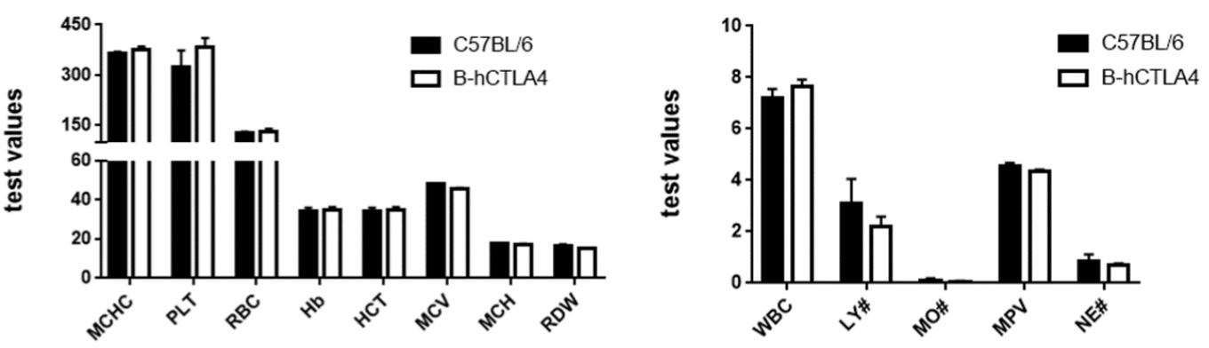
Hematology Analysis of CTLA4 Humanized Mice (B-hCTLA4)
Complete blood count (CBC) analysis of CTLA4 humanized mice (B-hCTLA4). Blood samples were collected from female wild-type C57BL/6 mice and CTLA4 humanized mice (B-hCTLA4) (n=3, 6-week-old) and analyzed for hematological parameters. No differences were observed across measured parameters between C57BL/6 and CTLA4 humanized mice (B-hCTLA4), indicating that replacement of mouse Ctla4 with human CTLA4 does not alter blood cell composition or morphology. Values are expressed as mean ± SEM.
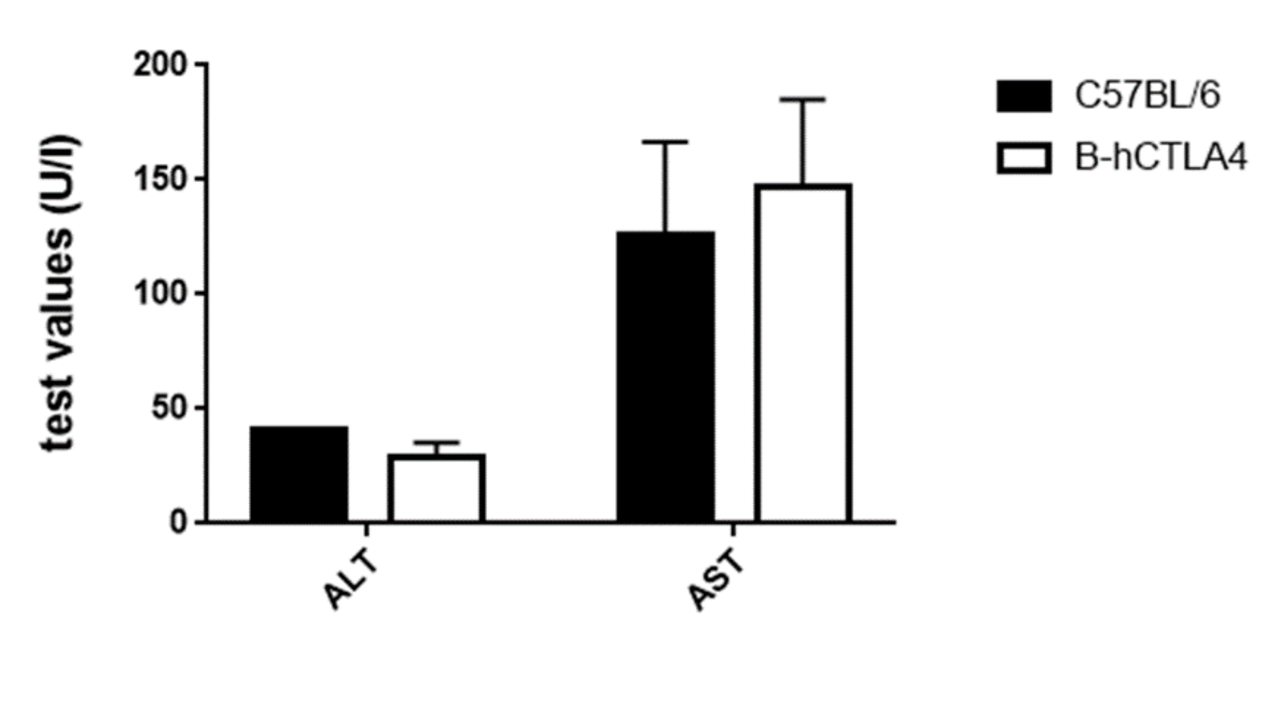
Biochemistry Analysis of CTLA4 Humanized Mice (B-hCTLA4)
Serum biochemistry analysis of CTLA4 humanized mice (B-hCTLA4). Serum samples were collected from wild-type C57BL/6 mice and CTLA4 humanized mice (B-hCTLA4) (n=3, 6-week-old) and analyzed for ALT and AST levels. No differences were observed in either parameter between C57BL/6 and CTLA4 humanized mice (B-hCTLA4), indicating that replacement of mouse Ctla4 with human CTLA4 does not alter ALT and AST levels or liver-related biochemical parameters. Values are expressed as mean ± SEM.
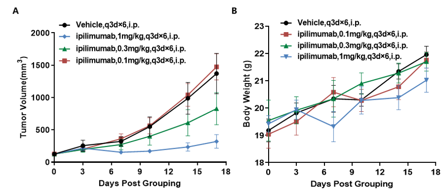
In Vivo Efficacy of Anti-Human CTLA4 Antibody in CTLA4 Humanized Mice (B-hCTLA4)
Antitumor activity of anti-human CTLA4 antibody in CTLA4 humanized mice (B-hCTLA4). Murine colon cancer MC38 cells were subcutaneously implanted into homozygous CTLA4 humanized mice (B-hCTLA4) (female, 6–7-week-old, n=5). Mice were grouped when tumor volume reached approximately 100 mm³, at which time they were treated with ipilimumab at the indicated doses and schedules. (A) Tumor growth curves following treatment. (B) Body weight changes during treatment. Ipilimumab (in house) inhibited MC38 tumor growth in CTLA4 humanized mice (B-hCTLA4), demonstrating that the CTLA4 humanized mice (B-hCTLA4) provide a powerful preclinical model for in vivo evaluation of anti-human CTLA4 antibody. Values are expressed as mean ± SEM.
In Vivo Efficacy of Anti-Human CTLA4 Antibodies in CTLA4 Humanized Mice (B-hCTLA4)
Antitumor activity of anti-human CTLA4 antibodies in CTLA4 humanized mice (B-hCTLA4). Murine colon cancer MC38 cells (5×10⁵) were subcutaneously implanted into homozygous CTLA4 humanized mice (B-hCTLA4) (female, 6–9-week-old, n=8). Mice were grouped when tumor volume reached approximately 100–150 mm³ and subsequently treated with anti-human CTLA4 antibodies as indicated. (A) Tumor growth curves following treatment with Tremelimumab analog and Ipilimumab analog at indicated doses and schedules. (B) Body weight changes during treatment. Anti-human CTLA4 antibodies inhibited MC38 tumor growth in CTLA4 humanized mice (B-hCTLA4) in a dose-dependent manner, demonstrating that CTLA4 humanized mice (B-hCTLA4) provide a powerful preclinical model for in vivo evaluation of anti-human CTLA4 antibodies. Values are expressed as mean ± SEM. The tumor take rate of this model was approximately 40%.
Frequently asked questions about CTLA4 humanized mice (B-hCTLA4 mice)
Q1: What are CTLA4 humanized mice (B-hCTLA4 mice)?
CTLA4 humanized mice (B-hCTLA4 mice) are genetically engineered mouse models in which the coding region of the mouse Ctla4 gene is replaced with the human CTLA4 gene. This enables physiological expression of human CTLA4 under endogenous regulatory elements, providing an in vivo platform for evaluating CTLA4-targeted immunotherapies.
Q2: Why are CTLA4 humanized mice (B-hCTLA4 mice) important for immuno-oncology research?
CTLA4 is a key immune checkpoint molecule that regulates T cell activation and immune tolerance. CTLA4 humanized mice (B-hCTLA4 mice) enable in vivo evaluation of anti-human CTLA4 antibodies by recapitulating human-specific target engagement, making them highly valuable for studying immune checkpoint blockade and tumor immunotherapy mechanisms.
Q3: How is human CTLA4 expression validated in CTLA4 humanized mice (B-hCTLA4 mice)?
Human CTLA4 expression in CTLA4 humanized mice (B-hCTLA4 mice) is validated using RT-PCR and flow cytometry. Human CTLA4 mRNA is detectable only in homozygous humanized mice, while mouse Ctla4 is present only in wild-type controls. At the protein level, flow cytometry of activated T cells confirms species-specific expression of human CTLA4.
Q4: Does CTLA4 humanization affect immune cell composition?
No. Flow cytometry analyses show that the frequency and distribution of major immune cell populations, including T cells, B cells, and NK cells, in spleen, blood, and lymph nodes are comparable between CTLA4 humanized mice (B-hCTLA4 mice) and wild-type mice.
Q5: What applications are supported by CTLA4 humanized mice (B-hCTLA4 mice)?
CTLA4 humanized mice (B-hCTLA4 mice) are widely used for in vivo efficacy and pharmacodynamic studies of anti-CTLA4 antibodies. In syngeneic tumor models such as MC38, CTLA4-targeting antibodies demonstrate dose-dependent tumor growth inhibition, supporting their use in preclinical evaluation of immuno-oncology therapies.
* When publishing results obtained using this animal model, please acknowledge the source as follows: The animal model [B-hCTLA4 mice] (Cat# 110011) was purchased from Biocytogen.