

C57BL/6JNifdc-Dmdtm1(mDmd Exon44-46 del; hDMD Exon44, Exon46 ins)Bcgen/Bcgen • 114088

DMD: Mutations in this gene lead to severe, progressive muscle-wasting disorders.

DMD

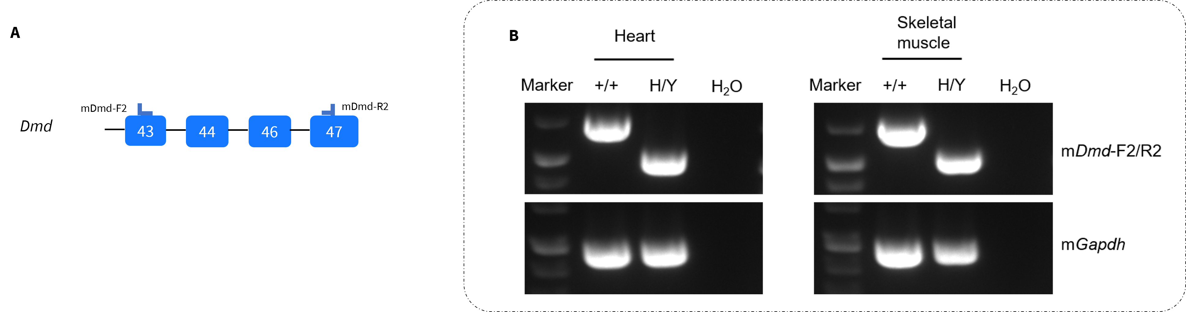
Strain specific analysis of DMD mRNA expression in wild-type C57BL/6JNifdc mice and homozygous B-hDMD(exon44-46, del45) mice by RT-PCR. Heart and skeletal muscle RNA were isolated from wild-type C57BL/6JNifdc mice (+/+) and homozygous B-hDMD(exon44-46, del45) mice (H/Y), then cDNA libraries were synthesized by reverse transcription, followed by PCR with mouse Dmd primers. (A) Primers were designed to detect the human exon 44 and exon 46. (B) The transcripts in homozygous mice are shorter than those in wild-type mice, and the sequences were confirmed via Sanger sequencing, demonstrating the successful exon splicing in B-hDMD(exon 44-46, del45) mice.
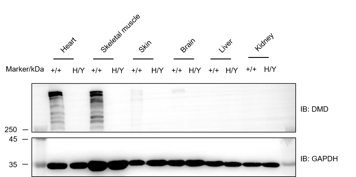
Western blot analysis of DMD protein expression in wild-type C57BL/6JNifdc mice and homozygous B-hDMD(exon44-46, del45) mice. Various tissue lysates were collected from wild-type C57BL/6JNifdc mice (+/+) and homozygous B-hDMD(exon44-46, del45) mice (H/Y), and then analyzed by western blot with anti-Dystrophin antibody (Sigma-Aldrich, D8168). 40 μg total proteins were loaded for western blot analysis. DMD was only detected in heart, skeletal muscle, skin and brain from wild-type mice but not in homozygous B-hDMD(exon44-46, del45) mice.
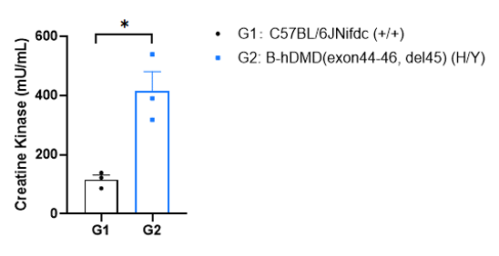
Serum creatine kinase activity analysis in wild-type C57BL/6JNifdc mice and homozygous B-hDMD(exon44-46, del45) mice by colorimetric. Serum was collected from wild-type C57BL/6JNifdc mice (+/+, n=3, 6-week-old male) and homozygous B-hDMD(exon44-46, del45) mice (H/Y, n=3, 6-week-old male). Creatine kinase activity was analyzed by colorimetric in a kinetic mode every 2 min for a total of 36 min at 37℃ (creatine kinase activity assay kit: Abcam, ab155901). OD450 readings were linear between T1 (at time 6) and T2 (12min for WT and homozygous mice). Creatine kinase activity was significantly greater in homozygous B-hDMD(exon44-46, del45) mice compared to that in wild-type mice. Significance was determined by unpaired t test. *P < 0.05.

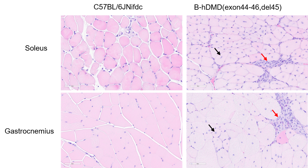
Representative histological images of skeletal muscle from wild-type C57BL/6JNifdc and homozygous B-hDMD(exon44-46, del45) mice. Gastrocnemius and soleus muscle sections from 4-month-old wild-type C57BL/6JNifdc and homozygous B-hDMD(exon44-46, del45) mice were presented to display histopathological phenotypes. Gastrocnemius and soleus muscle were stained with hematoxylin and eosin (H&E). Tissue histology was normal for wild-type control mice, but the muscle from homozygous B-hDMD(exon44-46, del45) mice displayed inflammation (red arrow) and centrally-located nuclei (black arrow).

Behavioral performance in wild-type C57BL/6JNifdc and homozygous B-hDMD(exon44-46, del45) mice. Grip strength tests were conducted to assay the behavioral performance in wild-type C57BL/6JNifdc and homozygous B-hDMD(exon44-46, del45) mice (male, 6-, 12-week-old, n=9-10). Grip strength produced by forelimb in homozygous B-hDMD(exon44-46, del45) mice was significant weaker than those in wild-type control mice at all the two time points. All grip strength measurements are normalized to the individual animal’s body weight. Values are expressed as mean ± SEM. At each time point, significance was determined by unpaired t test. *P < 0.05, **P < 0.01, ***P < 0.001.
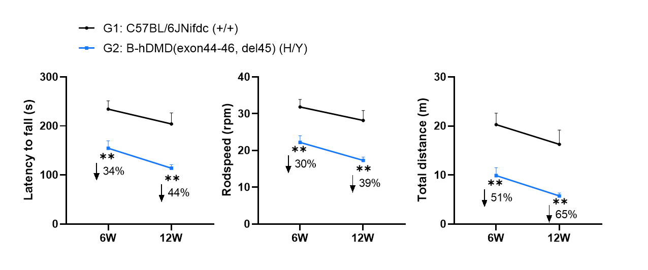
Behavioral performance in wild-type C57BL/6JNifdc and homozygous B-hDMD(exon44-46, del45) mice. Rotarod tests were conducted to assay the behavioral performance in wild-type C57BL/6JNifdc and homozygous B-hDMD(exon44-46, del45) mice (male, 6-, 12-week-old, n=9-10). Rotarod tests were performed to assay the motor coordination. The latency to fall, rodspeed and total distance were significantly decreased in homozygous B-hDMD(exon44-46, del45) mice at all the two time points, showing the impairment of motor coordination and balance. Values are expressed as mean ± SEM. At each time point significance was determined by unpaired t test. *P < 0.05, **P < 0.01, ***P < 0.001.
