Description
- Fah is the final enzyme in the tyrosine metabolism pathway, primarily expressed in the liver and kidneys. Deficiency of FAH is associated with Type 1 hereditary tyrosinemia (HT). As a result of the severe liver damage induced by Fah knockout (KO), this mouse model allows for the reconstruction of a functional human liver utilizing primary human hepatocytes.
- Gene editing strategy: The exons 2-14 of mouse Fah gene were knocked out in B-NDG Fah KO mice.
- mRNA expression analysis: Mouse Fah mRNA were detectable only in B-NDG mice but not in homozygous B-NDG Fah KO mice.
- Protein expression analysis: Fah was only detected in liver and kidney of B-NDG mice but not in homozygous B-NDG Fah KO mice.
- Application: This product is used for human hepatocytes transplantation and for evaluating the efficacy and safety of drugs associated with liver diseases.
Targeting strategy
Gene targeting strategy for B-NAG Fah KO mice. The exons 2-14 of mouse Fah gene were knocked out in B-NDG Fah KO mice.
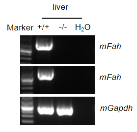
mRNA expression analysis in B-NDG Fah KO mice
Strain specific analysis of Fah mRNA expression in B-NDG mice and B-NDG Fah KO mice by RT-PCR. Liver RNA was isolated from B-NDG mice (+/+) and homozygous B-NDG Fah KO mice (-/-). cDNA libraries were synthesized by reverse transcription, followed by PCR with mouse Fah primers. Mouse Fah mRNA was detectable only in B-NDG mice but not in homozygous B-NDG Fah KO mice.
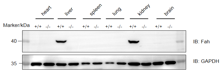
Protein expression analysis in B-NDG Fah KO mice
Western blot analysis of Fah protein expression in homozygous B-NDG Fah KO mice. Various tissue lysates were collected from B-NDG mice (+/+) and homozygous B-NDG Fah KO mice (-/-), and then analyzed by western blot with anti-Fah antibody (proteintech, 14928-1-AP). A total of 40 μg of protein was loaded for western blotting analysis. Fah was only detected in liver and kidney of B-NDG mice.
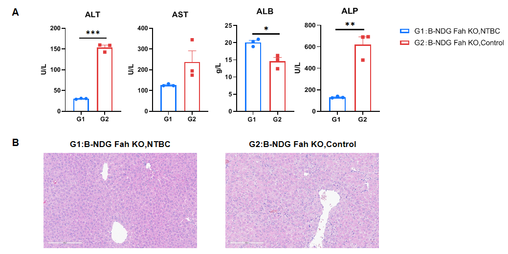
Liver damage analysis in B-NDG Fah KO mice
Liver damage analysis in B-NDG Fah KO mice. After removing NTBC drinking water from B-NDG Fah KO mice for three weeks, blood biochemical analysis revealed an increase in ALT, AST and ALP levels, while ALB levels decreased. Additionally, HE staining results indicated that the mice had developed liver damage. Values are expressed as mean ± SEM. Significance was determined by One-way or Two-way ANOVA test. *P < 0.05, **P < 0.01, ***P < 0.001.
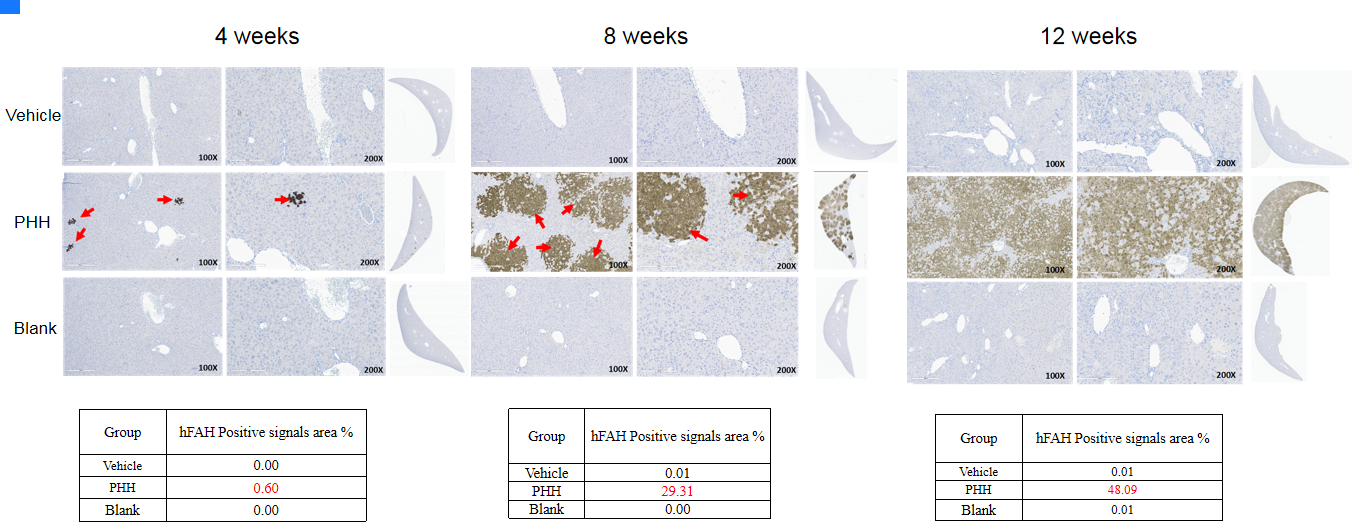
Human FAH in liver
Note: Vehicle (Sham-operated group); PHH (Primary Human Hepatocytes); Blank (Without any treatment). The levels of human FAH gradually increased in the livers of B-NDG Fah KO mice following the transplantation of human hepatocytes. The sections were scanned with a Leica Aperio GT450 scanner, and the staining positive area was analyzed using the HALO area quantification algorithm.
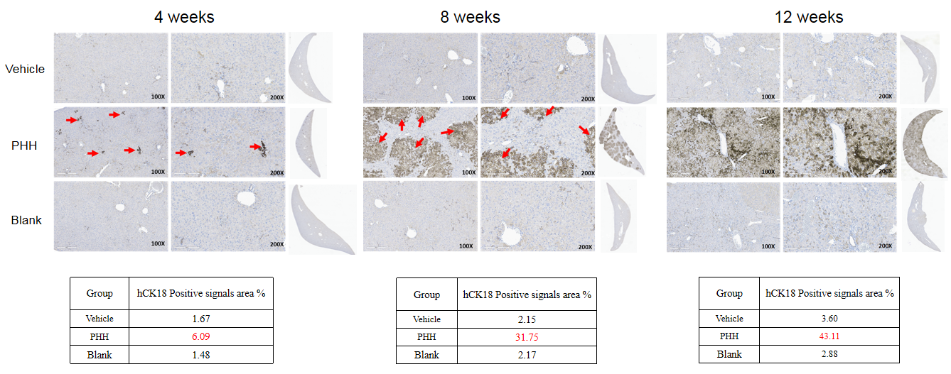
Human CK18 in liver
Note: Vehicle (Sham-operated group); PHH (Primary Human Hepatocytes); Blank (Without any treatment). The levels of human CK18 gradually increased in the livers of B-NDG Fah KO mice following the transplantation of human hepatocytes. The sections were scanned with a Leica Aperio GT450 scanner, and the staining positive area was analyzed using the HALO area quantification algorithm.
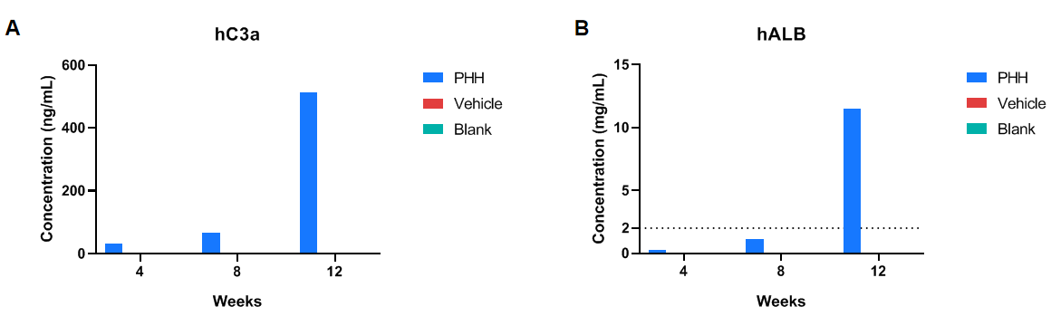
Expression of human C3a and human ALB in serum
Detection of human proteins in the blood of transplanted animals. (A) Human C3a detected in the serum. (B) Human albumin detected in the serum. Blood samples used in A and B were obtained from the same animals at the same time points. Both human C3a and albumin proteins in PHH group significantly increased over time compared with control animals (Vehicle and Blank groups).
* When publishing results obtained using this animal model, please acknowledge the source as follows: The animal model [B-NDG Fah KO mice] (Cat# 112959) was purchased from Biocytogen.