

C57BL/6-Cd40tm1(CD40)Bcgen Cd40lgtm1(CD40LG)Bcgen/Bcgen • 121335

CD40/CD40L: A key costimulatory axis in adaptive immunity and its therapeutic intervention

CD40
CD40L
B-hCD40/hCD40L Mice were obtained by mating B-hCD40 Mice (110009) and B-hCD40L Mice (110756)

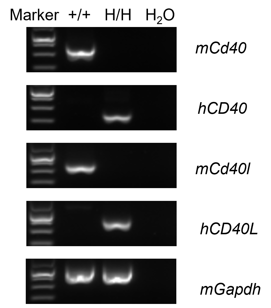
Strain specific analysis of CD40 and CD40L gene expression in wild-type C57BL/6 mice and B-hCD40/CD40L mice by RT-PCR. Mouse Cd40 and Cd40l mRNA were detectable in splenocytes of wild-type C57BL/6 mice (+/+). Human CD40 and CD40L mRNA were detectable only in homozygous B-hCD40/CD40L (H/H), but not in wild-type C57BL/6 mice (+/+).
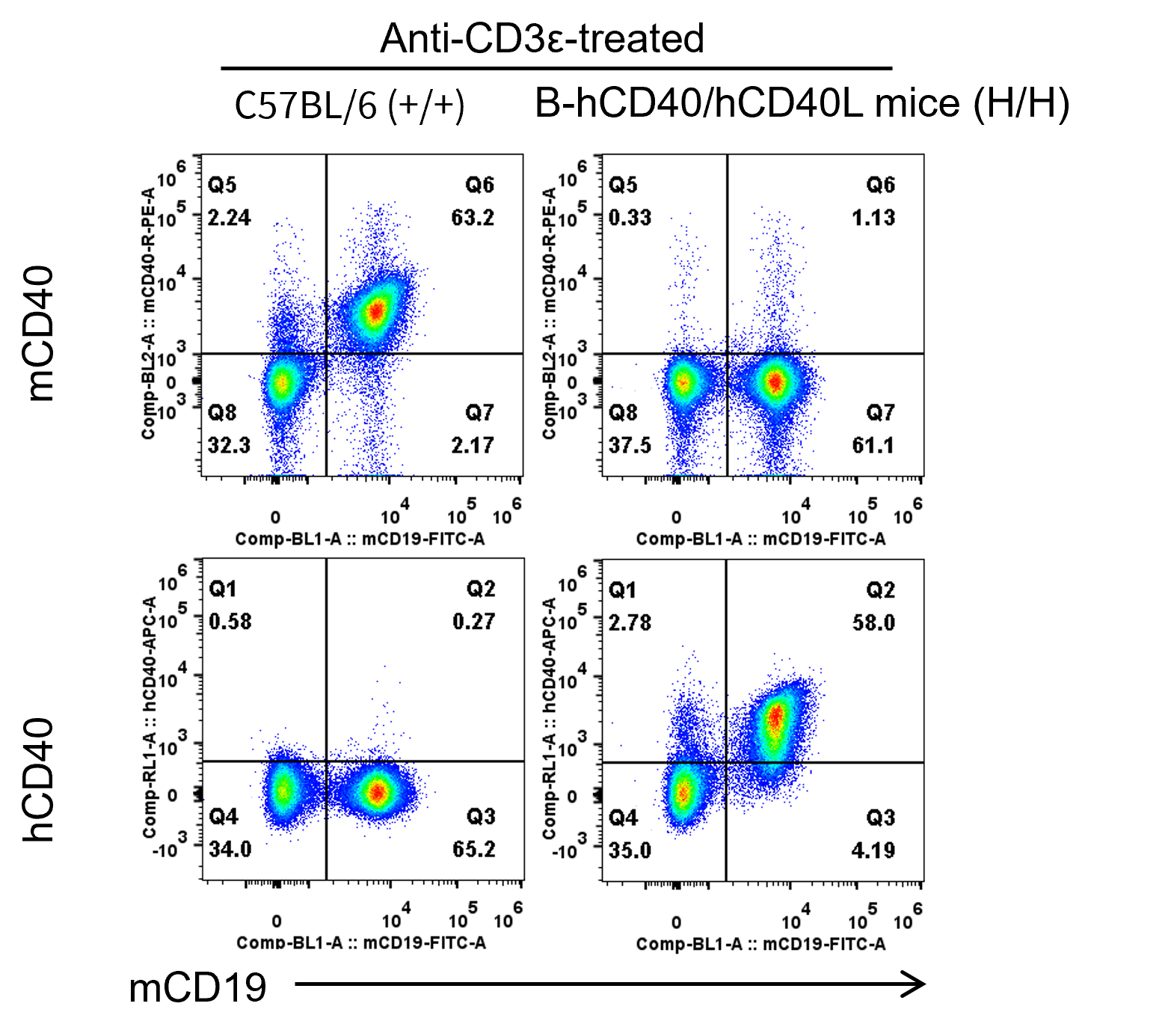
Strain specific CD40 expression analysis in homozygous B-hCD40/hCD40L mice by flow cytometry. Splenocytes were collected from wild-type C57BL/6 mice (+/+) and homozygous B-hCD40/hCD40L mice (H/H) stimulated with anti-CD3ε in vivo (7.5 μg/mice, stimulation for 24 hours, i.p.), and analyzed by flow cytometry with anti-mouse CD40 antibody (Biolegend, 124609) and anti-human CD40 antibody (Biolegend, 313008). Mouse CD40 was detectable in wild-type C57BL/6 mice. Human CD40 was exclusively detectable in homozygous B-hCD40/hCD40L but not in wild-type C57BL/6 mice.
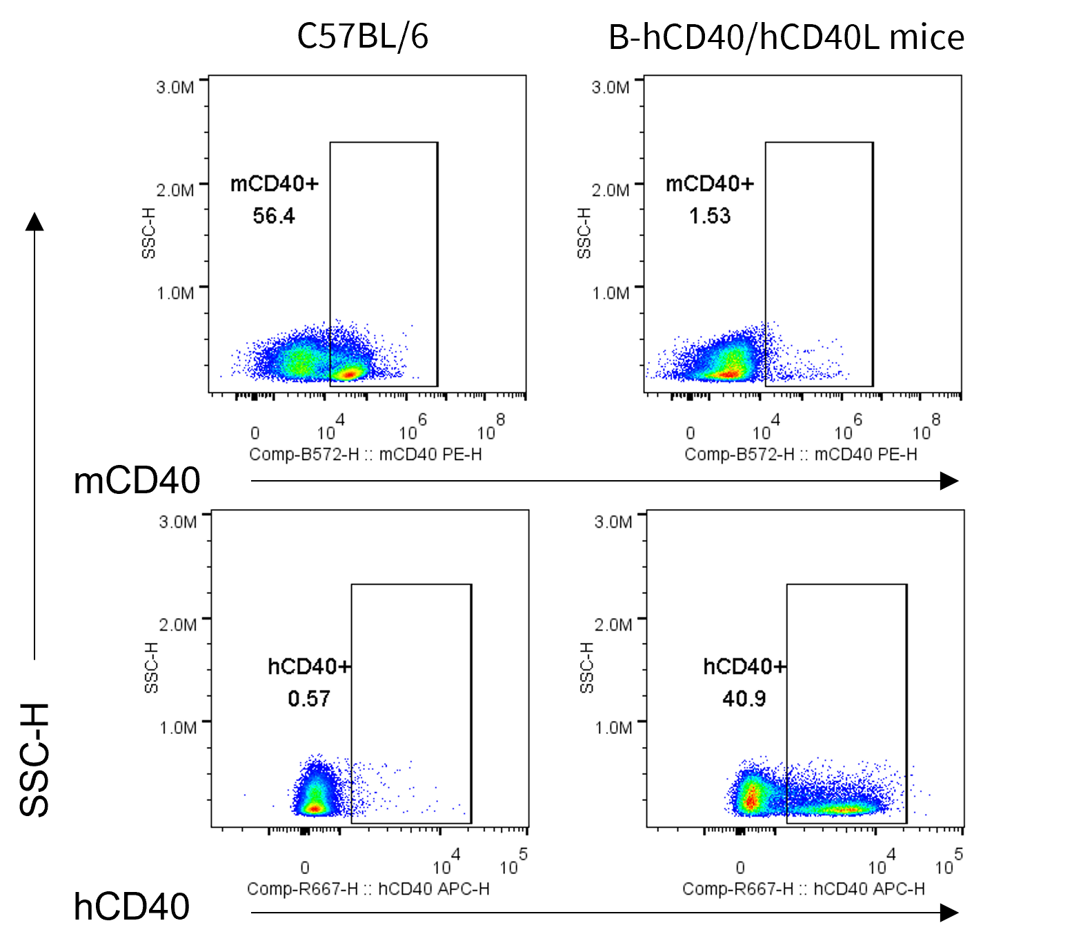
Strain specific CD40 expression analysis in wild-type C57BL/6 mice and homozygous humanized B-hCD40/hCD40L by flow cytometry. Bone marrow cells were collected from wild-type C57BL/6 mice and homozygous B-hCD40/hCD40L mice. Protein expression was analyzed with anti-mouse CD40 antibody (Biolegend, 124609) and anti-human CD40 antibody (Biolegend, 334309) by flow cytometry. Mouse CD40 was only detectable on megakaryocytes from bone marrow of wild-type C57BL/6 mice. Human CD40 was only detectable on megakaryocytes from bone marrow of homozygous B-hCD40/hCD40L mice.
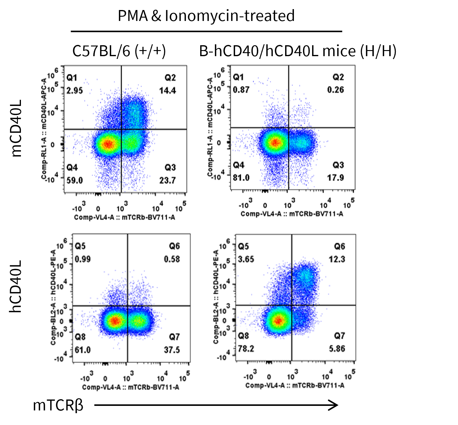
Strain specific CD40L expression analysis in homozygous B-hCD40/hCD40L mice by flow cytometry. Thymocytes were collected from wild-type C57BL/6 mice (+/+) and homozygous B-hCD40/hCD40L mice (H/H) and stimulated with PMA & Ionomycin, then analyzed by flow cytometry with anti-mouse CD40L antibody (Biolegend, 106509) and anti-human CD40L antibody (Biolegend, 310805). Mouse CD40L was detectable in wild-type C57BL/6 mice. Human CD40L was exclusively detectable in homozygous B-hCD40/hCD40L but not in wild-type C57BL/6 mice.

Strain specific mouse and human soluble CD40L (sCD40L) expression analysis in wild-type C57BL/6JNifdc mice and homozygous humanized B-hCD40/hCD40L mice by ELISA. Splenocytes were collected from wild-type C57BL/6JNifdc mice (+/+) (male, 8-week-old) and homozygous B-hCD40/hCD40L mice (H/H; H/H) (male, 8-week-old), and cultured in 96-well plated at 2E6 cells/well, then stimulated with PMA & ionomycin (Biolegend, 423301) for 5 or 24 h. Then cell culture supernatants were collected for ELISA analysis of mouse sCD40L (Abcam, ab275105) and human sCD40L (Abcam, ab99991). Mouse sCD40L was detectable in cell culture supernatant of splenocytes from C57BL/6JNifdc mice after PMA & ionomycin stimulation. Human sCD40L was detectable in cell culture supernatant of splenocytes from homozygous B-hCD40/hCD40L mice after PMA & ionomycin stimulation.
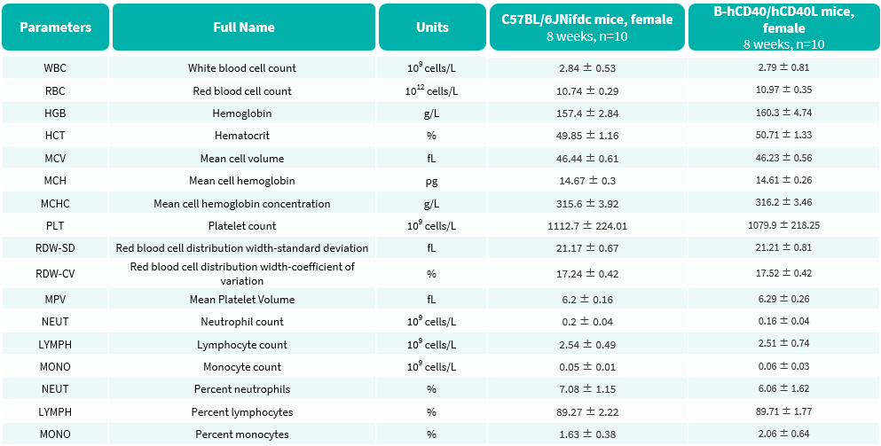
Complete blood count (CBC) of B-hCD40/hCD40L mice. Values are expressed as mean ± SD.
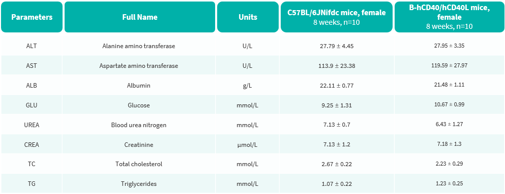
Blood biochemical parameters of B-hCD40/hCD40L mice. Values are expressed as mean ± SD.

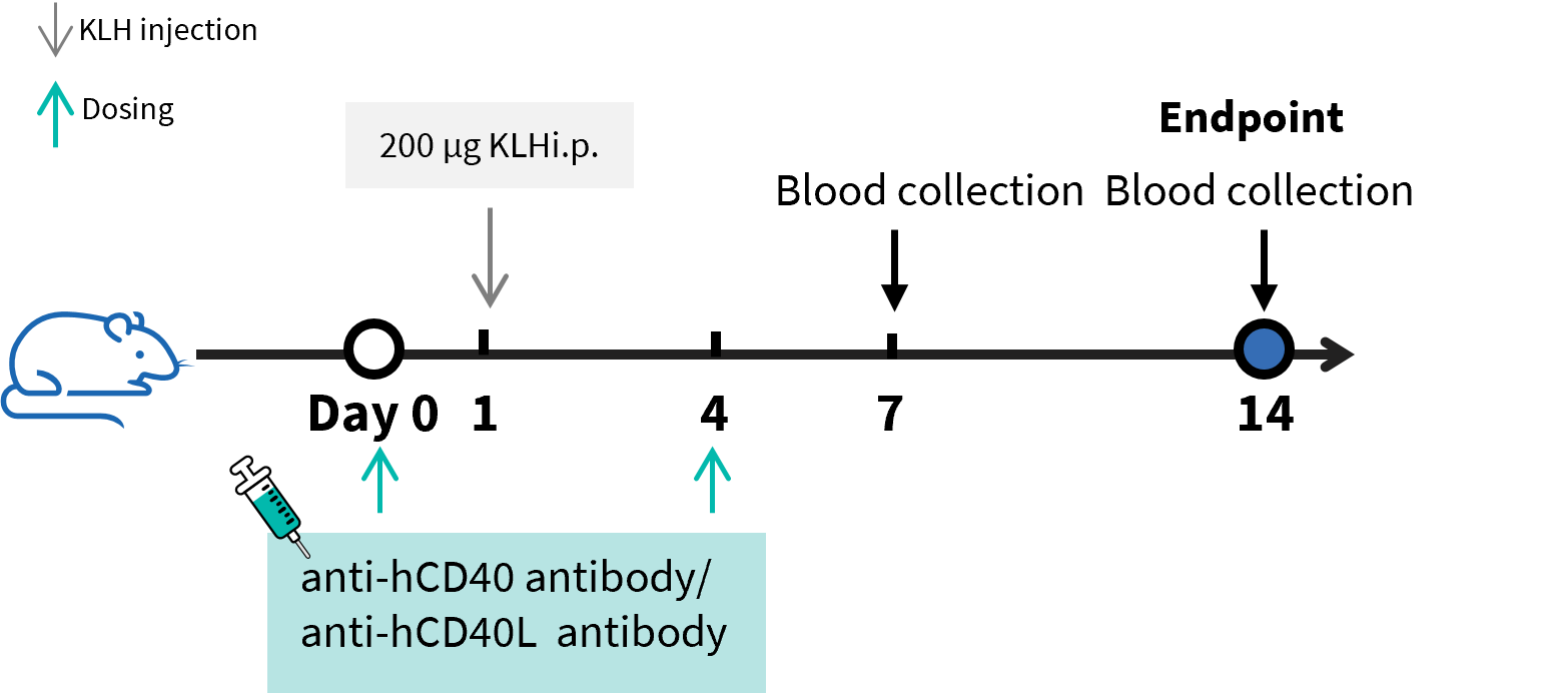
B-hCD40/hCD40L mice were used to establish T cell-Dependent Antibody Response assay (TDAR assay) and evaluate the efficacy of anti-CD40 antibody and anti-CD40L antibody. B-hCD40/hCD40L mice (n=5) were intraperitoneally immunized with 200 μg KLH on day 1 and treated with anti-CD40L antibody (provided by the client) or anti-CD40 antibody bleselumab analog (in house) on day 0 and day 4. Blood was collected on day 7 and day 14 and analyzed by ELISA with KLH specific IgG antibody and KLH specific IgM antibody.

B-hCD40/hCD40L mice was used to establish The T-Dependent Antibody Response assay (TDAR assay) and evaluate the efficacy of anti-CD40 antibody and anti-CD40L antibody. B-hCD40/hCD40L mice (n=5) were intraperitoneally immunized with 200 μg KLH on Day 1 and treated with anti-CD40L antibody (provided by the client) or anti-CD40 antibody bleselumab analog (in house). Blood was collected on Day 7 and Day 14 and analyzed by ELISA with KLH specific IgG antibody and KLH specific IgM antibody. (A) Body weight of B-hCD40/CD40L mice increased steadily; (B, C) Concentration of mouse KLH specific IgM and IgG were significantly increased after immunization. But the concentration of KLH specific IgG and IgM in the groups treated with anti-CD40L antibody or anti-CD40 antibody bleselumab analog (in house) were significantly decreased when compared to that in the control group, demonstrating that the B-hCD40/hCD40L mice provide a powerful preclinical model for in vivo evaluation of anti-CD40 antibody and anti-CD40L antibody.
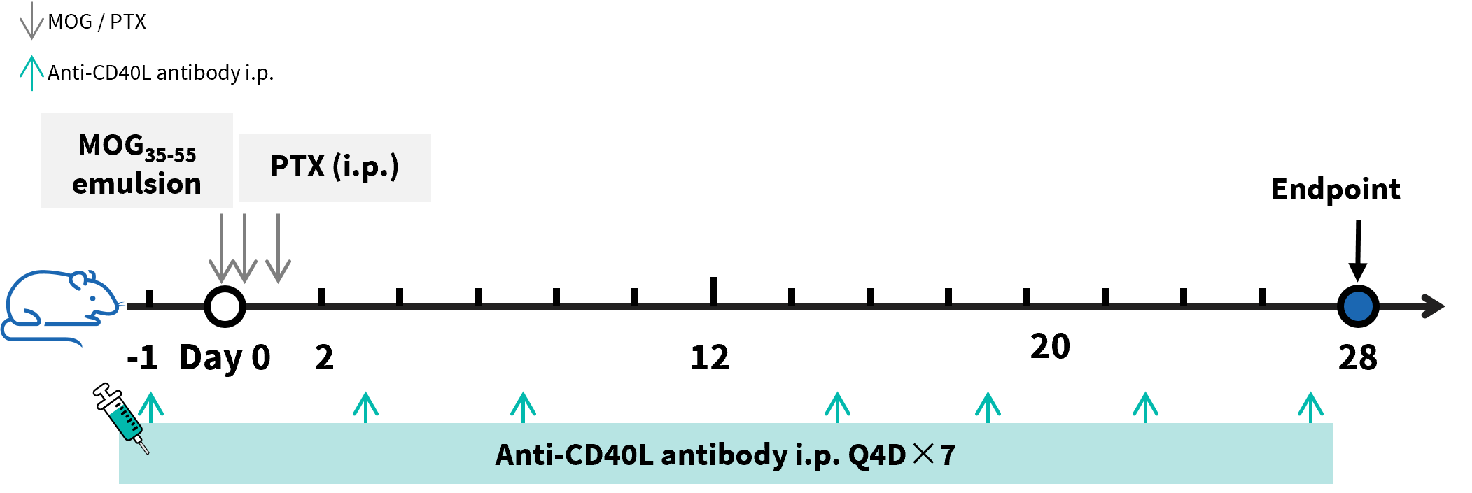
Effects of anti-CD40L antibody (in-house) on MOG35-55 induced EAE. B-hCD40/hCD40L mice (female, 11-week-old, n=6) received MOG35-55 emulsion injection (s.c.) on neck and buttock on day 0. PTX (i.p.) were given 2 and 24 hour after MOG injection. Anti-CD40L antibody dapirolizumab analog (in house) or SAR441344 analog (in house) were treated on day -1, 3, 7, 15, 19, 23, 27. Values are expressed as mean ± SEM. MOG: myelin-oligodendrocyte glycoprotein; PTX: pertussis toxin.

Effects of anti-CD40L antibody (in-house) on MOG35-55 induced EAE. Body weight (A) and clinical score (B) were recorded every two days. The results showed that compared with the control group (G1), MOG35-55 immunized mice (G2) exhibited symptoms such as tail weakness, limping, hind limb paralysis and other symptoms, with a significant increase in clinical scores. This indicates that the EAE disease model was successfully induced in B-hCD40/hCD40L mice. After treating anti-CD40L antibody dapirolizumab analog (in house) or SAR441344 analog (in house), a significant alleviation of clinical symptoms was observed. Values are expressed as mean ± SEM.
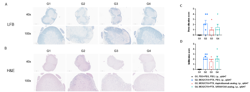
Effects of anti-CD40L antibody (in-house) improves EAE clinical signs and controls inflammation and demyelination. Spinal cords were removed from B-hCD40/hCD40L mice on day 30 and stained with Luxol fast blue (LFB) (A) or hematoxylin and eosin (H&E) (B). Representative sections are shown. The score of inflammatory cells and demyelination of spinal cord (C&D).
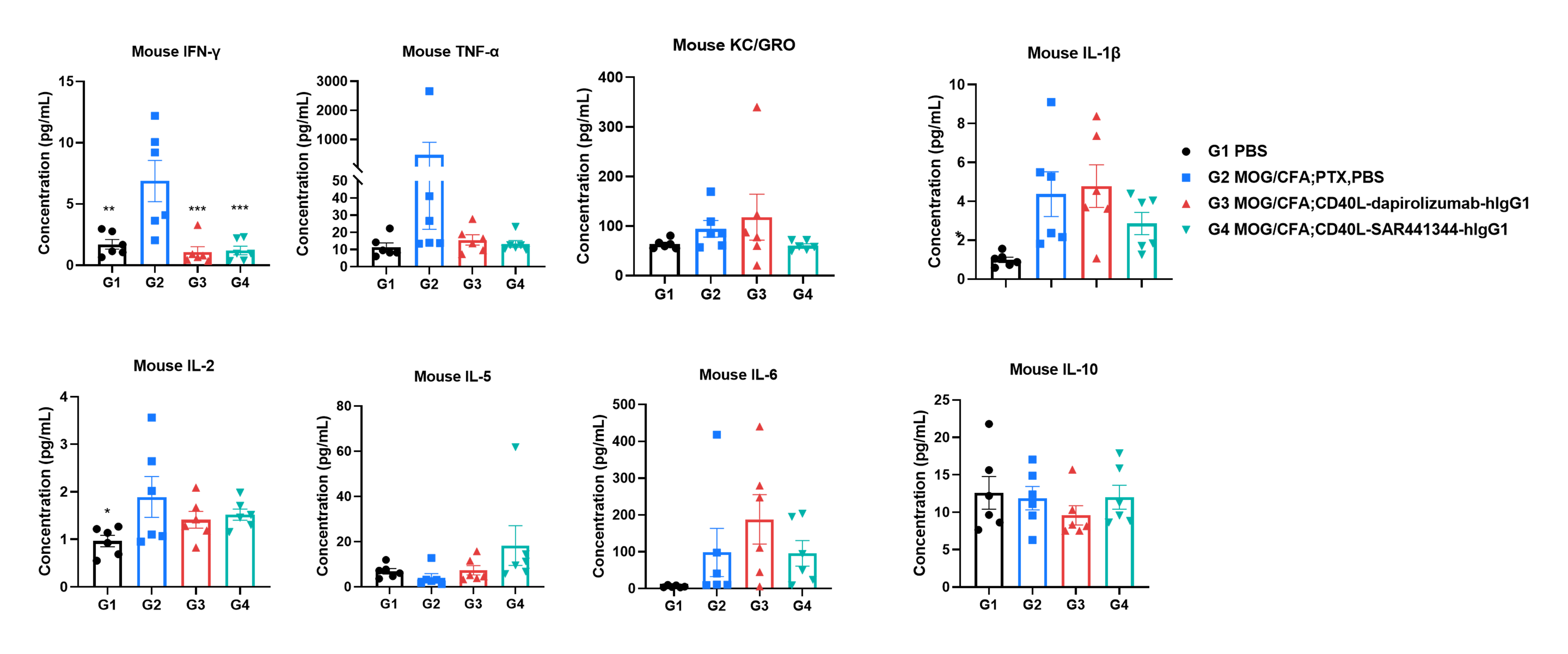
Effects of anti-CD40L antibody (in-house) on MOG35-55 induced EAE. Mice received MOG35-55 emulsion injection (s.c.) on neck and buttock (red point) on day 0. PTX (i.p.) were given 2 and 24 hour after MOG injection. Anti-CD40L antibody dapirolizumab analog (in house) or SAR441344 analog (in house) were administered Q4D for seven times. Serum were collected at study endpoint and the cytokine levels were assessed. Values are expressed as mean ± SEM, n=6, compared with G2, *p<0.05, **p<0.01, *** p<0.001, **** p<0.0001.
