

C57BL/6-Cd40tm1(CD40)Bcgen/Bcgen • 110009

CD40: a key costimulatory receptor in regulating immune response and its therapeutic intervention

CD40

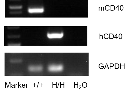
Strain specific analysis of CD40 gene expression in WT and B-hCD40 mice by RT-PCR. Spleen tissue was isolated from wild-type C57BL/6 mice (+/+) and homozygous B-hCD40 mice (H/H).Mouse Cd40 mRNA was detectable in splenocytes of wild-type (+/+). Human CD40 mRNA was detectable only in homozygous B-hCD40 mice (H/H) but not in wild-type C57BL/6 mice.
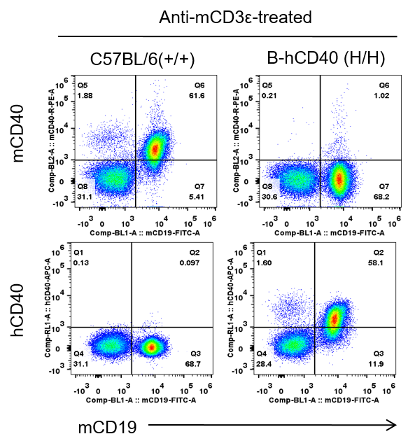
Strain specific CD40 expression analysis in homozygous B-hCD40 mice by flow cytometry. Splenocytes were collected from C57BL/6 and homozygous B-hCD40 (H/H) mice stimulated with anti-CD3ε in vivo (7.5 μg/mice), and analyzed by flow cytometry with species-specific anti-CD40 antibody. Mouse CD40 was exclusively detectable in C57BL/6 mice. Human CD40 were exclusively detectable in homozygous B-hCD40 mice but not C57BL/6 mice.
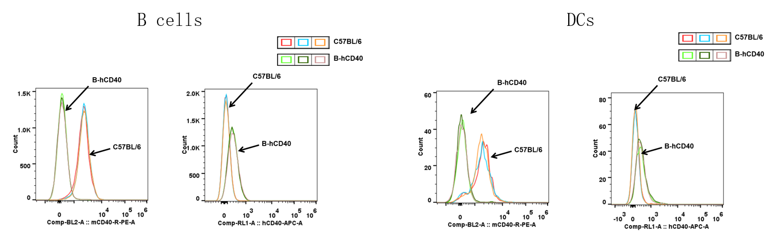
Strain specific CD40 expression analysis in homozygous B-hCD40 mice by flow cytometry. Splenocytes were collected from C57BL/6 and homozygous B-hCD40 (H/H) mice, and analyzed by flow cytometry with species-specific anti-CD40 antibody. Mouse CD40 was exclusively detectable in C57BL/6 mice. Human CD40 was exclusively detectable in homozygous B-hCD40 mice but not C57BL/6 mice.
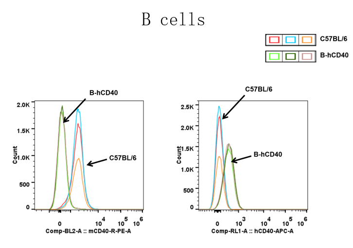
Strain specific CD40 expression analysis in homozygous B-hCD40 mice by flow cytometry. Blood cells were collected from C57BL/6 and homozygous B-hCD40 (H/H) mice, and analyzed by flow cytometry with species-specific anti-CD40 antibody. Mouse CD40 was exclusively detectable in C57BL/6 mice. Human CD40 was exclusively detectable in homozygous B-hCD40 mice but not C57BL/6 mice.
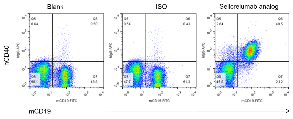
Analysis of splenocytes of B-hCD40 mice by FACS. Splenocytes were isolated from female B-hCD40 mice (n=3). Flow cytometry analysis of the splenocytes was performed to assess human CD40 expression on B cells. Single live cells were gated for CD45 population and used for further analysis as indicated here. Human CD40 expression was detectable on CD19 B cells in B-hCD40 mice as evidenced by selicrelumab analog (in house) binding vs isotype control.
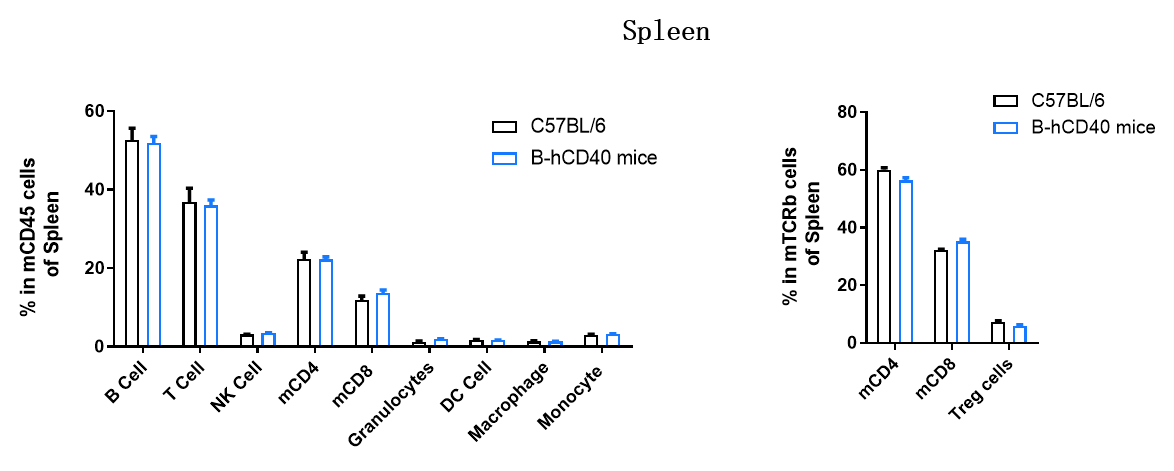
Analysis of leukocyte subpopulations by flow cytometry in spleen. Splenocytes were isolated from C57BL/6 and B-hCD40 mice (female, 9-week-old, n = 3). Single live cells were gated on the CD45⁺ population and analyzed by flow cytometry as indicated. Values are expressed as mean ± SEM.
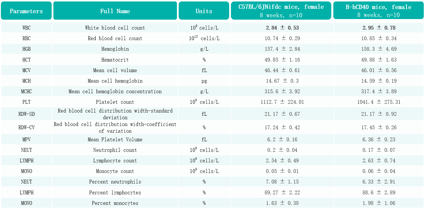
Complete blood count (CBC) of B-hCD40 mice. Values are expressed as mean ± SD.
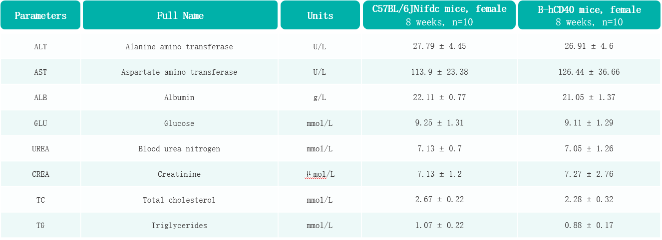
Blood biochemical parameters of B-hCD40 mice. Values are expressed as mean ± SD.

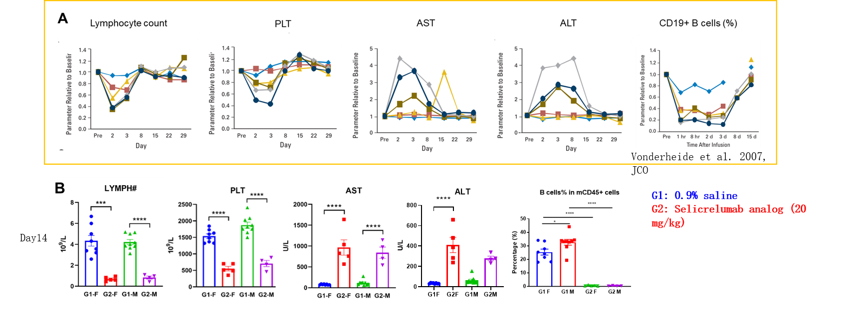
Immune-related adverse event comparison between patients and humanized B-hCD40 mice.(A) After injection of selicrelumab analog, the clinical patients had obvious hematotoxicity, and the number of peripheral lymphocytes and platelets in the blood decreased. The increase of ALT and AST suggested the occurrence of hepatotoxicity. In addition, CD19+ B cells decreased significantly. (B) B-hCD40 mice of different genders were treated with selicrelumab analog, which showed adverse reactions consistent with clinical symptoms.
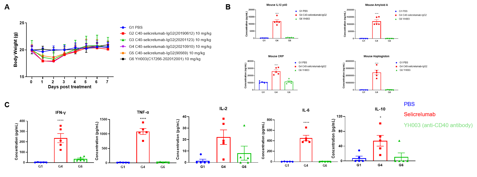
Toxicological profiles of selicrelumab analog (in house) in B-hCD40 mice. (A) The body weight of mice in selicrelumab analog treatment group decreased significantly. (B) Acute phase proteins such as CRP, Amyloid A, Haptoglobin and IL12P40 is increased in response to selicrelumab analog in B-hCD40 mice. (C) The mice treated with selicrelumab had obvious cytokine release syndrome, IFN- γ, TNF- α, IL-2, IL-6, IL-10 increased obviously.

Antitumor activity of anti-human CD40 antibodies in B-hCD40 mice. (A) Anti-human CD40 antibodies inhibited MC38 tumor growth in B-hCD40 mice. Murine colon cancer MC38 cells (5ⅹ105) were subcutaneously implanted into heterozygous B-hCD40 mice (female, 4-week-old, n=5). Mice were grouped when tumor volume reached approximately 100 mm3, at which time they were treated with three anti-human CD40 antibodies with doses and schedules indicated in panel. (B) Body weight changes during treatment. As shown in panel A, anti-human CD40 antibodies were efficacious in controlling tumor growth in B-hCD40 mice, demonstrating that the B-hCD40 mice provide a powerful preclinical model for in vivo evaluation of anti-human CD40 antibodies. Values are expressed as mean ± SEM.

Antitumor activity of anti-human CD40 antibody in B-hCD40 mice. (A) Anti-human CD40 antibody inhibited MC38 tumor growth in B-hCD40 mice. Murine colon cancer MC38 cells(5ⅹ105) were subcutaneously implanted into homozygous B-hCD40 mice (female,4-week-old, n=5). Mice were grouped when tumor volume reached approximately 100 mm3, at which time they were treated with anti-human CD40 antibody with doses and schedules indicated in panel A. (B) Body weight changes during treatment. As shown in panel A, anti-human CD40 antibody was efficacious in controlling tumor growth in B-hCD40 mice, demonstrating that the B-hCD40 mice provide a powerful preclinical model for in vivo evaluation of anti-human CD40 antibodies. Values are expressed as mean ± SEM.

Antitumor activity of anti-human CD40 antibody in B-hCD40 mice. (A) Anti-hCD40 antibody selicrelumab analog (in house) inhibited MC38 tumor growth in B-hCD40 mice. Murine colon cancer MC38 cells(5ⅹ105) were subcutaneously implanted into homozygous B-hCD40 mice (female,8-9 week-old, n=5). Mice were grouped when tumor volume reached approximately 150±50 mm3, at which time they were treated with anti-human CD40 antibody selicrelumab analog (in house) with doses and schedules indicated in panel A. (B) Body weight changes during treatment. As shown in panel A, anti-human CD40 antibody selicrelumab analog (in house) was efficacious in controlling tumor growth in B-hCD40 mice, demonstrating that the B-hCD40 mice provide a powerful preclinical model for in vivo evaluation of anti-human CD40 antibodies. Values are expressed as mean ± SEM.
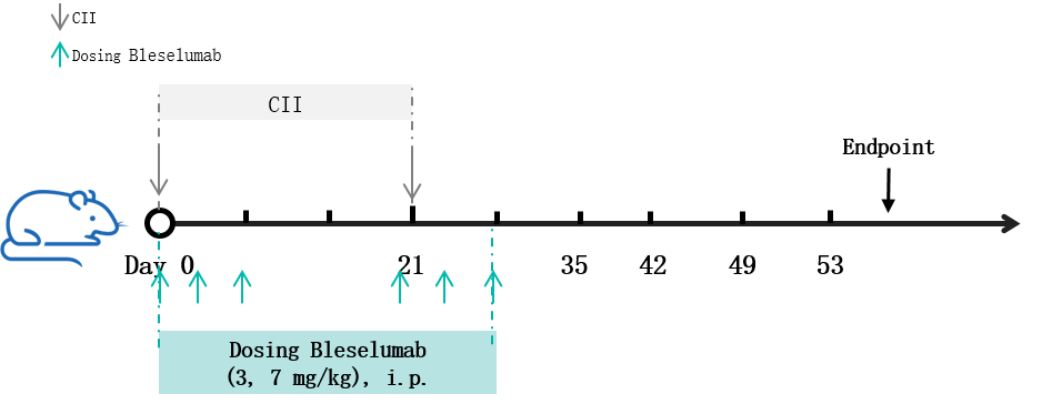
Experimental schedule for the induction of collagen-induced arthritis (CIA) and in vivo efficacy of anti-human CD40 antibody in B-hCD40 mice. Mice were subcutaneously injected with CII emulsion on day 0 and 21. The anti-human CD40 antibody bleselumab analog (in-house) was administered by intraperitoneal injection (n = 10). CII, collagen.

Efficacy of the anti-human CD40 antibody in a mouse arthritis model. (A) Clinical score; (B) Incidence of disease; (C) Change in mouse body weight. The results showed that in the modeling group (G1&G3), the clinical scores of the mice significantly increased, indicating successful establishment of the CIA model, while body weight fluctuations were more pronounced compared to the non-model group (G2). After the administration of anti-human CD40 antibody (bleselumab analog, synthesized in-house), the average clinical score of the treatment group (G4&G5) was significantly lower than that of the model group, suggesting that the condition of mice was effectively controlled. Additionally, the maximum incidence in the treatment group also showed significantly lower than the model group, indicating that the anti-human CD40 antibody has a therapeutic effect on this disease.
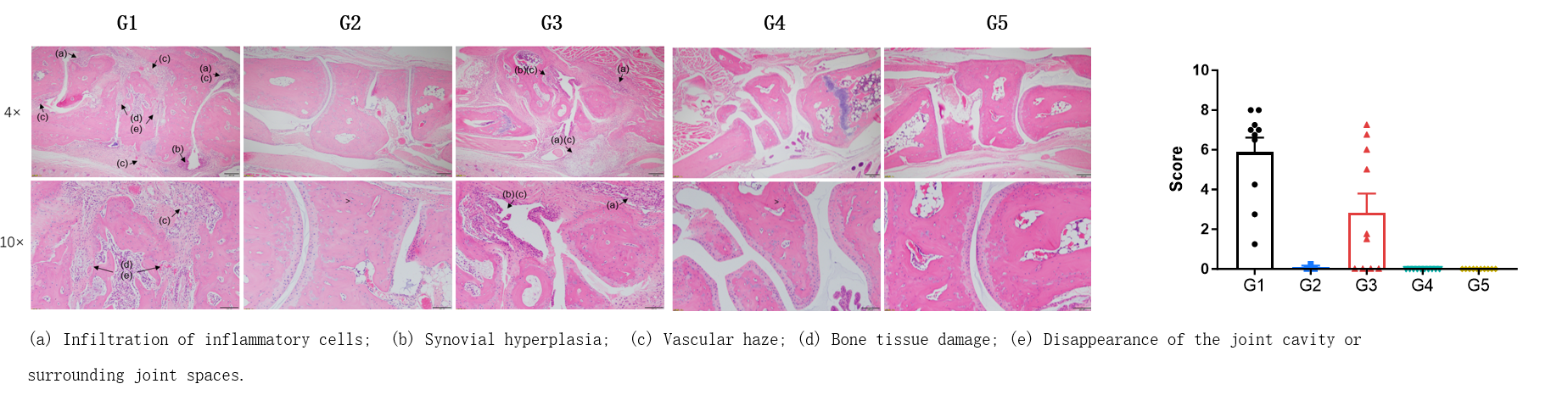
The pathological analysis of the efficacy of the anti-human CD40 antibody bleselumab analog (in-house) in CIA model.(A) Pathological sections stained with H&E; (B) Pathological scores. The results showed that in the non-model group (G2), there were no significant abnormal changes observed, with a smooth cartilage surface in the ankle joint and a clearly defined joint cavity. In the modeling group (G1&G3), the animals exhibited the infiltration of inflammatory cells (a), synovial hyperplasia (d) and vascular haze (e). Compared to the model group, the bleselumab treatment groups (G4&G5) significantly alleviated the disease symptoms in the mice. This suggests that bleselumab analog has a therapeutic effect on arthritis in mice.


Establishment of SLE Mouse Model based on B-hCD40 Mice.Content of mouse Anti-dsDNA IgG in serum was decreased after treatment with anti-CD40 antibody in pristane-induced SLE B-hCD40 mice.

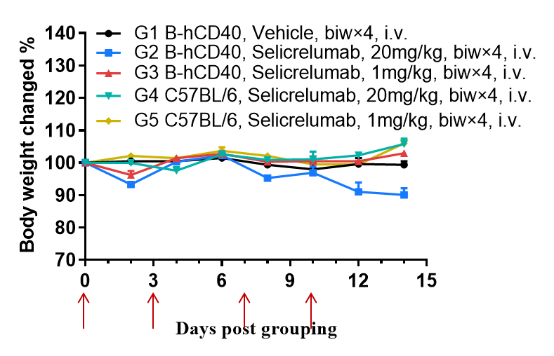
High does of selicrelumab (anti human CD40) moderately reduced body weight in B-hCD40 mice but not in wild-type C57BL/6 mice. Homozygous B-hCD40 mice and C57BL/6 mice were treated with vehicle or selicrelumab analog (in house) on days indicated by red arrows (n=3). Selicrelumab analog at 20 mg/kg caused significant body weight reduction compared with vehicle control in B-hCD40 mice, while the same treatment had no effect in C57BL/6 mice. Values are expressed as mean ± SEM.
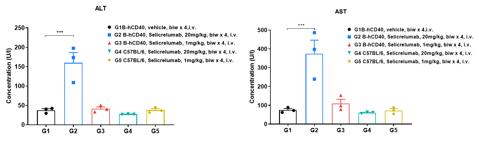
High-dose human CD40 antibody caused significant changes in AST and ALT in B-hCD40 mice. Homozygous B-hCD40 mice and C57BL/6 mice were treated with PBS or selicrelumab analog (in house) as indicated on the dosing schedule (n=3). The blood biochemical analysis was conducted on B-hCD40 mice treated with anti-human CD40 antibody 24 hours after the termination of efficacy evaluation experiment. Selicrelumab analog at 20 mg/kg caused significant increase of AST and ALT compared with PBS control in B-hCD40 mice, while the same treatment had no effect in C57BL/6 mice. Values are expressed as mean ± SEM.
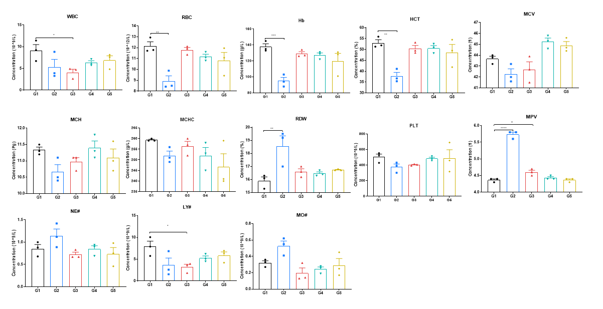
High-dose human CD40 antibody caused significant changes of complete blood count in B-hCD40 mice. Homozygous B-hCD40 mice and C57BL/6 mice were treated with PBS or selicrelumab analog (in house) as indicated on the graph (n=3). The complete blood count (CBC) was performed 24 hours after last dosing on day 11. Selicrelumab analog at 20 mg/kg caused significant reduction of WBC, RBC, Hb, HCT and LY#, and increase of RDW and MPV compared with PBS control in B-hCD40 mice, while the same treatment had no effect in C57BL/6 mice. Values are expressed as mean ± SEM.
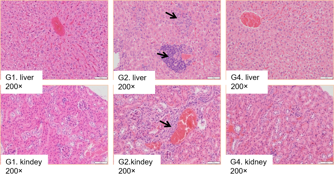
High dose selicrelumab caused liver and kidney pathological changes in B-hCD40 mice. Homozygous B-hCD40 mice and C57BL/6 mice were treated with PBS or selicrelumab analog (n=3). The tissue pathology analysis was performed on day 14. Selicrelumab analog at 20mg/kg caused increasing lymphocyte infiltration (as indicated by arrows) into liver and kidney in B-hCD40 mice, while the same treatment had no effect on C57BL/6 mice.
