you may also like
With an estimated 850 million people worldwide affected by kidney disease, nephrology is rapidly shifting toward more mechanism- and target-driven therapies. Recent FDA approvals reflect this momentum, including BAFF/APRIL-targeting Trutakna™ (atacicept) for IgA nephropathy, ETA/AT1 dual antagonist Filspari® (sparsentan) for FSGS, and complement-targeted therapies such as Fabhalta® (iptacopan) and Empaveli® (pegcetacoplan) for complement 3 glomerulopathy (C3G). Continued progress in APOL1- and complement-directed pipelines further underscores the growing momentum behind precision nephrology.
As kidney therapeutics become increasingly target- and mechanism-specific, Biocytogen’s portfolio of target-humanized and disease-relevant mouse models provides fit-for-purpose in vivo systems for evaluating human target biology, renal disease phenotypes, and therapeutic response across emerging kidney drug programs.
♦ IgA Nephropathy — IGHA1/CD89 humanized mice (B-hIGHA1/hCD89 plus mice): Develop progressive renal IgA deposition and enable evaluation of therapeutic responses to BAFF/APRIL pathway modulation.
♦ Complement-Mediated Kidney Disease — C3 R102G humanized mice (B-hC3*R102G mice plus): Provide a human C3-based in vivo system for studying complement dysregulation and evaluating human C3-targeted therapeutic strategies.
♦ Autosomal Dominant Polycystic Kidney Disease (ADPKD) — B-Pkd1 flox, CAG-iCreERT2 mice: Develop renal cyst formation and tubular dilation following induction, providing a disease-relevant model of genetically driven cystic kidney pathology.
♦ APOL1-Mediated Kidney Disease — C3 R102G humanized mice (B-hAPOL1-G1 mice): Enable evaluation of APOL1-targeting nucleic acid therapeutics under basal and IFN-γ-stimulated conditions.
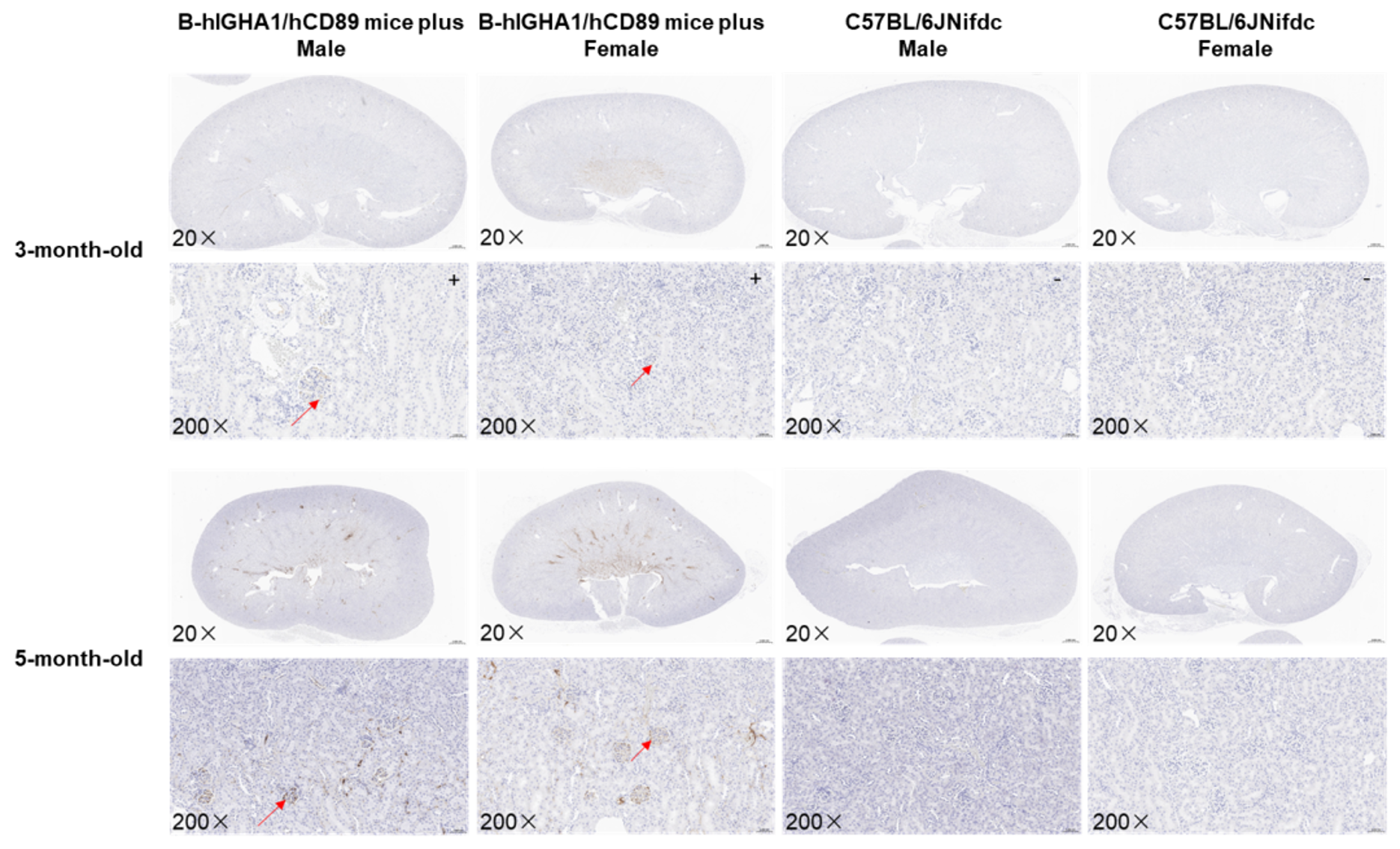
Analysis of glomerular IgA deposition in B-hIGHA1/hCD89 plus mice. Kidney tissues were analyzed by immunohistochemistry (IHC) for immunoglobulin A (IgA). Compared with wild-type controls, B-hIGHA1/hCD89 plus mice showed prominent glomerular IgA deposition that progressively increased with age.
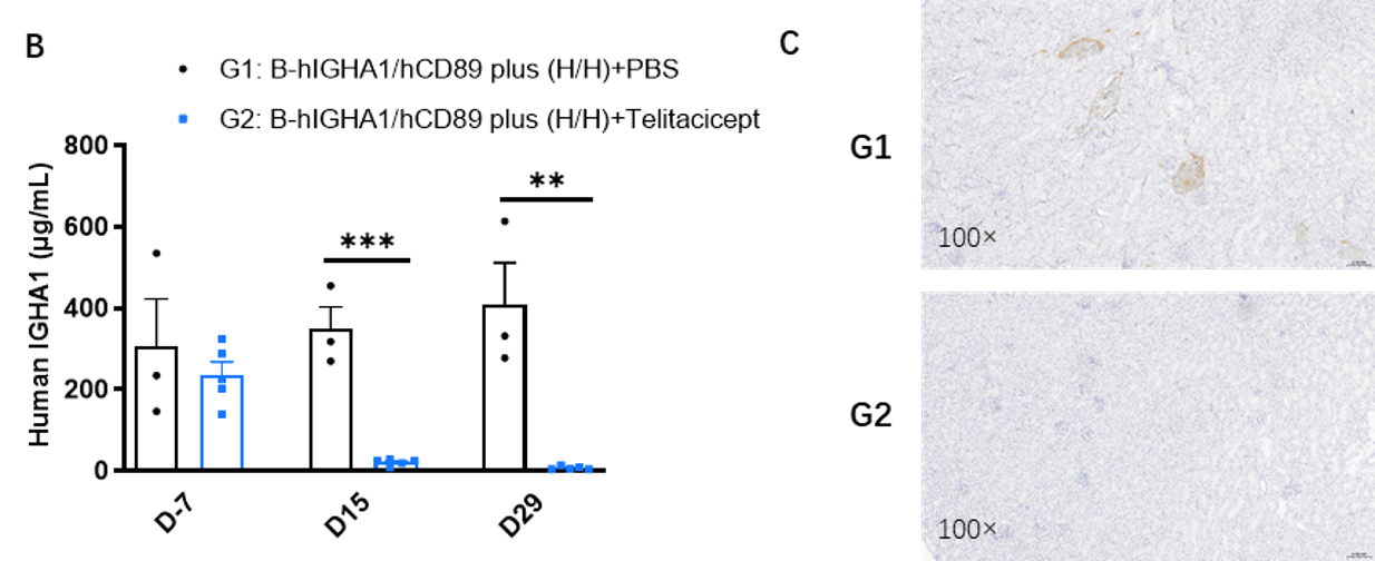
Inhibitory effect of telitacicept on human IGHA1 in B-hIGHA1/hCD89 plus mice. B-hIGHA1/hCD89 plus mice were treated with PBS (n=3) or telitacicept (n=5) every other day for 14 doses. Serum human IGHA1 levels measured on Days −7, 15, and 29 were significantly reduced with telitacicept. Kidney immunohistochemistry showed prominent IgA deposition in control mice but no detectable deposition after treatment.
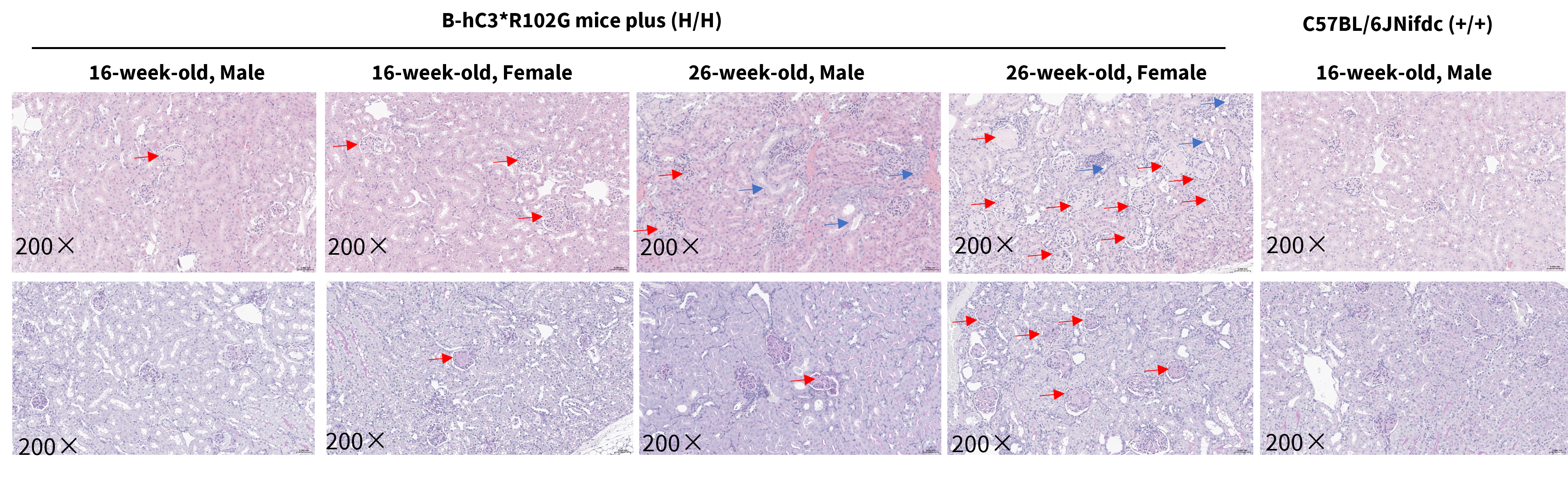
Histopathological analysis in B-hC3*R102G mice plus. The kidneys of homozygous B-hC3*R102G mice plus (H/H) were isolated at 16 weeks old and 26 weeks old and analyzed with HE staining and PAS staining. The kidney of B-hC3*R102G mice plus showed glomerular cell proliferation, renal tubular basophilic changes, and dilation. Compared with wild-type mice, B-hC3*R102G mice plus exhibited increased PAS-positive staining indicative of glomerular mesangial matrix expansion. And there were no obvious abnormalities in wild-type C57BL/6JNifdc mice. Red arrows: Glomerular lesions, Blue arrows: Tubular lesions.
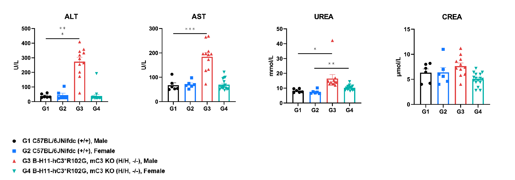
Analysis of blood biochemicals in B-hC3*R102G mice plus and wild-type C57BL/6JNifdc mice. Serum was collected from wild-type C57BL/6JNifdc mice (+/+) and and homozygous B-hC3*R102G mice plus (H/H) (male n=11, female n=16, 16 weeks old) and analyzed for biochemistry. The ALT, AST, and UREA were increased in male B-hC3*R102G mice plus.
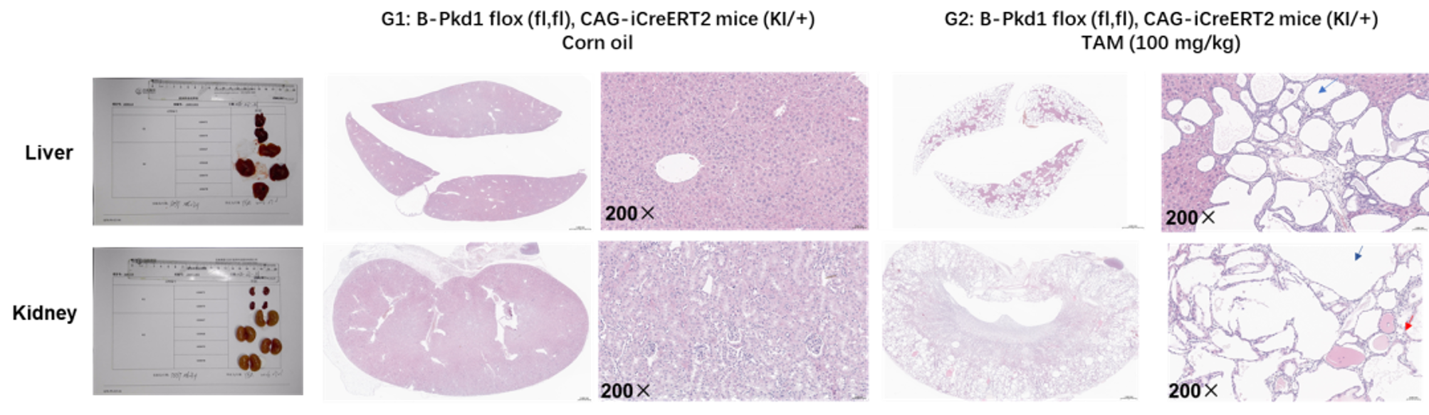
Tamoxifen-induced cystic pathology in B-Pkd1 flox, CAG-iCreERT2 mice. At 16 weeks post-tamoxifen, H&E staining showed extensive hepatic cysts and bile duct proliferation, along with renal cysts, tubular dilation, and intratubular calcific deposits compared with controls. Red arrows indicate tubular calcification; blue arrows indicate cysts.
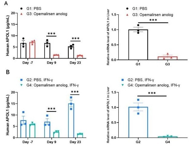
Inhibitory effect of an APOL1-targeting nucleic acid therapeutic in homozygous B-hAPOL1-G1 mice. Eight-week-old females were treated with an Opemalirsen analog or PBS, followed by IFN-γ or PBS challenge. Serum APOL1 measured at baseline and on Days 9 and 23 was markedly reduced by the Opemalirsen analog under both basal and IFN-γ–stimulated conditions, despite IFN-γ–induced APOL1 upregulation.
|
As kidney drug development becomes more focused on defined disease mechanisms and molecular targets, the choice of preclinical model becomes increasingly important. Models that reflect both human target biology and relevant renal pathology can help researchers better understand treatment response and generate more informative in vivo data.
Biocytogen offers a broad range of target-humanized and disease-relevant mouse models for kidney research, covering immune-mediated, complement-associated, cystic, and genetically driven kidney diseases. These models can be used to assess target engagement, renal pathology, pharmacodynamic response, therapeutic activity, and safety across different stages of preclinical development.
A target-humanized model is designed primarily to reproduce human target biology, while a disease model is designed to reproduce relevant disease pathology. For example, B-hAPOL1-G1 and B-hIGHA1/hCD89 plus mice enable studies involving human therapeutic targets, whereas B-Pkd1 flox, CAG-iCreERT2 mice reproduce genetically induced cystic kidney pathology.
Target-humanized mouse models enable human target biology to be studied directly in vivo, making them especially valuable for therapeutics whose activity depends on human-specific target sequence, expression, or pharmacology. In kidney research, these models can support the evaluation of target engagement, pharmacodynamic response, renal pathology, therapeutic activity, and safety, helping connect molecular mechanisms with relevant in vivo outcomes during preclinical development.
B-hIGHA1/hCD89 plus mice provide a disease-relevant model for IgA nephropathy (IgAN). These mice develop progressive glomerular IgA deposition with age, and treatment with telitacicept reduced serum human IGHA1 levels and renal IgA deposition, enabling pharmacodynamic evaluation of therapies that modulate the BAFF/APRIL pathway.
B-hC3*R102G Mice plus provide a human C3-based in vivo system for investigating C3-associated renal pathology and human C3 biology. The model is particularly relevant to research on complement-associated kidney disease and may provide a platform for evaluating therapeutics directed at human C3 when appropriate pharmacology studies are incorporated.
Following induction, B-Pkd1 flox, CAG-iCreERT2 mice develop focal renal cysts, tubular dilation, and intratubular calcific deposits. The model also shows extensive liver cyst formation and bile duct proliferation, providing a system for investigating the multi-organ cystic pathology associated with loss of Pkd1.
In the available B-hAPOL1-G1 study, IFN-γ stimulation increased APOL1-G1 levels, creating a higher-expression condition in which APOL1-targeted therapy could be evaluated. The opemalirsen analog maintained APOL1 suppression under both basal and IFN-γ-stimulated conditions, providing a pharmacodynamic readout for APOL1-directed drug development.
The two model types address different translational questions. Target-humanized models, such as B-hIGHA1/hCD89 plus, B-hC3*R102G Mice plus, and B-hAPOL1-G1 mice, can enable direct evaluation of therapeutics acting on human molecular targets, while disease-relevant genetic models, such as B-Pkd1 flox, CAG-iCreERT2 mice, reproduce defined renal pathology. Used together, these approaches can connect human target engagement, renal phenotype, pharmacodynamic response, and therapeutic evaluation across different kidney disease mechanisms.
Jager KJ, Kovesdy C, Langham R, Rosenberg M, Jha V, Zoccali C. A single number for advocacy and communication—worldwide more than 850 million individuals have kidney diseases. Kidney International. 2019;96(5):1048–1050. doi:10.1016/j.kint.2019.07.012.
Lv J, Liu L, Wang W, et al.; TELIGAN Investigators. Telitacicept for IgA Nephropathy—Interim Analysis of a Phase 3 Trial. New England Journal of Medicine. 2026;394(19):1916–1924. doi:10.1056/NEJMoa2514415.
Fakhouri F, Bomback AS, Ariceta G, et al.; VALIANT Trial Investigators Group. Trial of Pegcetacoplan in C3 Glomerulopathy and Immune-Complex MPGN. New England Journal of Medicine. 2025;393(22):2210–2220. doi:10.1056/NEJMoa2501510.
Ibrahim ST, Chinnadurai R, Ali I, et al. Genetic polymorphism in C3 is associated with progression in chronic kidney disease (CKD) patients with IgA nephropathy but not in other causes of CKD. PLoS ONE. 2020;15(1):e0228101. doi:10.1371/journal.pone.0228101.
Piontek K, Menezes LF, Garcia-Gonzalez MA, Huso DL, Germino GG. A critical developmental switch defines the kinetics of kidney cyst formation after loss of Pkd1. Nature Medicine. 2007;13(12):1490–1495. doi:10.1038/nm1675.
Egbuna O, Zimmerman B, Manos G, et al. Inaxaplin for Proteinuric Kidney Disease in Persons with Two APOL1 Variants. New England Journal of Medicine. 2023;388(11):969–979. doi:10.1056/NEJMoa2202396.