you may also like
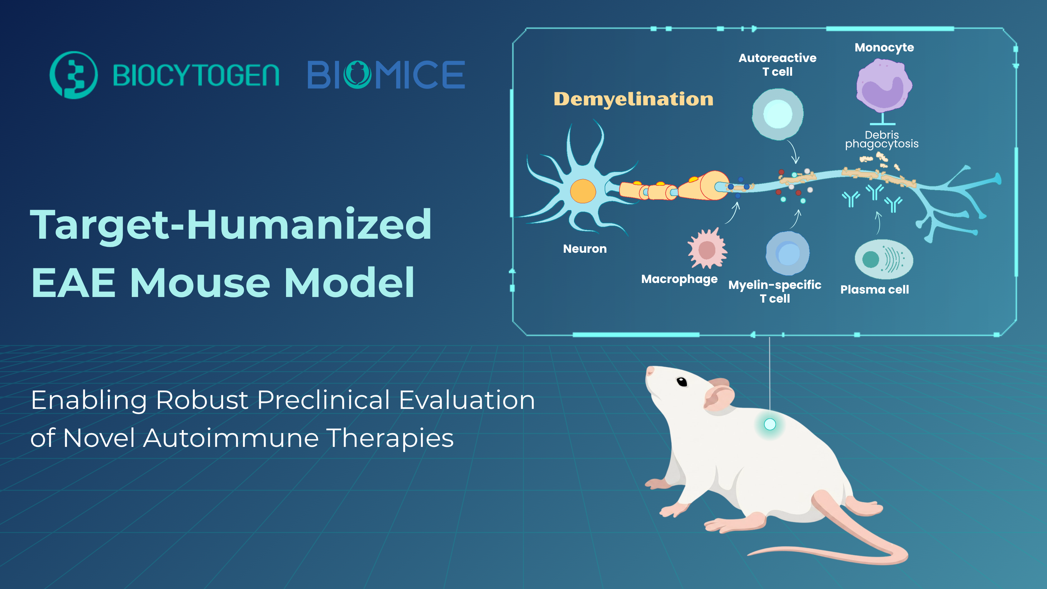
For nearly a century, Experimental Autoimmune Encephalomyelitis (EAE) has reigned as the cornerstone of Multiple Sclerosis (MS) research, serving as a versatile preclinical platform for dissecting neuroinflammation. Driven by genetic and environmental factors, MS is a chronic autoimmune disease characterized by immune-mediated demyelination of the central nervous system. This damage disrupts communication between the brain and the rest of the body, resulting in a highly variable clinical course that ranges from relapsing-remitting episodes to progressive neurological decline.
Because no single model can capture the full complexity of human MS, researchers rely on a diverse portfolio of application-driven EAE models to target specific pathological angles. Whether the research focuses on the gut-brain axis using germ-free models, blood-brain barrier integrity via connexin knockouts, or mimicking relapsing-remitting MS in SJL/J mice or progressive phenotypes in C57BL/6 and NOD strains—EAE offers an adaptable platform. This unparalleled versatility is why EAE remains the primary model of choice for MS therapeutic discovery, having successfully paved the way for major FDA-approved disease-modifying therapies (DMTs).
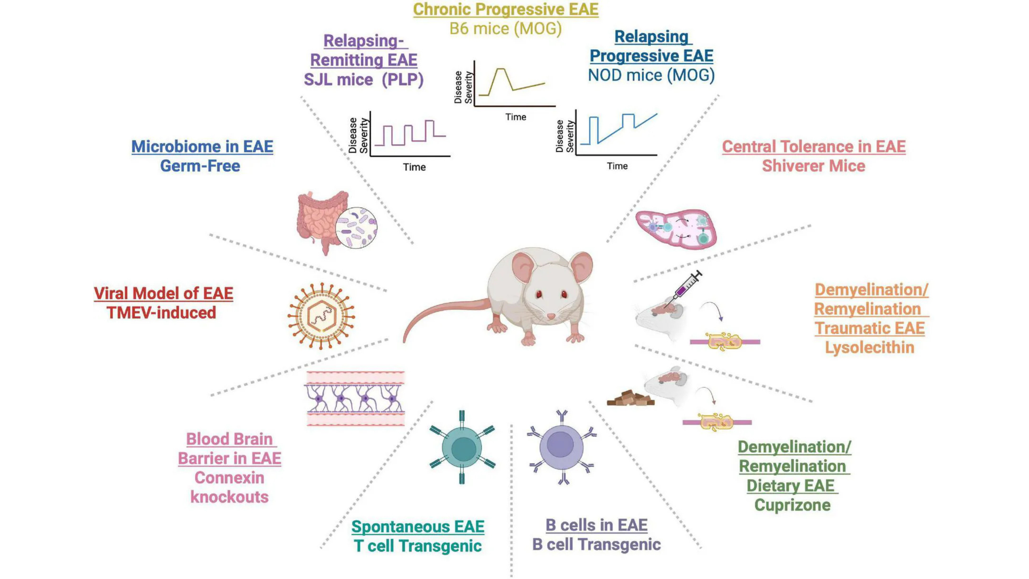
Diversity of EAE Mouse Model Mimicking Clinical Aspects of Multiple Sclerosis (Melamed et al. 2022)
While standard rodent models remain invaluable for MS research, they fall short when evaluating modern, human-specific biologics. Many next-generation biologics are designed to recognize human epitopes and often exhibit little or no cross-reactivity with wild-type rodent targets, limiting their translational relevance.
To overcome this challenge, Biocytogen has developed target-humanized EAE models that preserve authentic disease pathology while introducing human target biology, enabling direct in vivo evaluation of human-specific therapeutics. Our MOG-induced EAE model platform can be readily applied across our comprehensive portfolio of target-humanized mouse models, such as CD40/CD40L, CD3/CD20, and CD40/FcRn humanized models. Compared with conventional EAE models, our target-humanized EAE models provide validated, enhanced translational relevance for efficacy, toxicology, and mechanism-of-action studies.
► MOG1-125 Recombinant Protein Activation (T & B Cell Co-Driven Pathway): Specifically designed for evaluating therapies targeting B cells, pathogenic autoantibodies, or complement-mediated mechanisms, this model is induced using the full extracellular domain of myelin oligodendrocyte glycoprotein (MOG), a key central nervous system autoantigen. By recapitulating antibody-mediated demyelination, it provides a valuable platform for evaluating human antibodies and other biologics.
► MOG35-55 Peptide Activation (T Cell-Driven Pathway): Induced using the immunodominant MOG35–55 peptide, this classic EAE model selectively elicits T cell-mediated neuroinflammation and is well suited for evaluating therapies targeting autoreactive T-cell responses, including Th1- and Th17-associated pathways. It provides a robust platform for studying chronic neuroinflammation and immune cell infiltration into the central nervous system.
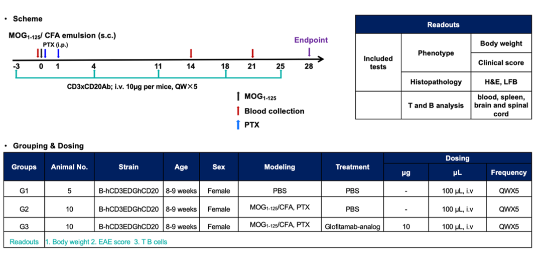
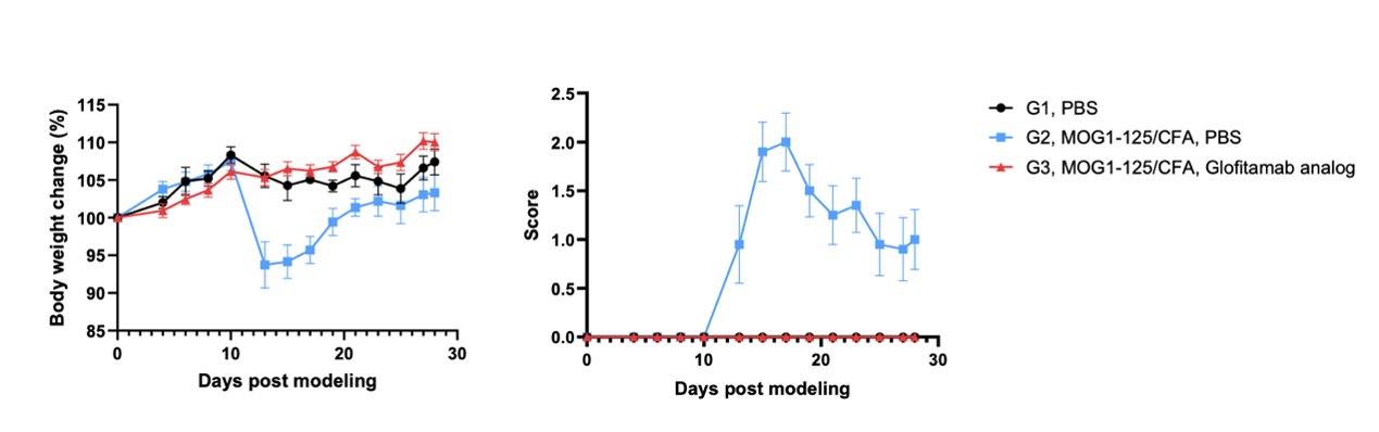
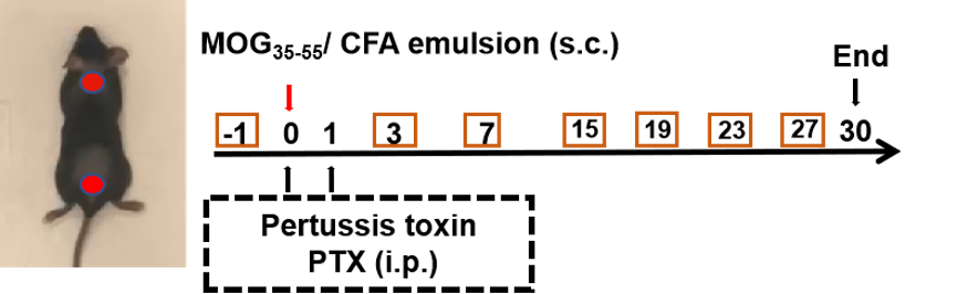
Effects of the anti-CD3×CD20 antibody (Glofitamab analog) on MOG1-125-induced EAE in B-hCD3EDG/hCD20 mice. Mice were subcutaneously injected with MOG1-125 emulsion in the neck and buttock regions on day 0. Pertussis toxin (PTX) was administered intraperitoneally at 2 and 24 hours after MOG immunization (G2, G3). The control group (G2) and the anti-CD3×CD20 antibody-treated group (G3) were administered QW. Body weight (A) and clinical scores (B) were recorded every other day. Data are presented as mean ± SEM.
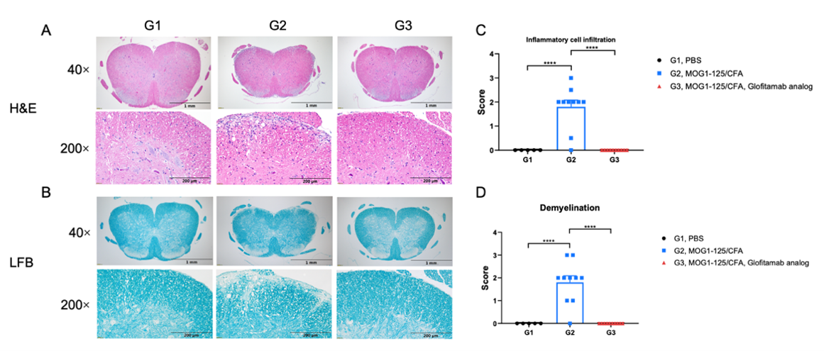
Effects of anti-CD3×CD20 antibody (Glofitamab analog) on inflammatory cell infiltration and demyelination. Spinal cords were removed from B-hCD3EDG/hCD20 mice on day 28 and stained with Hematoxylin and eosin (H&E) (A) or Luxol fast blue (LFB) (B). Representative sections are shown. The score of inflammatory cells and demyelination of the spinal cord (C, D). Values are expressed as mean ± SEM, Versus G2, **** p<0.0001.
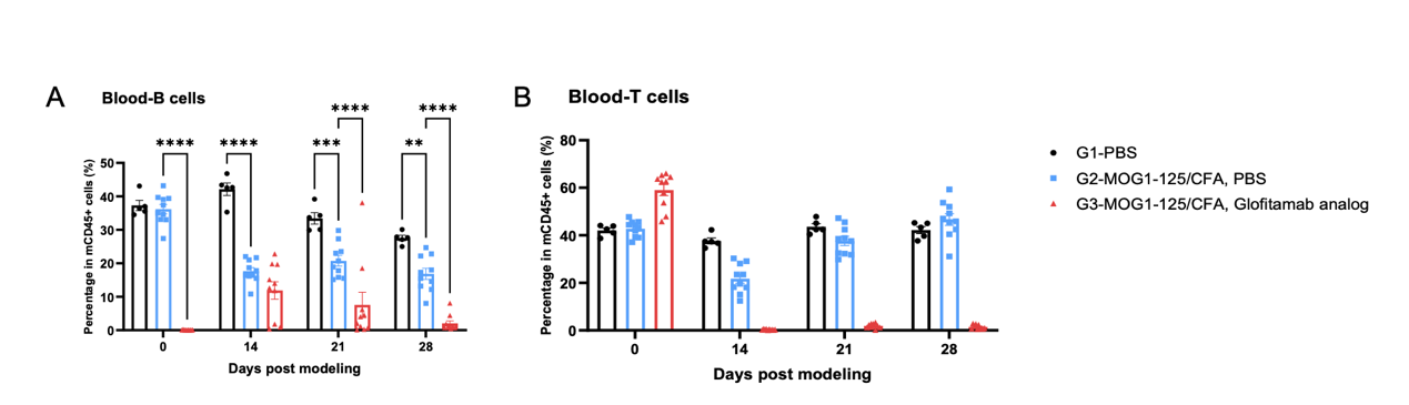
Effects of anti-CD3×CD20 antibody on the level of T and B cells on MOG1-125-induced EAE in B-hCD3EDG/hCD20 mice. Blood was collected on days 0, 14, 21, and the endpoint was 28. Levels of T and B cells were evaluated by flow cytometry. Values are expressed as mean ± SEM. Two-way ANOVA with Dunnett’s test. n=5-10, *P<0.05, **P<0.01, ***P<0.001, ****P<0.001 Versus G2.
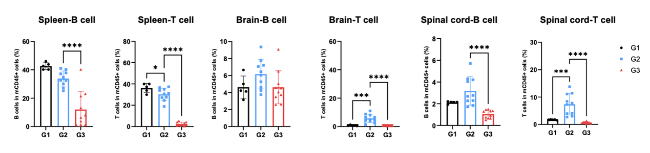
Effects of anti-CD3×CD20 antibody on the level of T and B cells on MOG1-125-induced EAE in B-hCD3EDG/hCD20 mice. Spleen, brain, and spinal cord were collected at the endpoint of 28. Levels of T and B were evaluated by flow cytometry. Values are expressed as mean ± SEM. Two-way ANOVA with Dunnett’s test. n=5-10, *P<0.05, **P<0.01, ***P<0.001, ****P<0.001 Versus G2.
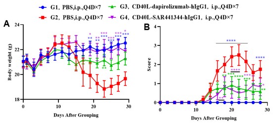
Effects of anti-CD40L on MOG35-55-induced EAE in B-hCD40/CD40L mice. Mice received MOG35-55 emulsion injection (s.c.) on neck and buttock (red point) on day 0. PTX (i.p.) were given 2 and 48 hour after MOG injection (G2-G4). Isotype control group(G3) or anti-CD40 antibodies group(G4) were administered BIW. Body weight (A) and clinical score (B) were recorded every two days. Values are expressed as mean ± SEM, compared with G2, *p<0.05, **p<0.01, *** p<0.001, **** p<0.0001.
• LFB and H&E Staining in MOG35-55-Induced EAE in CD40/CD40L Humanized Mice
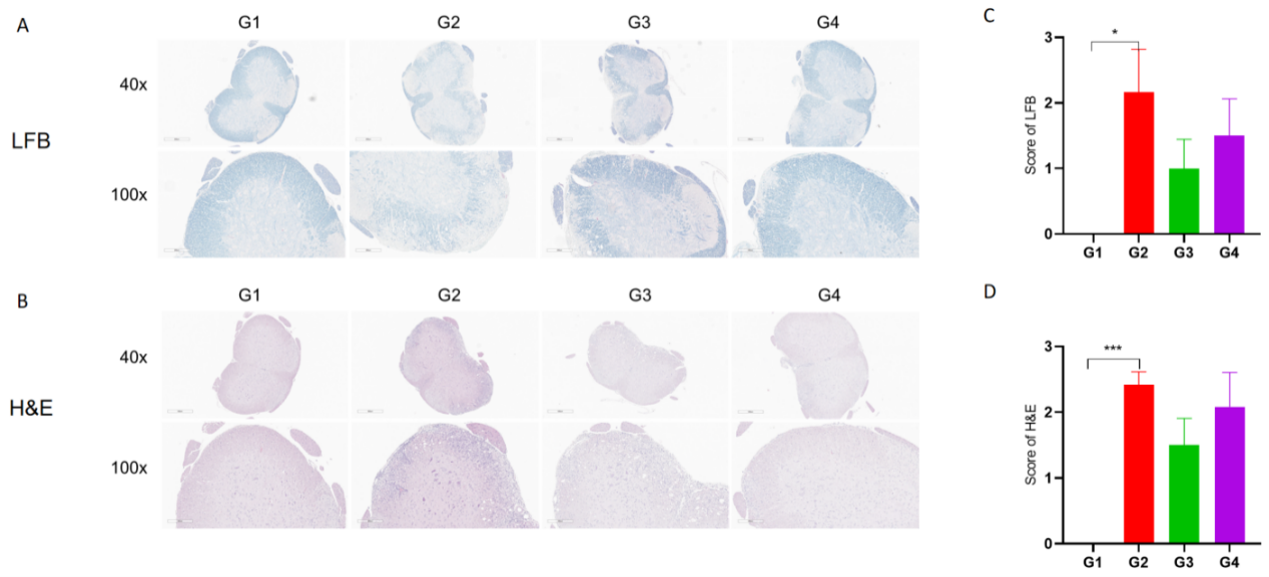
Anti-CD40L antibodies (analog, in-house) improves EAE clinical signs and controls inflammation and demyelination. Spinal cords were removed from B-hCD40/hCD40L mice on day 30 and stained with Luxol fast blue (LFB) (A) or Hematoxylin and eosin (H&E) (B). Representative sections are shown. The score of inflammatory cells and demyelination of spinal cord (C&D). Values are expressed as mean ± SEM, compared with G2, *p<0.05, **p<0.01, *** p<0.001.
• Cytokine Analysis in MOG35-55-Induced EAE in CD40/CD40L Humanized Mice
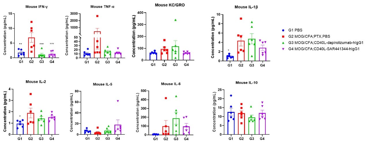
Effects of anti-CD40L on MOG35-55-induced EAE in B-hCD40/CD40L mice. Mice received MOG35-55 emulsion injection (s.c.) on neck and buttock (red point) on day 0. PTX (i.p.) were given 2 and 48 hour after MOG injection. Isotype control or anti-CD40 antibodies were administered BIW. Serum were collected at study endpoint and the cytokine levels were assessed. Values are expressed as mean ± SEM, n=6, compared with G2, *p<0.05, **p<0.01, *** p<0.001, **** p<0.0001.
As autoimmune and inflammatory drug discovery increasingly shifts toward highly targeted therapies, researchers need advanced translational models that connect human target biology directly to therapeutic outcomes. Biocytogen supports this need through a comprehensive matrix of target-humanized mouse models, advanced disease induction capabilities, validated efficacy testing and safety evaluation platforms. By aligning model design with specific therapeutic mechanisms, Biocytogen helps researchers build tailored preclinical strategies for next-generation autoimmnue breakthroughs. 👉Contact us to learn more!
Q1: Why is the EAE model considered the cornerstone of MS research?
EAE serves as a highly versatile toolkit for dissecting neuroinflammation by targeting specific pathological angles like the gut-brain axis or blood-brain barrier integrity. This unparalleled adaptability has made it the primary engine for discovering major FDA-approved disease-modifying therapies for MS.
Q2: What are the primary translational limitations of standard rodent models?
Standard rodent models present a translational bottleneck for modern biologics because these precisely engineered therapies are designed to recognize human epitopes and often exhibit little or no cross-reactivity with wild-type rodent proteins. As a result, standard mice are unable to support meaningful in vivo evaluation of therapeutic efficacy and mechanisms of action.
Q3: What is the difference between MOG1-125 and MOG35-55 activation pathways?
The MOG1-125 recombinant protein pathway is a T & B cell co-driven platform designed to validate advanced therapies targeting B cells, autoantibodies, or complement cascades. In contrast, the MOG35-55 peptide pathway is a classic T cell-driven model optimized for evaluating therapeutics targeting T cell-mediated autoimmunity and chronic-progressive neuroinflammation.
Q4: What data readouts does Biocytogen’s EAE model deliver?
The platform captures a comprehensive set of efficacy and pharmacodynamic readouts, such as continuous body weight and clinical score monitoring, histopathological evaluation of spinal cord inflammation and demyelination, and in-depth immunological profiling and cytokine analysis. Together, these endpoints support efficacy evaluation, safety assessment, and mechanism-of-action studies.