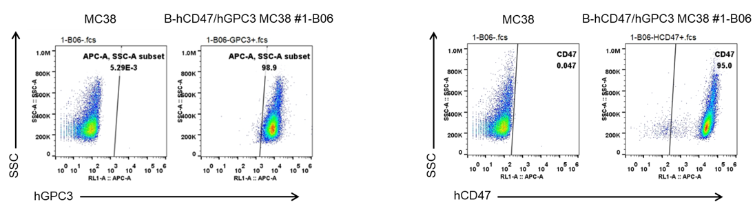
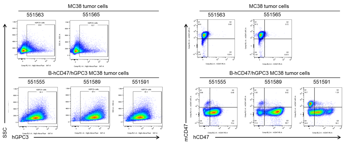
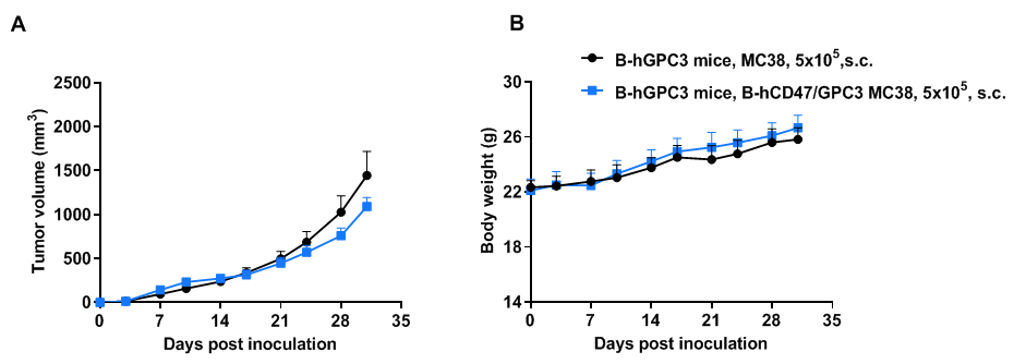
B-hCD47/hGPC3 MC38

• 322406

B-hCD47 MC38
B-hCD56 MC38

B-hCD47/hGPC3 MC38

Product nameB-hCD47/hGPC3 MC38
Catalog number322406
Strain backgroundC57BL/6
AliasesIA3; DGSX, GTR2-2, MXR7, OCI-5, SDYS, SAP, MER6, OGB, SGBS, SGBS1
TissueColon
DiseaseColon carcinoma
SpeciesMouse

on this page

  • Description
  • Phenotypic analysis
  • Physiological data
Description
  • Origin: The MC38 cell line is derived from C57BL/6 murine colon adenocarcinoma cells. The cell line is a commonly used murine model for colorectal carcinoma.
  • Background Information: CD47, also known as integrin-associated protein (IAP), is a member of the immunoglobulin superfamily containing a five-pass transmembrane attachment. CD47 is heavily glycosylated and widely expressed by hematopoietic and nonhematopoietic cells. CD47 interacts with several membrane integrins and also acts as a receptor for thrombospondin (THBS1). Glypicans (GPCs) are a family of glycosylphosphatidylinositol (GPI)-anchored heparan sulphate proteoglycans (HSPGs) that may play a role in the control of cell division and growth regulation.
  • Gene targeting strategy: The exogenous promoter and human GPC3 coding sequence was inserted to murine Rosa26 site. The exogenous promoter and human CD47 coding sequence was inserted to replace part of murine exon 2 and all of exon 3. The insertion disrupts the endogenous murine Cd47 gene, resulting in a non-functional transcript.
  • Tumorigenicity: Confirmed in B-hGPC3 mice. 
  • Application: B-hCD47/hGPC3 MC38 tumor models can be used for preclinical evaluation of bispecific antibody drugs targeting human CD47 and GPC3.
  • Notes: The B-hCD47/hGPC3 MC38 cell line fails to form tumors in B-hSPRIA/hCD47 mice, suggesting that this cell line may also not form tumors in wild-type C57BL/6 mice. We recommend using the B-hCD47/hGPC3 MC38 #1-B06 cell line for tumor formation or efficacy studies in GPC3-related humanized mice.
Protein expression analysis

GPC3 and CD47 expression analysis in B-hCD47/hGPC3 MC38 cells by flow cytometry. Single cell suspensions from wild-type MC38 and B-hCD47/hGPC3 MC38 #1-B06 cultures were stained with species-specific anti-GPC3 antibody (Invitrogen, MA5-40988) and species-specific anti-CD47 antibody (Biolegend, 323124). Human GPC3 and CD47 were detected on the surface of B-hCD47/hGPC3 MC38 cells but not wild-type MC38 cells. 

Protein expression analysis of tumor tissue

GPC3 and CD47 expression evaluated on B-hCD47/hGPC3 MC38 tumor cells by flow cytometry. B-hCD47/hGPC3 MC38 cells were subcutaneously transplanted into B-hGPC3 mice (n=6). Upon conclusion of the experiment, tumor cells were harvested and assessed for human GPC3 and CD47 expression by flow cytometry (Invitrogen, MA5-40988; Biolegend, 323124). As shown, human GPC3 and CD47 were highly expressed on the surface of tumor cells. Therefore, B-hCD47/hGPC3 cells can be used for in vivo efficacy studies evaluating novel GPC3 and CD47 therapeutics.

Tumor growth curve & body weight changes

Subcutaneous tumor growth of B-hCD47/hGPC3 MC38 cells. B-hCD47/hGPC3 MC38 cells (5×10⁵) and wild-type MC38 cells (5×10⁵) were subcutaneously implanted into B-hGPC3 mice (H/H) (female, 7-week-old, n=6). Tumor volume and body weight were measured twice a week. (A) Average tumor volume. (B) Body weight. Volume was expressed in mm3 using the formula: V=0.5 × long diameter × short diameter2. Results indicate that B-hCD47/hGPC3 MC38 cells were able to establish tumors in vivo and can be used for efficacy studies. Values are expressed as mean ± SEM.