

C57BL/6-Cd3etm1(CD3E)Bcgen Cd3dtm1(CD3D)Bcgen Cd3gtm1(CD3G)Bcgen Ighetm1(IGHE)Bcgen Fcer1atm1(FCER1A)Bcgen /Bc • 113954

CD3: An Important Therapeutic Target in Immunotherapy
Protein Expression: CD3 is expressed on nearly all mature T cells, including CD4⁺ T cells, CD8⁺ T cells, and γδ T cells. It is generally absent from B cells, NK cells, and myeloid cells.
Signaling Pathway: CD3 mediates TCR signaling through intracellular ITAM motifs. After antigen recognition, CD3 recruits signaling molecules such as ZAP-70, leading to activation of the NFAT, NF-κB, and MAPK/AP-1 pathways.
Therapeutic Inhibition: CD3 is an important therapeutic target for modulating T-cell activity. Anti-CD3 antibodies, such as Teplizumab, can suppress or regulate T-cell responses and are used in autoimmune diseases. CD3 is also widely used in bispecific T-cell engagers, which redirect T cells to kill tumor cells, as seen with Blinatumomab.
IgE/FCER1A: Key Drivers of Allergic Inflammation
Gene Information: IgE: The constant region of the IgE heavy chain is encoded by the IGHE gene, located on chromosome 14q32.33. FCER1A: The alpha subunit of the high-affinity IgE receptor (FCER1A) is encoded by the FCER1A gene, located on chromosome 1q23.2.
Protein Expression: IgE is an antibody isotype produced and secreted by plasma cells (differentiated B cells). FCER1A is a transmembrane protein primarily expressed on the surface of mast cells and basophils (as a tetrameric α\β\γ2 complex) and on antigen-presenting cells like dendritic cells (as a trimeric αγ2 complex). Notably, the surface expression of FCER1A is dynamically upregulated in proportion to ambient serum IgE levels.
Signaling Pathway: The FCER1A subunit serves as the specific docking site for the Fc portion of IgE. Under normal conditions, IgE binds to FCER1A in a monomeric form without activating the cell. However, when multivalent allergens cross-link these pre-bound IgE-FCER1A complexes, it triggers intracellular signaling via the ITAMs (immunoreceptor tyrosine-based activation motifs) on the receptor’s β and γ subunits. This drives immediate mast cell/basophil degranulation and the release of inflammatory mediators..
Therapeutic Inhibition: Therapies targeting this axis disrupt the interaction between IgE and FCER1A to prevent downstream allergic cascades: Anti-IgE Biologics (e.g., Omalizumab): Bind to free serum IgE at the exact site where it would normally attach to FCER1A. This neutralizes free IgE and indirectly leads to a massive downregulation of FCER1A receptors on effector cells due to ligand deprivation. Anti-FCER1A Inhibitors: Directly target or block the alpha subunit to prevent IgE docking. Clinical Outcomes: Both approaches effectively suppress the release of histamine, leukotrienes, and Th2 cytokines, significantly reducing airway eosinophils, tissue inflammation, and clinical exacerbations in severe asthma and chronic spontaneous urticaria (CSU).

CD3EDG
IgE
FCER1A
Exons 1-5 of mouse Fcer1a gene that encode extracellular domain are replaced by human counterparts in B-hCD3EDG/hIgE/hFCER1A mice.
B-hCD3EDG/hIgE/hFCER1A mice were obtained by breeding B-hCD3EDG mice with B-hIgE/hFCER1A mice.

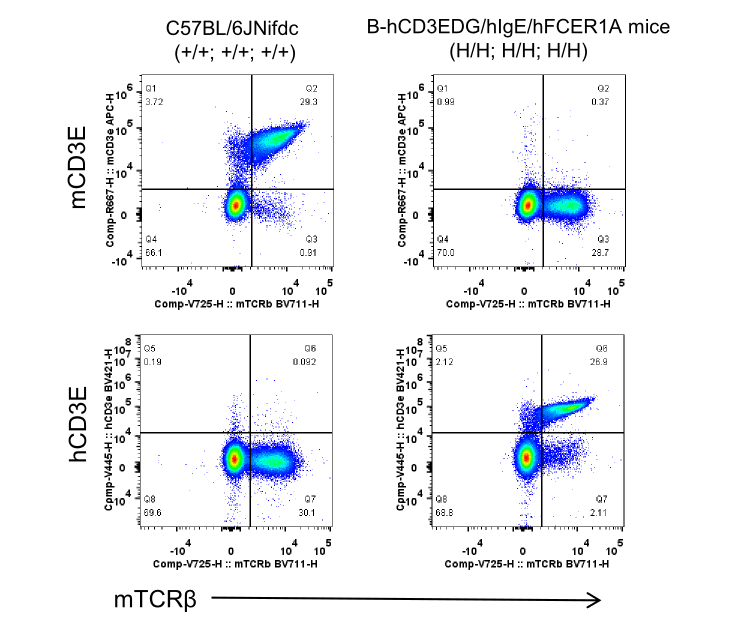
Strain specific CD3E expression analysis in wild-type C57BL/6JNifdc mice and homozygous B-hCD3EDG/hIgE/hFCER1A mice by flow cytometry. Splenocytes were collected from C57BL/6JNifdc mice (+/+; +/+; +/+) and homozygous B-hCD3EDG/hIgE/hFCER1A mice (H/H; H/H; H/H). Protein expression was analyzed with anti-mouse CD3E antibody (Biolegend, 100312) and anti-human CD3E (BD Horizon , 562426) by flow cytometry.
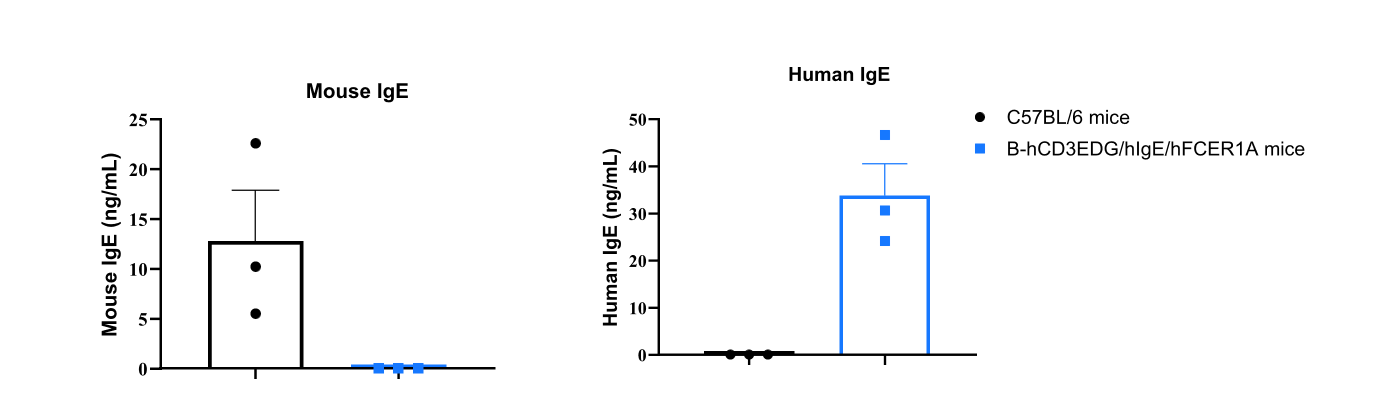
Strain-specific IgE expression evaluated by ELISA in wild-type C57BL/6JNifdc mice and homozygous B-hCD3EDG/hIgE/hFCER1A mice. Serum was collected from C57BL/6JNifdc mice (+/+; +/+; +/+) and B-hCD3EDG/hIgE/hFCER1A mice (H/H; H/H; H/H) (Female, n=3, 10-week-old). Mouse and human IgE levels in serum were quantified by ELISA (mouse IgE ELISA kit: Biolegend, 432404; human IgE ELISA kit: abcam, ab195216).
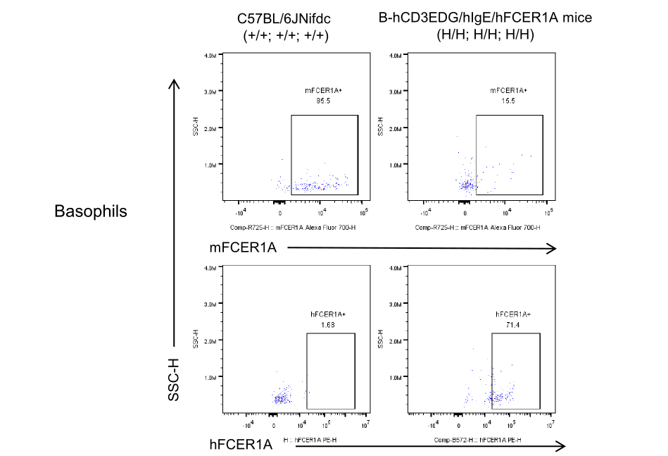
Mouse and human FCER1A expression analysis in bone marrow. Basophils of the bone marrow were collected from wild-type C57BL/6 mice (+/+; +/+; +/+) and homozygous B-hCD3EDG/hIgE/hFCER1A mice (H/H; H/H; H/H), and analyzed by flow cytometry with species-specific anti-FCER1A antibody. (Anti-Human FCER1A: Biolegend, 334609; Anti-Mouse FcεRIA: Biolegend, 134315).
