Description
Biological Features and Therapeutic Potential of CD3 and MSLN
- Gene Information: CD3 is a multi-subunit T-cell receptor complex essential for T-cell signaling, while MSLN (Mesothelin) is a cell-surface tumor-associated antigen. Both are established targets for T-cell redirecting therapies.
- Protein Expression: CD3 is specifically expressed on T cells, whereas MSLN is normally expressed at low levels on mesothelial tissues and highly overexpressed in several solid tumors, including mesothelioma, pancreatic, and ovarian cancers.
- Signaling Pathway: CD3 engagement activates the TCR signaling cascade, leading to T-cell activation, proliferation, cytokine production, and cytotoxicity. MSLN binding provides tumor-cell targeting, enabling CD3-mediated T-cell redirection and tumor killing.
- Therapeutic Inhibition: CD3×MSLN bispecific antibodies recruit and activate T cells by simultaneously binding CD3 on T cells and MSLN on tumor cells, resulting in targeted killing of MSLN-positive cancer cells. Additional therapeutic strategies include monoclonal antibodies, CAR-T cells, antibody drug conjugates, and cancer vaccines targeting MSLN.
Targeting Strategy
CD3EDG
- The chimeric human CD3EDG was expressed, while mouse Cd3edg were knocked out in B-hCD3EDG/hMSLN mice.
MSLN
- The mouse Msln gene sequences that encode the full length of coding region were replaced by human MSLN coding gene sequence in B-hCD3EDG/hMSLN mice.
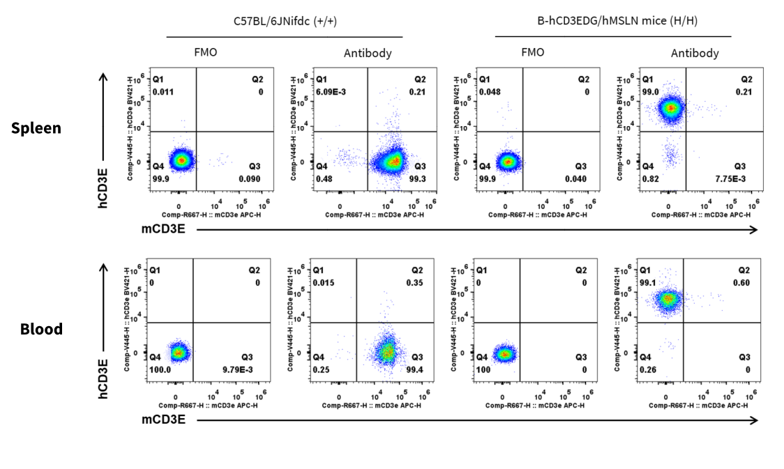
CD3E Protein Expression in Spleen
Mouse and human CD3E expression analysis in splenocytes. Splenocytes and blood were collected from wild-type C57BL/6 mice and homozygous B-hCD3EDG/hMSLN mice (Female, 8-week-old, n = 1). CD3E expression on T cells was analyzed by flow cytometry using species-specific anti-CD3E antibodies (anti-human CD3E antibody, BD Horizon, 562426; anti-mouse CD3E antibody, Biolegend, 100312).
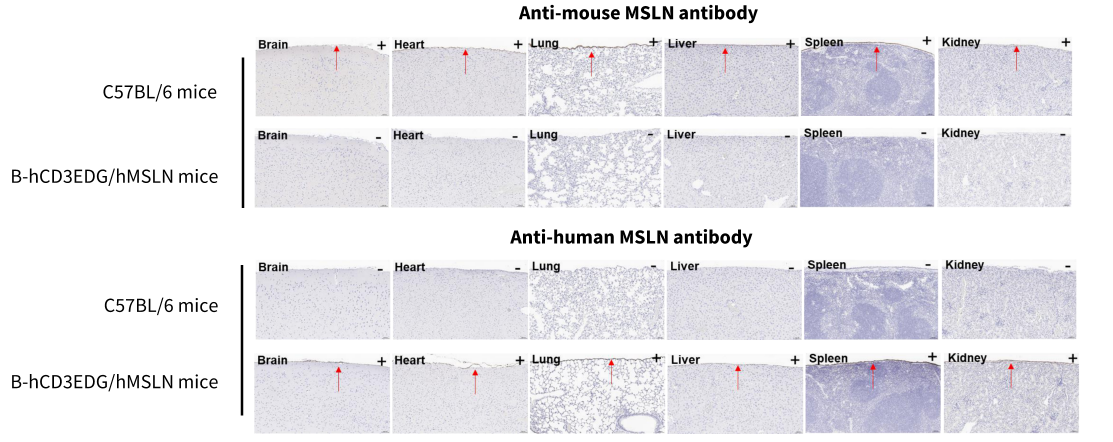
Immunohistochemical analysis of organs in B-hCD3EDG/hMSLN mice
Major organs were collected from wild-type C57BL/6 mice and homozygous B-hCD3EDG/hMSLN mice (female, 8 weeks old), and analyzed by IHC using anti-mouse MSLN antibody (Abcam, ab213174) and anti-human MSLN antibody (Abcam, ab196235) . The arrow indicates tissue cells with positive MSLN staining (brown). "+" and "-" indicate positive and negative tissues, respectively.
* When publishing results obtained using this animal model, please acknowledge the source as follows: The animal model [B-hCD3EDG/hMSLN mice] (Cat# 114368) was purchased from Biocytogen.