

C57BL/6N-Crbntm2(CRBN)Bcgen/Bcgen • 113236


CRBN

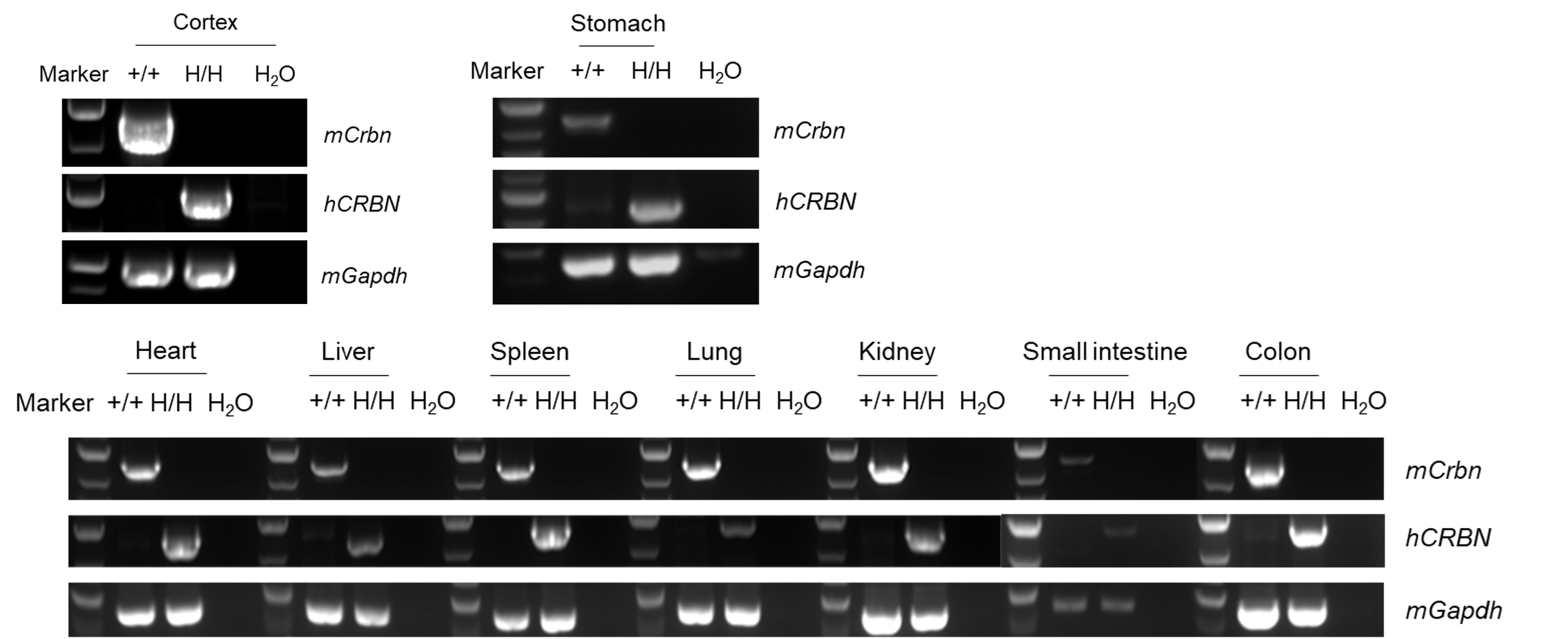
Strain-specific analysis of CRBN mRNA expression in wild-type C57BL/6 mice and CRBN humanized mice by RT-PCR. Total RNA was isolated from the heart, liver, spleen, lung, kidney, stomach, small intestine, colon, and cortex of wild-type C57BL/6 mice (+/+) and homozygous CRBN humanized mice (H/H). cDNA was synthesized by reverse transcription, followed by PCR amplification using mouse- or human-specific CRBN primers. Sequencing of the short PCR products confirmed that the amplified sequences matched the corresponding database reference sequences.
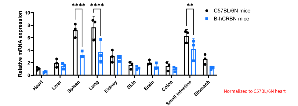
qPCR analysis of CRBN/Crbn mRNA expression in wild-type C57BL/6N mice and CRBN humanized mice. Total RNA was isolated from various tissues of wild-type C57BL/6N mice (+/+) (female, 7 weeks old, n = 3) and homozygous CRBN humanized mice (H/H) (female, 7 weeks old, n = 3). cDNA was synthesized by reverse transcription, followed by quantitative PCR (qPCR) using primers targeting an identical sequence shared by human CRBN and mouse Crbn in wild-type C57BL/6N and homozygous CRBN humanized mice. Relative mRNA expression levels were normalized to those in the heart of wild-type C57BL/6N mice. Values are expressed as mean ± SEM. Statistical significance was determined by two-way ANOVA. *p < 0.05, **p < 0.01, ***p < 0.001, ****p < 0.0001.
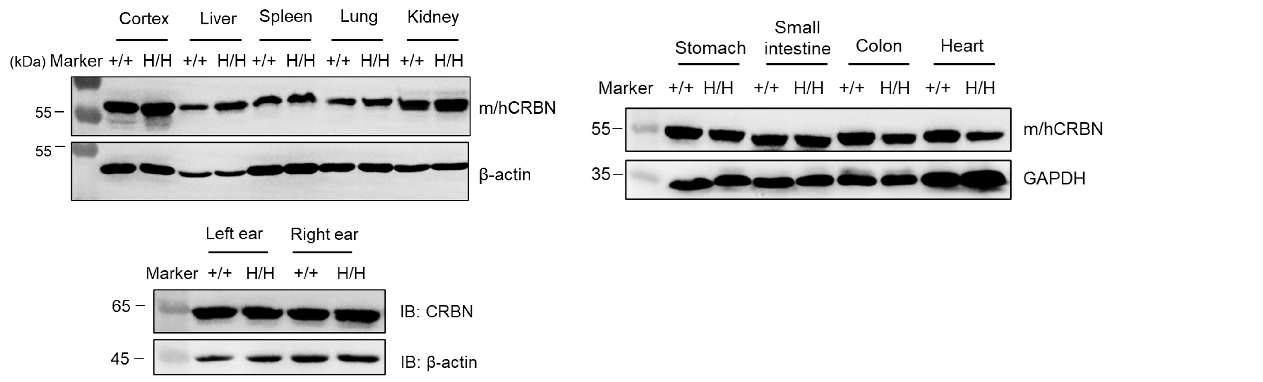
Strain-specific analysis of CRBN protein expression in homozygous CRBN humanized mice by Western blot. Cortex, liver, spleen, lung, kidney, stomach, small intestine, colon, heart, and ear tissues were collected from wild-type C57BL/6 mice (+/+) and homozygous CRBN humanized mice (H/H). CRBN protein expression was analyzed by Western blot using an anti-CRBN antibody.
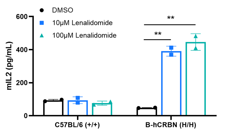
Naive CD4⁺ T cells derived from CRBN humanized mice exhibited increased IL-2 secretion after treatment with lenalidomide. Naive CD4⁺ T cells were collected from wild-type C57BL/6 mice (+/+) and homozygous CRBN humanized mice (H/H), then stimulated with DMSO or lenalidomide in vitro for 24 hours. Supernatants were collected, and IL-2 production was measured by ELISA.
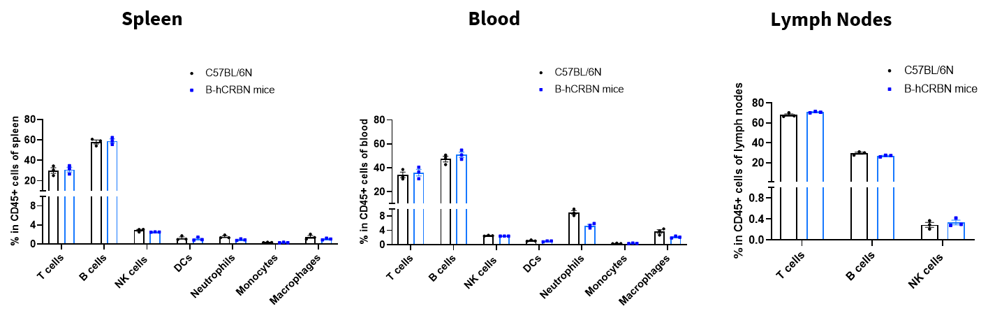
Flow cytometry analysis of leukocyte subpopulations in immune organs and peripheral blood of CRBN humanized mice. Splenocytes, peripheral blood, and lymph nodes were isolated from female C57BL/6N and CRBN humanized mice (8 weeks old, n = 3). Single live cells were gated on the CD45⁺ population and analyzed by flow cytometry as indicated. Values are expressed as mean ± SEM.
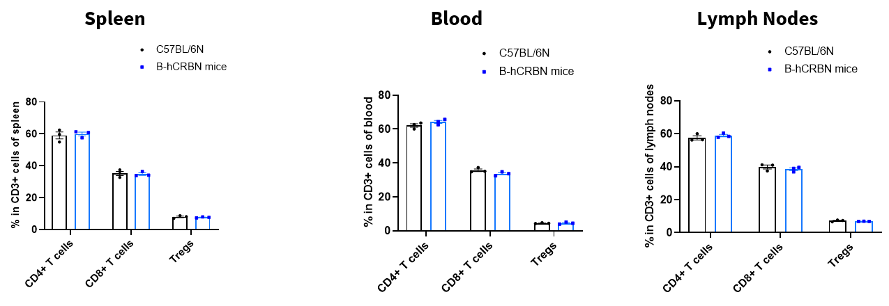
Flow cytometry analysis of T-cell subpopulations in immune organs and peripheral blood of CRBN humanized mice. Splenocytes, peripheral blood, and lymph nodes were isolated from female C57BL/6N and CRBN humanized mice (8 weeks old, n = 3). Single live cells were gated on the CD3⁺ T-cell population and analyzed by flow cytometry as indicated. Values are expressed as mean ± SEM.
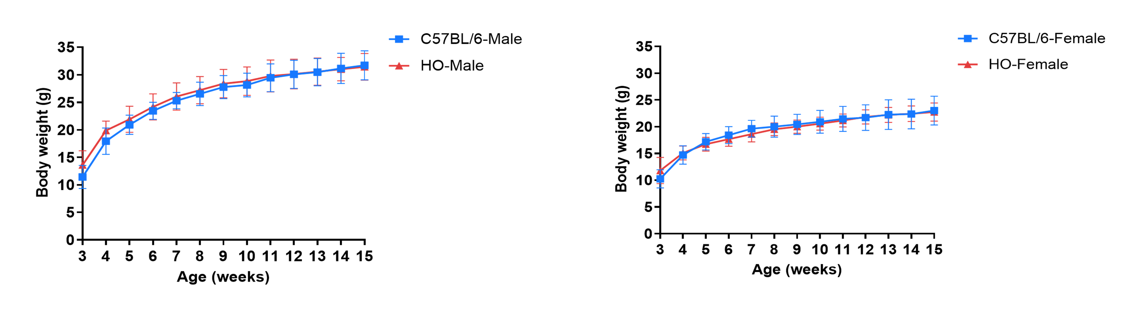
Growth curve analysis of CRBN humanized mice. Three-week-old CRBN humanized mice were grouped by sex (10 males and 10 females). Body weight was measured weekly on the same day for 13 consecutive weeks. The minimum and maximum body weights presented in the table were calculated as mean ± SD. The body weight data followed an approximately normal distribution, with approximately 68% of values falling within one standard deviation (± SD) of the mean.
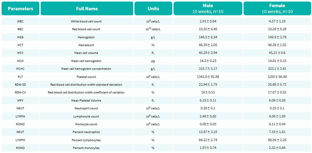
Complete blood count (CBC) analysis of CRBN humanized mice. Values are expressed as mean ± SD.
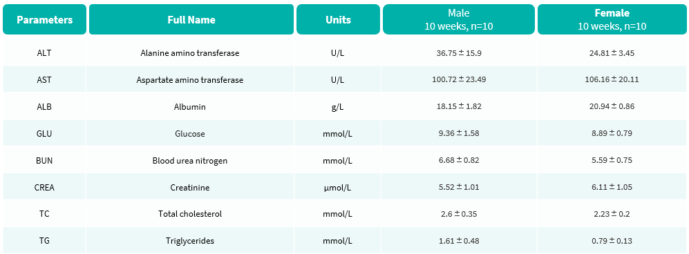
Blood biochemical analysis of CRBN humanized mice. Values are expressed as mean ± SD.
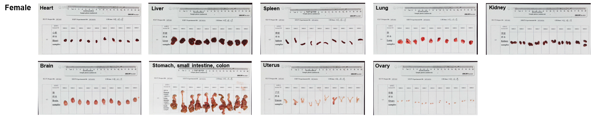
Organs of female CRBN humanized mice (10 weeks old, n = 10).
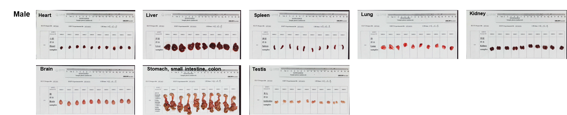
Organs of male CRBN humanized mice (10-week-old, n = 10).
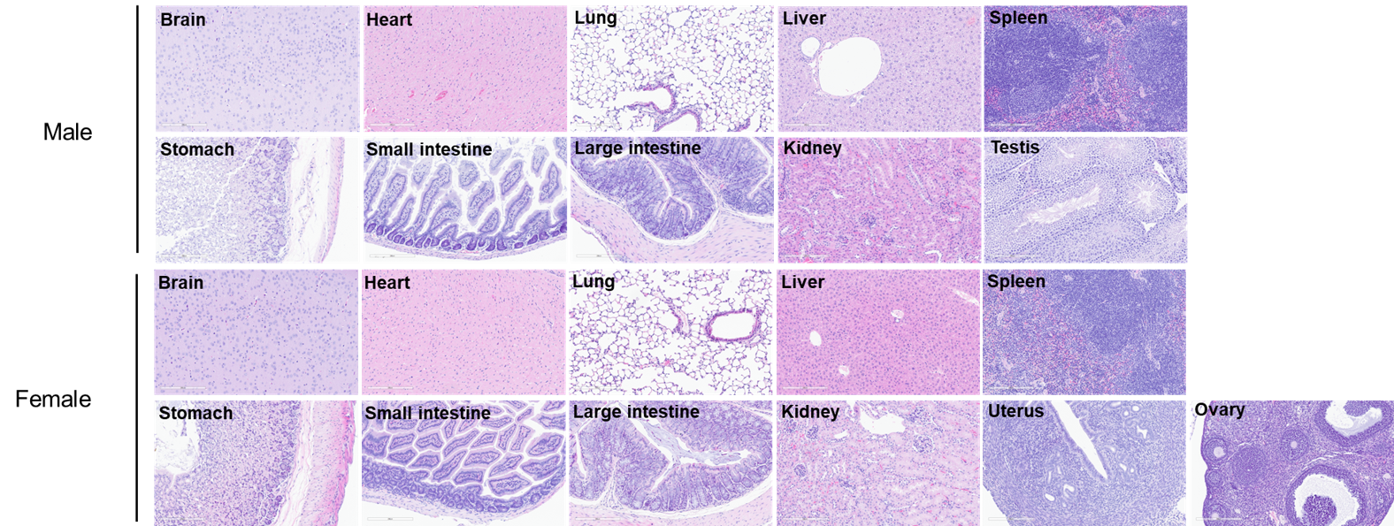
Histopathological analysis of major organs in CRBN humanized mice. Major organs were collected from 10-week-old CRBN humanized mice (male, n = 10; female, n = 10) and analyzed by hematoxylin and eosin (H&E) staining.


In vivo efficacy of NEK7-molecular glue (by the client) in an LPS- and nigericin-induced mouse peritonitis model using CRBN humanized mice. CRBN humanized mice were orally dosed either vehicle or the test compound twice daily (BID), starting 24 hours prior to stimulation. On the day of stimulation, mice received a final dose, followed by lipopolysaccharide (LPS, 1 mg/kg, i.p.) 1 hour later and nigericin (3 mg/kg, i.p.) 3 hours after LPS administration. Two hours later, spleens and peritoneal lavage fluid were collected for analysis.
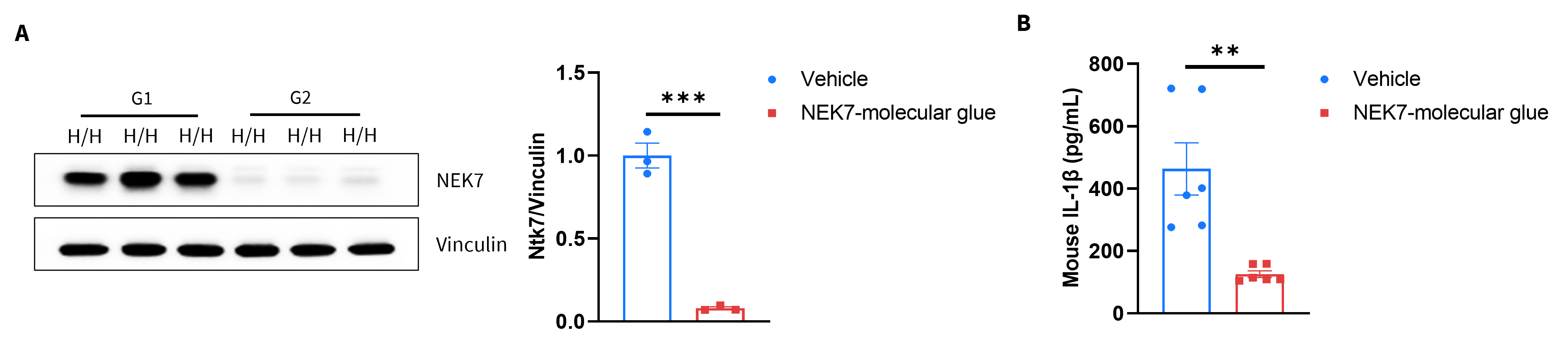
Analysis of of spleen and peritoneal lavage fluid by western blot and ELISA in CRBN humanized mice. (A) Spleen tissue lysates were collected and analyzed by Western blot using an anti-NEK7 antibody. Relative NEK7 protein expression levels were quantified by densitometric analysis and are presented. (B) Peritoneal lavage fluid was collected, and mouse IL-1β levels were measured by ELISA. Values are expressed as mean ± SEM. Statistical significance was determined using an unpaired t-test. *p < 0.05, **p < 0.01, ***p < 0.001.
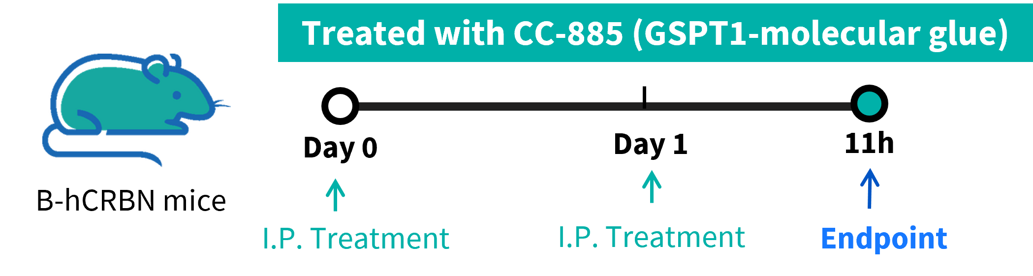
In vivo toxicity evaluation of CC-885 in CRBN humanized mice. (A) CRBN humanized mice and wild-type C57BL/6N mice were administered CC-885 intraperitoneally once daily. Mice were euthanized 11 hours after the final dose on the following day. Lymph nodes, liver, and large intestine tissues were collected for analysis.
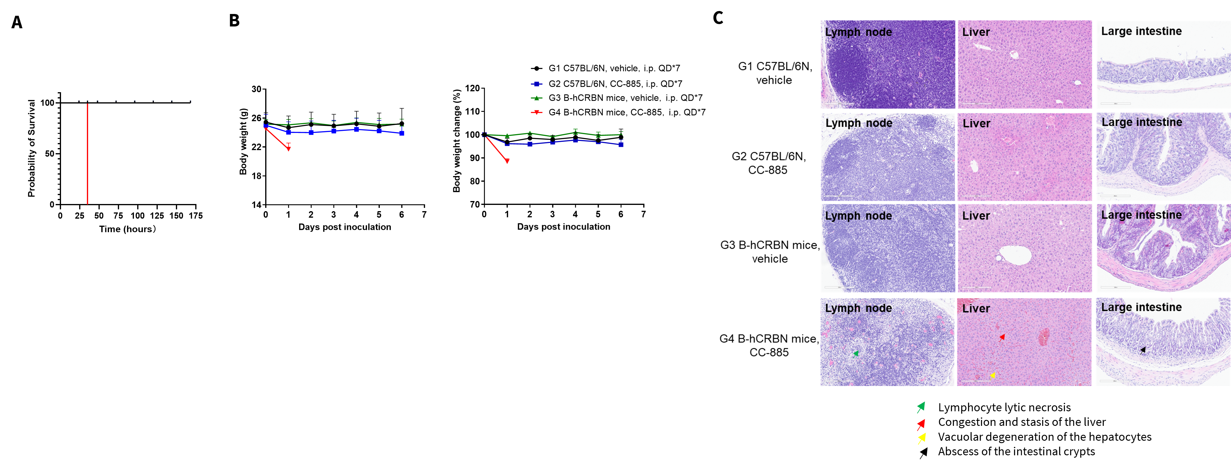
In vivo toxicity evaluation of CC-885 in CRBN humanized mice. Lymph nodes, liver, and large intestine tissues were collected for analysis. (A) Survival rate. (B) Body weight and body weight changes during treatment. (C) Histopathological analysis. Scale bar: 100 μm.
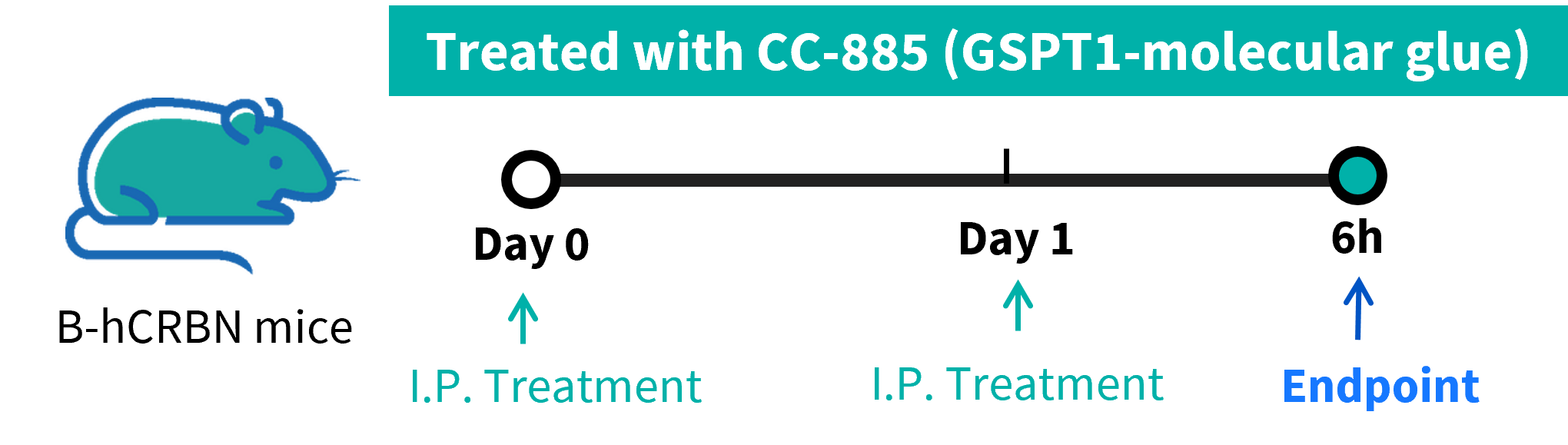
In vivo toxicity evaluation of CC-885 in CRBN humanized mice. CRBN humanized mice and wild-type C57BL/6N mice were administered CC-885 intraperitoneally once daily. Mice were euthanized 6 hours after the final dose on the following day. Skeletal muscle, heart, and blood samples were collected for analysis.
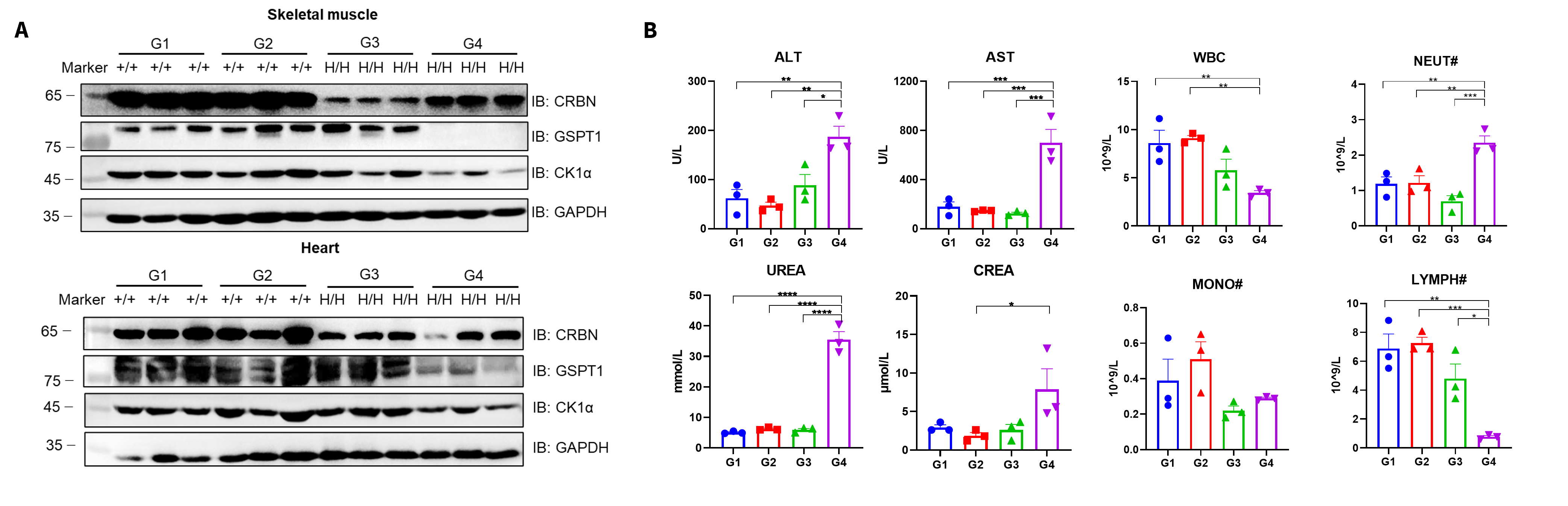
Skeletal muscle, heart and blood of CRBN humanized mice were collected for analysis. Skeletal muscle and heart tissue lysates were collected from all group, then analyzed by western blot with anti-CRBN antibody , anti-GSPT1 antibody and anti-CK1α antibody. 40 μg total proteins were loaded for western blotting analysis. (A) Western blot analysis of GSPT1 protein expression. (B) Biochemical test and complete blood count (Additional hematological parameters are not shown.)

Q1: What are CRBN humanized mice used for?
They are designed for evaluating human CRBN-binding drugs, including small molecules, molecular glues, and PROTAC-based degraders, under physiologically relevant human CRBN expression.
Q2: Do CRBN humanized mice exhibit normal baseline physiology?
Yes. CBC, biochemistry, organ weight, and histopathology show no abnormalities, supporting their suitability for long-term studies.
Q3: Why do CRBN humanized mice respond differently to CC-885?
CC-885 selectively binds human CRBN, resulting in degradation of substrates such as GSPT1. This on-target effect occurs only in CRBN humanized mice, not WT animals.
Q4: Can these mice be used for immunomodulatory drug testing?
Yes. Lenalidomide-induced IL-2 upregulation is observed only in CRBN humanized mice, reflecting the human-specific CRBN dependence of its immunomodulatory activity.
Q5: Are CRBN humanized mice suitable for oncology studies?
Absolutely. Tumor cell line inoculation combined with CRBN-dependent therapeutics allows in vivo evaluation of efficacy, safety, and mechanism of action.
