Description
- Gene Information: Human DMPK locates on chromosome 19q13.3; normal alleles carry 5–34 CTG repeats in its 3′UTR. Pathogenic expansion over 50 repeats triggers autosomal dominant DM1 via toxic RNA gain-of-function, with repeat size correlating with disease severity and anticipation.
- Protein Expression: DMPK serine/threonine kinase highly expresses in skeletal and cardiac muscle, and also express in various tissues.
- Signaling Pathway: Toxic DMPK-CUG RNA disturbs two core pathways: sequestration blocks MBNL-mediated splicing, and hyperactivated CUGBP1/GSK3β plus impaired insulin signaling disrupt muscle contraction, metabolism and ion channel function underlying DM1 myotonia
- Therapeutic Inhibition: Current inhibitory strategies include ASO gapmers, RNAi and CRISPRi to degrade or silence mutant DMPK transcripts, dissolving nuclear RNA foci, restoring free MBNL and correcting global splicing defects to relieve multisystem DM1 manifestations preclinically.
Targeting strategy
- The exons 1~15 of mouse Dmpk gene that encode the full-length protein were replaced by human DMPK exons 1~15 in B-hDMPK mice.
- The promoter, 5’UTR and 3’UTR regions were also replaced by human counterparts, allowing human DMPK expression to be driven by the human DMPK promoter, while endogenous mouse Dmpk transcription and translation are abolished.
mRNA Expression Analysis
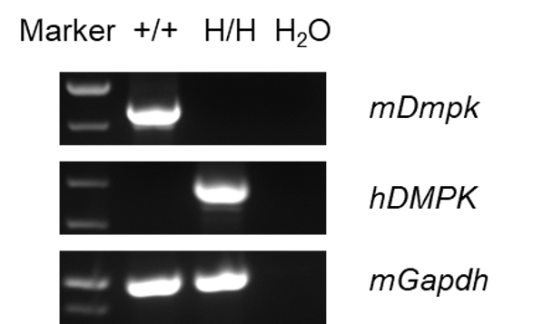
- Mouse Dmpk mRNA was only detectable in wild-type C57BL/6JNifdc mice but not in homozygous B-hDMPK mice.
- Human DMPK mRNA was exclusively detectable in homozygous B-hDMPK mice. Human DMPK sequences were confirmed by Sanger-sequencing.
Strain specific analysis of DMPK mRNA expression in wild-type C57BL/6JNifdc mice and homozygous B-hDMPK mice by RT-PCR. Muscle RNA was isolated from wild-type C57BL/6JNifdc mice (+/+) and homozygous B-hDMPK mice (H/H), then cDNA libraries were synthesized by reverse transcription, followed by PCR with mouse or human DMPK primers.
Relative mRNA Expression Analysis
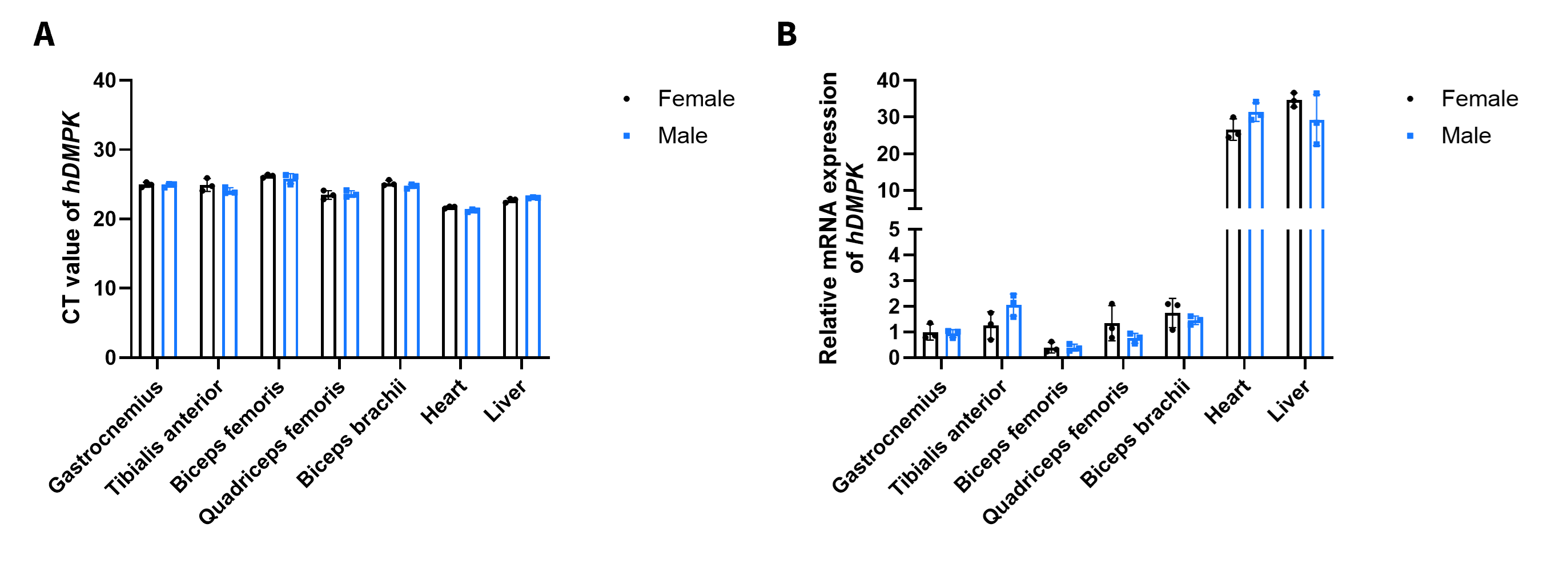
- No significant sex-dependent differences in DMPK expression in skeletal muscle, heart and liver tissues were observed between male and female B-hDMPK mice.
Strain specific analysis of DMPK mRNA expression in B-hDMPK mice by RT-qPCR. Various skeletal muscle tissues, including gastrocnemius, tibialis anterior, quadriceps femoris, biceps femoris, Biceps brachii, heart and liver tissues were collected from homozygous B-hDMPK mice (H/H) (female and male, 8-week-old, n=3), followed by RNA isolation. Then cDNA libraries were synthesized by reverse transcription, followed by qPCR with human DMPK primers. (A) CT value of human DMPK. (B) Relative mRNA expression levels of DMPK. Gene expression was normalized to the reference gene GAPDH and calculated using the 2^(−ΔΔCt) method.
Protein Expression Analysis
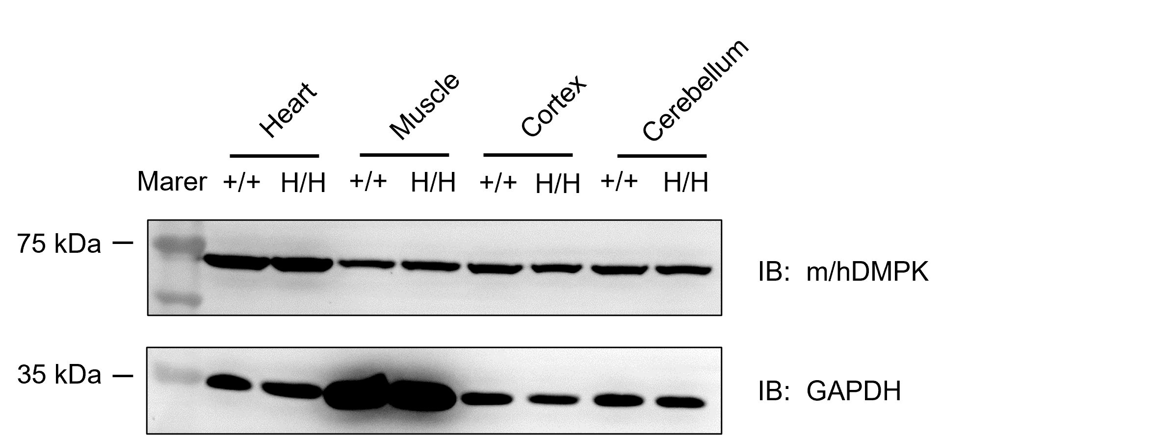
- DMPK was detectable in heart, muscle, cortex and cerebellum of both homozygous B-hDMPK mice and wild-type C57BL/6JNifdc mice, as the antibody was cross-reactive between human and mouse.
Western blot analysis of DMPK protein expression in homozygous B-hDMPK mice. Various tissue lysates were collected from wild-type C57BL/6JNifdc mice (+/+) and homozygous B-hDMPK mice (H/H), and then analyzed by western blot with species-specific anti-human DMPK antibody (abcam, ab102804). 50 μg total proteins were loaded for western blotting analysis.
Inhibitory efficiency of the antibody oligonucleotide conjugates drug against the human DMPK
- The human DMPK mRNA in the drug treatment groups (G3) were significantly reduced compared to the control groups (G1 and G2) in tibialis anterior muscle, demonstrating that TFR1 antibody can deliver siRNA drugs into skeletal muscle.
- B-hTFR1/hDMPK mice provide a powerful preclinical model for in vivo evaluation of human DMPK targeted antibody oligonucleotide conjugates drugs.
The inhibitory efficiency of the antibody oligonucleotide conjugates (AOC) drug against human DMPK in heterozygous B-hTFR1/hDMPK mice. The antibody oligonucleotide conjugates drug (AOC, produced in-house), naked antibody (produced in-house) and PBS were administered to the heterozygous B-hTFR1/hDMPK mice individually on day 0. The mice were sacrificed on day 7, and the liver, gastrocnemius muscle and tibialis anterior muscle were collected to detect the expression level of human DMPK mRNA by qPCR. Values are expressed as mean ± SEM.
* When publishing results obtained using this animal model, please acknowledge the source as follows: The animal model [B-hDMPK mice] (Cat# 113278) was purchased from Biocytogen.