

C57BL/6-Il21rtm1(IL21R)Bcgen/Bcgen • 110766
on this page

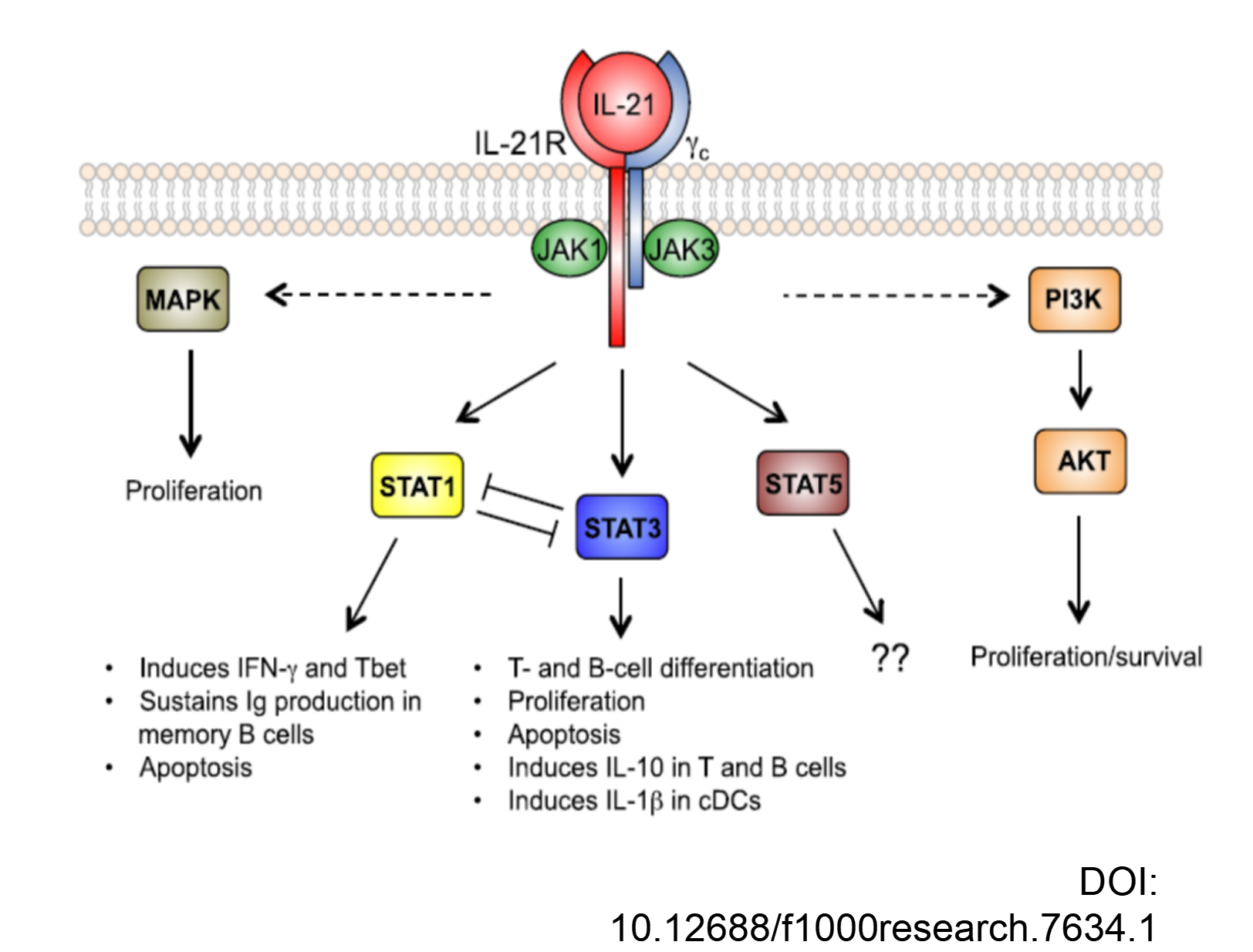
IL21R: A cytokine receptor driving immune cell proliferation and differentiation.

IL21R encodes a cytokine receptor for interleukin 21 (IL21). It belongs to the type I cytokine receptors, and has been shown to form a heterodimeric receptor complex with the common gamma-chain, a receptor subunit also shared by the receptors for interleukin 2, 4, 7, 9, and 15. This receptor transduces the growth promoting signal of IL21, and is important for the proliferation and differentiation of T cells, B cells, and natural killer (NK) cells. The ligand binding of this receptor leads to the activation of multiple downstream signaling molecules, including JAK1, JAK3, STAT1, and STAT3. Knockout studies of a similar gene in mouse suggest a role for this gene in regulating immunoglobulin production.


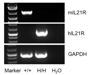
Species specific analysis of IL21R gene expression in wild-type C57BL/6 mice and homozygous humanized B-hIL21R mice by RT-PCR. Spleen RNA was isolated from wild-type C57BL/6 mice (+/+) and homozygous B-hIL21R mice (H/H), and then cDNA libraries were synthesized by reverse transcription, followed by PCR with mouse Il21r primers and human IL21R primers.
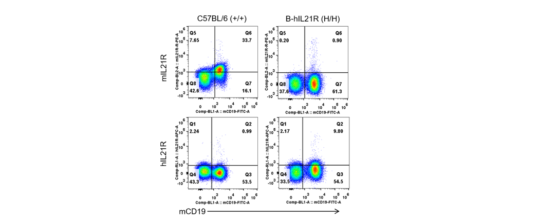
Strain specific IL21R expression analysis in wild-type C57BL/6 mice and homozygous humanized B-hIL21R mice by flow cytometry. Splenocytes were collected from wild-type C57BL/6 mice and homozygous B-hIL21R mice. Protein expression was analyzed with anti-mouse IL21R antibody (Biolegend, 131905) and anti-human IL21R antibody (Biolegend, 347807) by flow cytometry.
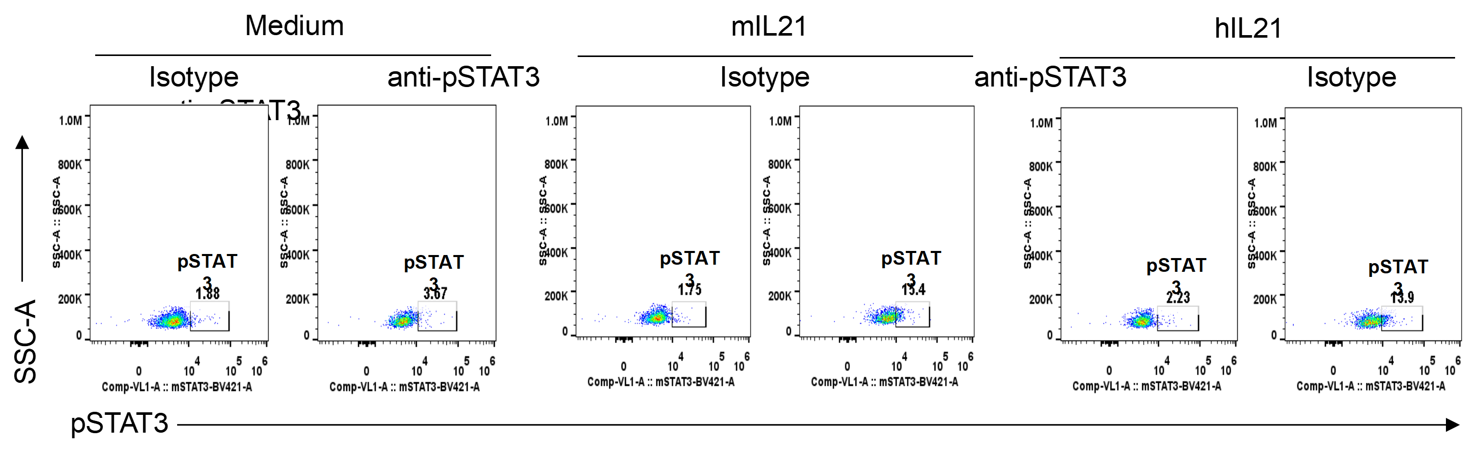
Mouse pSTAT3 was induced with mouse IL21 and human IL21 in homozygous B-hIL21R mice analyzed by flow cytometry. Splenocytes were collected from homozygous B-hIL21R mice (H/H), and stimulated with culture medium, mouse IL21 or human IL21. The induction of STAT5 phosphorylation on CD8+ T cells with the indicated stimulators was assayed by flow cytometry.

Analysis of leukocyte subpopulations by flow cytometry in immune organs and blood. Splenocytes, peripheral blood, and lymph nodes were isolated from C57BL/6 mice and homozygous B-hIL21R mice (female, 6-week-old, n = 3). Single live cells were gated on the CD45⁺ population and analyzed by flow cytometry as indicated. Values are expressed as mean ± SEM.

Analysis of T-cell subpopulations by flow cytometry in immune organs and blood. Splenocytes, peripheral blood, and lymph nodes were isolated from C57BL/6 mice and homozygous B-hIL21R mice (female, 6-week-old, n = 3). Single live cells were gated on the TCRβ⁺ T-cell population and analyzed by flow cytometry as indicated. Values are expressed as mean ± SEM.

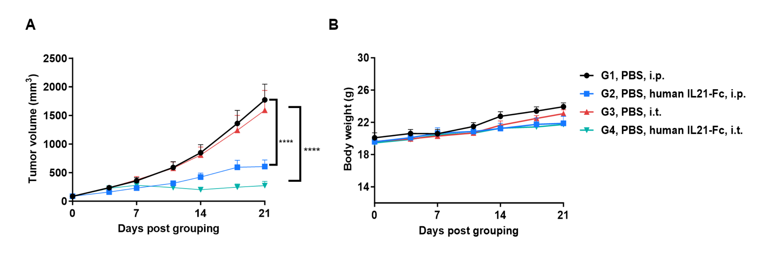
Antitumor activity of human IL21-Fc (in-house) in B-hIL21R mice. (A) Tumor growth curves. (B) Body weight changes during treatment. Values are expressed as mean ± SEM. Significance was determined by two-way ANOVA test. *P < 0.05, **P < 0.01, ***P < 0.001.
