

C57BL/6-Il4tm2(IL4)Bcgen Il4ratm1(IL4RA)Bcgen Il13tm1(IL13)BcgenTslptm1(TSLP)Bcgen Crlf2tm2(CRLF2)Bcgen/Bcgen • 113622

IL-4/IL-4Rα: The Core Signaling Pathway in Type 2 Inflammation and Therapeutic Intervention
Protein Expression: IL-4 is primarily secreted by activated immune cells driving allergic responses, including Th2 cells, mast cells, basophils, and eosinophils. IL-4Rα is widely and constitutively expressed across a diverse range of cell types. It is found on hematopoietic cells (B cells, T cells, macrophages) as well as non-hematopoietic cells (airway epithelial cells, smooth muscle cells, and fibroblasts), making it a ubiquitous sensor for type 2 inflammation.
Signaling Pathway: Type I Receptor Complex (IL-4 Specific): Formed by the assembly of IL-4Rα and the common gamma chain (γc). This complex is predominantly expressed on hematopoietic cells. Type II Receptor Complex (IL-4 / IL-13 Shared): Formed by the assembly of IL-4Rα and IL-13Rα1. This complex is widely expressed on non-hematopoietic barrier tissues. When IL-4 binds to IL-4Rα, it recruits either γc or IL-13Rα1, cross-activating downstream JAK1/JAK3 (Type I) or JAK1/TYK2 (Type II) kinases. This triggers the phosphorylation, dimerization, and nuclear translocation of STAT6 to drive the transcription of pro-inflammatory genes.
Therapeutic Inhibition: Because IL-4Rα is the shared and indispensable subunit for both IL-4 and IL-13 signaling, it represents a master therapeutic target. By utilizing monoclonal antibodies such as dupilumab to specifically block IL-4Rα, therapies can simultaneously shut down both Type I and Type II receptor signaling. This dual inhibition effectively halts downstream type 2 inflammation, leading to profound clinical improvements including: Reduced serum IgE levels (by blocking B cell class switching) Decreased tissue eosinophilia (by suppressing eotaxin and VCAM-1 expression) Diminished airway remodeling and mucus hypersecretion Lowered systemic secretion of downstream type 2 cytokines.
TSLP: A key cytokine in inflammation and its therapeutic intervention
Gene Information: Thymic stromal lymphopoietin (TSLP) is a protein-coding gene located on chromosome 5q22.1. It encodes a hemopoietic cytokine that is a member of the interleukin 7-like cytokine family.
Protein Expression: TSLP is primarily expressed by activated epithelial cells (lung, gut), skin keratinocytes, and fibroblasts. Two main isoforms exist: the short form (sfTSLP) is constitutively expressed and plays a homeostatic role, while the long form (lfTSLP) is induced during inflammation.
Signaling Pathway: TSLP exerts its effects by binding to a high-affinity heterodimeric receptor complex composed of the TSLP receptor chain (TSLPR) and the IL-7 receptor alpha chain (IL-7Rα).
Therapeutic Inhibition: By blocking TSLP binding to its receptor, tezepelumab inhibits downstream inflammation and improves clinical outcomes, including reduced serum IgE levels, decreased airway eosinophils, reduced mucus production, and lowered cytokine secretion.

IL4
IL4RA
TSLP
TSLPR
Note: B-hIL4/hIL4RA/hTSLP/hTSLPR mice (C) were obtained by breeding B-hIL4/hIL4RA mice (C) with B-hTSLP/hTSLPR mice (C).

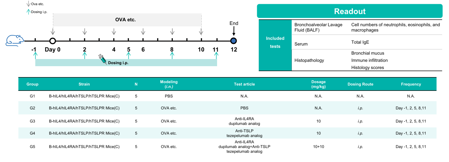
Experimental schedule for the induction of asthma and in vivo efficacy of anti-human IL4RA antibody and anti-human TSLP antibody in B-hIL4/hIL4RA/hTSLP/hTSLPR mice(C). In the OVA etc.-induced model, animals are administered OVA etc. intranasally to induce asthma models. The anti-human IL4RA antibody and anti-human TSLP antibody were administered by intraperitoneal injection (n = 5).
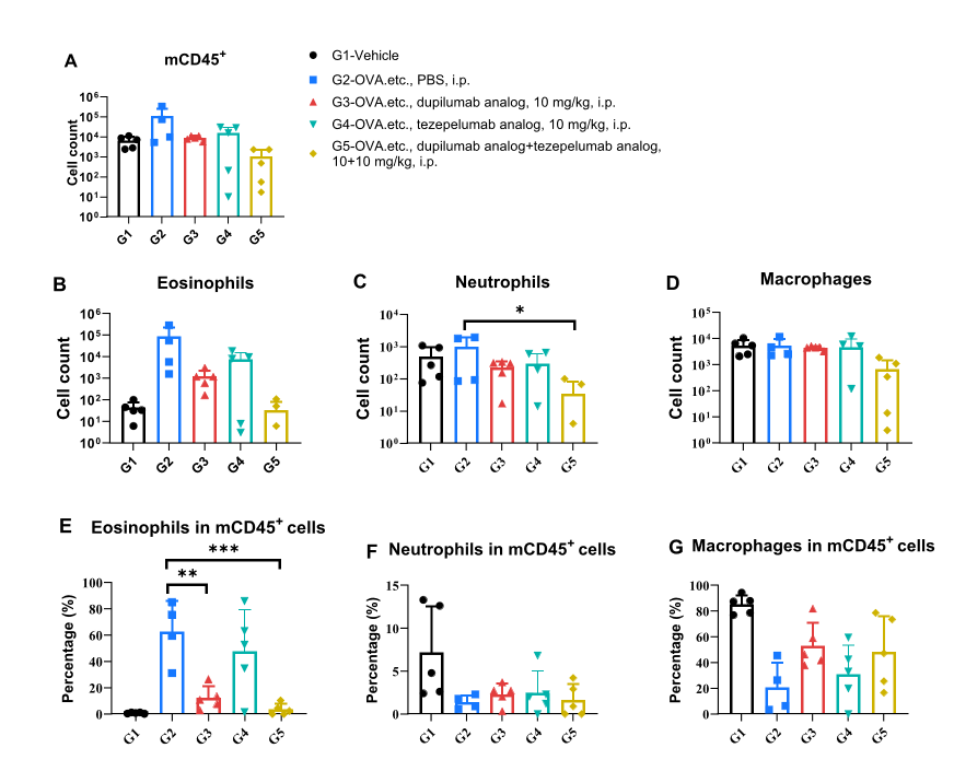
Analysis of immune cells in BALF. B-hIL4/hIL4RA/hTSLP/hTSLPR mice (C) (female, 11-week-old, n = 5) were immunized with OVA etc. to induce asthma. Anti-human IL4RA antibody and anti-human TSLP antibody were intraperitoneally injected to B-hIL4/hIL4RA/hTSLP/hTSLPR mice (C). (A&B) The percentage of eosinophils in CD45+ of bronchoalveolar lavage fluid (BALF) in the dupilumab analog (in house) and the combination therapy group of dupilumab analog and tezepelumab analog (in house) treated groups decreased significantly compared with the OVA etc.-induced PBS treated group. Values were expressed as mean ± SEM. Significance was determined by two-way ANOVA test. *P < 0.05, P < 0.01, ***P < 0.001.
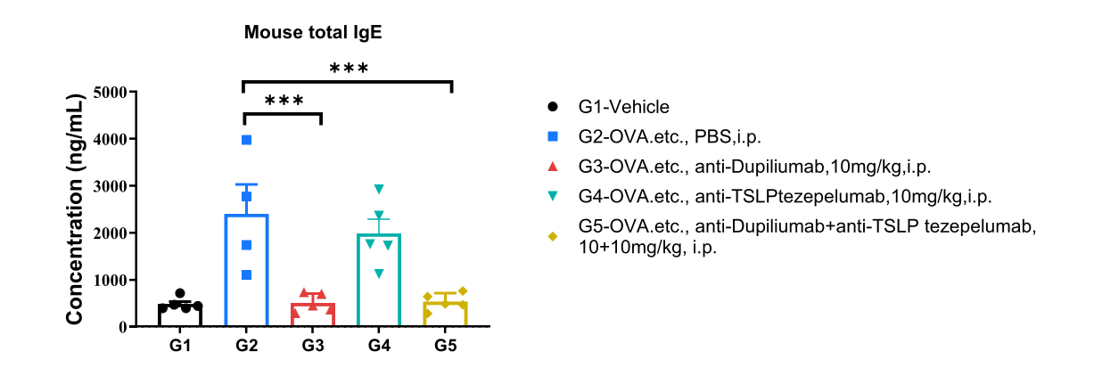
Analysis of mouse total IgE in serum. B-hIL4/hIL4RA/hTSLP/hTSLPR mice(C) (female, 11-week-old, n=5) were immunized with OVA etc. to induce asthma. Anti-human IL4RA antibody and anti-human TSLP antibody were intraperitoneally injected to B-hIL4/hIL4RA/hTSLP/hTSLPR mice(C). Serum was collected at the study endpoint. IgE level was analyzed by ELISA. The results showed that the levels of total IgE in mice treated with dupilumab analog, the combination therapy group of dupilumab and tezepelumab analog treated groups showed a significant reduction compared with untreated mice. Values were expressed as mean ± SEM. Significance was determined by two-way ANOVA test. *P < 0.05, **P < 0.01, ***P < 0.001.
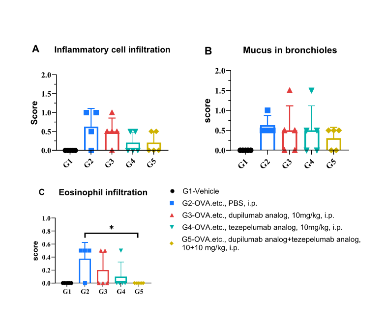
H&E staining of asthma model in B-hIL4/hIL4RA/hTSLP/hTSLPR mice (C). Lung tissues were collected at the study endpoint and analyzed with H&E staining. The results showed that the group of mice the combination therapy group of dupilumab analog and tezepelumab analog in eosinophil infiltration in lung tissue was lower than that in untreated mice, indicating that B-hIL4/hIL4RA/hTSLP/hTSLPR mice (C) provided a powerful preclinical model for in vivo evaluation of anti-human IL4RA antibodies and anti-human TSLP antibodies. Values are expressed as mean ± SEM. Significance was determined by unpaired t-test. *P < 0.05, P < 0.01, ***P < 0.001.
