Description
PD-1: An immune-checkpoint receptor and therapeutic target for restoring T-cell anti-tumor activity
- Gene Information: PD-1 (PDCD1/CD279) is an inhibitory immune-checkpoint receptor expressed mainly on activated T cells. PD-L1 (CD274/B7-H1) is a major PD-1ligand expressed by antigen-presenting cells and many tumor cells.
- Protein Expression: PD-1 is induced after antigen-specific T-cell stimulation and can be sustained during chronic activation or T-cell exhaustion. PD-L1 expression may be constitutive in tumor cells or induced by inflammatory cytokines such as IFN-γ.
- Signaling Pathway: PD-1/PD-L1 engagement suppresses TCR/CD28 downstream signaling, reducing T-cell activation, cytokine production, cytotoxicity, and tumor-cell killing.
- Therapeutic Inhibition: Anti-human PD-1 antibodies block PD-1-mediated inhibitory signaling and restore cytotoxic T-cell activity. B-hPD-1 mice plus provide an immune-competent in vivo platform for evaluating anti-human PD-1 antibodies and combination therapies.
CD2: A vital co-stimulatory molecule in the activation, proliferation and intercellular interactions of immune cells
- Gene Information: CD2 is a transmembrane glycoprotein and is located on chromosome 1p13.1. CD2 is positioned adjacent to the TCR complex in the immunological synapse, supporting adhesion and signaling.
- Protein Expression: CD2 is a transmembrane glycoprotein mainly expressed on the surface of lymphoid cells. It has high specificity for cell lineages and is one of the key phenotypic markers used in immunology for identifying T cells and NK cells.
- Signaling Pathway: CD2 binds to the ligand CD58 (LFA-3) on the surface of target cells or antigen-presenting cells, exerting both physical adhesion and signal transduction functions: it can not only establish and stabilize the physical contact of the immune synapse, but also cooperate with the TCR/CD3 complex to transmit co-stimulatory signals, thereby promoting the efficient activation, proliferation and secretion of cytokines of T cells and NK cells.
- Therapeutic Inhibition: The therapeutic effect of blocking CD2 mainly relies on breaking the binding between CD2 and its ligand CD58 (LFA-3), thereby destabilizing the immune synapse and depriving T cells and NK cells of the key co-stimulatory signals necessary for activation, and directly inhibiting the excessive proliferation and functional activation of immune cells.
Targeting strategy
PD-1
- A chimeric CDS including human PD-1 gene encoding the signal peptide and extracellular region, mouse PD-1 gene encoding the transmembrane and cytoplasmic region, a stop sequence WPRE were inserted after the initiation codon ATG of mouse PD-1 gene in B-hPD-1 plus/hCD2 mice.
- The endogenous mouse promoter and 5′ UTR are retained. The chimeric PD-1 expression is driven by the endogenous mouse PD-1 promoter, while endogenous mouse PD-1 transcription and translation are disrupted.
CD2
- The exons 1-4 of mouse Cd2 gene that encode signal peptide and extracellular domain were replaced by human counterparts in B-hPD-1 plus/hCD2 mice.
- The genomic region of mouse Cd2 gene that encodes transmembrane domain and cytoplasmic portion was retained. The promoter, 5’UTR and 3’UTR region of the mouse gene were also retained. The chimeric CD2 expression was driven by endogenous mouse Cd2 promoter, while mouse Cd2 gene transcription and translation will be disrupted.
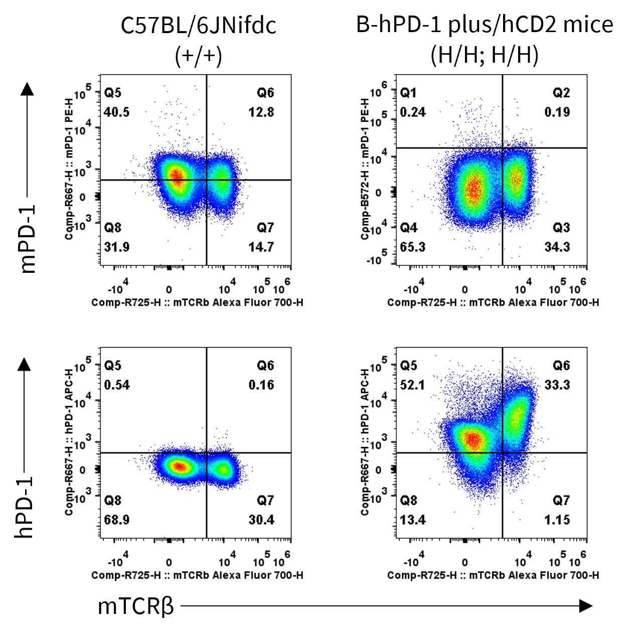
PD-1 Protein Expression Analysis in Spleen
- Human PD-1 was exclusively detectable in homozygous B-hPD-1 plus/hCD2 mice but not wild-type C57BL/6JNifdc mice.
Strain specific PD-1 expression analysis in wild-type C57BL/6JNifdc mice and homozygous B-hPD-1 plus/hCD2 mice by flow cytometry. Splenocytes were collected from wild-type C57BL/6JNifdc mice (+/+) and homozygous B-hPD-1 plus/hCD2 mice (H/H;H/H), stimulated with anti-CD3ε in vivo for 24 hours. The protein expression was analyzed with anti-mouse PD-1 antibody (Biolegend, 109104) and anti-human PD-1 antibody (Biolegend, 329908) by flow cytometry. Mouse PD-1 was only detectable in wild-type C57BL/6JNifdc mice. Human PD-1 was only detectable in homozygous B-hPD-1 plus/hCD2 mice.
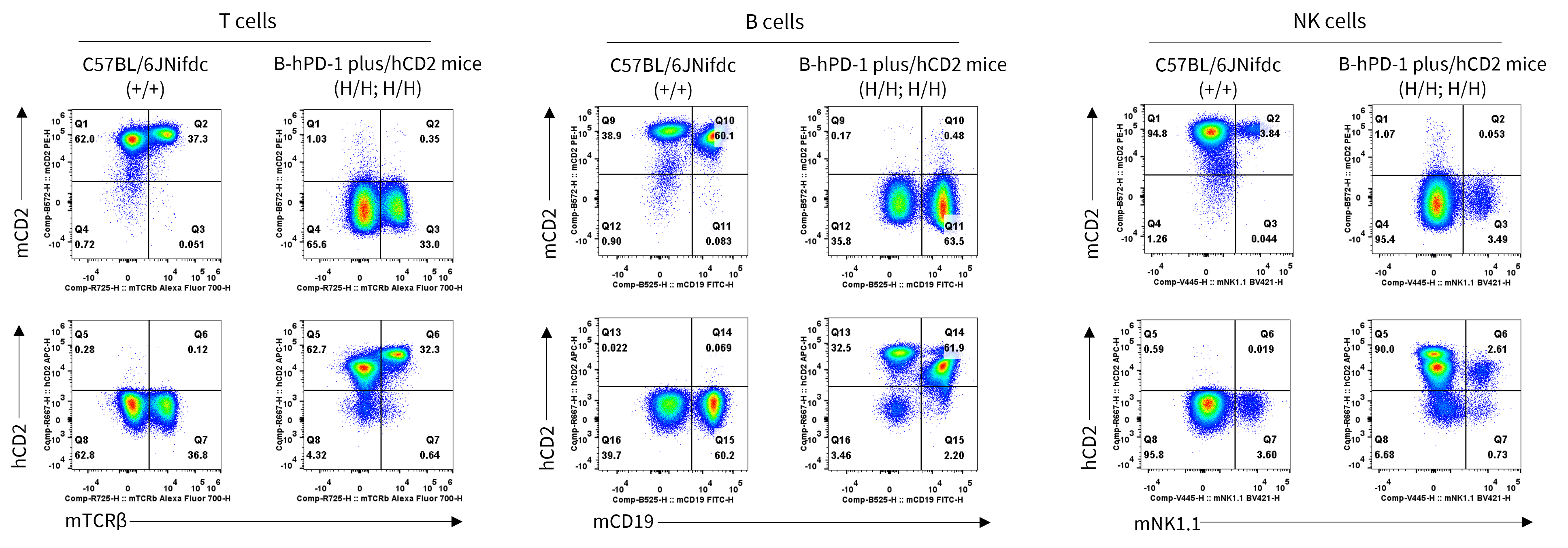
CD2 Protein Expression Analysis in Spleen
- Human CD2 was exclusively detectable in homozygous B-hPD-1 plus/hCD2 mice but not wild-type C57BL/6JNifdc mice.
Strain specific CD2 expression analysis in wild-type C57BL/6JNifdc mice and homozygous B-hPD-1 plus/hCD2 mice by flow cytometry. Splenocytes were collected from wild-type C57BL/6JNifdc mice (+/+) and homozygous B-hPD-1 plus/hCD2 mice (H/H;H/H). The protein expression was analyzed with anti-mouse CD2 antibody (Biolegend, 100107) and anti-human CD2 antibody (Biolegend, 300213) by flow cytometry. Mouse CD2 was only detectable in T cells, B cells, and NK cells of wild-type C57BL/6JNifdc mice. Human CD2 was only detectable in T cells, B cells, and NK cells of homozygous B-hPD-1 plus/hCD2 mice.
* When publishing results obtained using this animal model, please acknowledge the source as follows: The animal model [B-hPD-1 plus/hCD2 mice] (Cat# 114035) was purchased from Biocytogen.