

C57BL/6N-Tnfsf15tm2(TNFSF15)Bcgen Tnfrsf25tm3(TNFRSF25)Bcgen/Bcgen • 113082
on this page

Key Advantages
Validation
Applications

TL1A
DR3

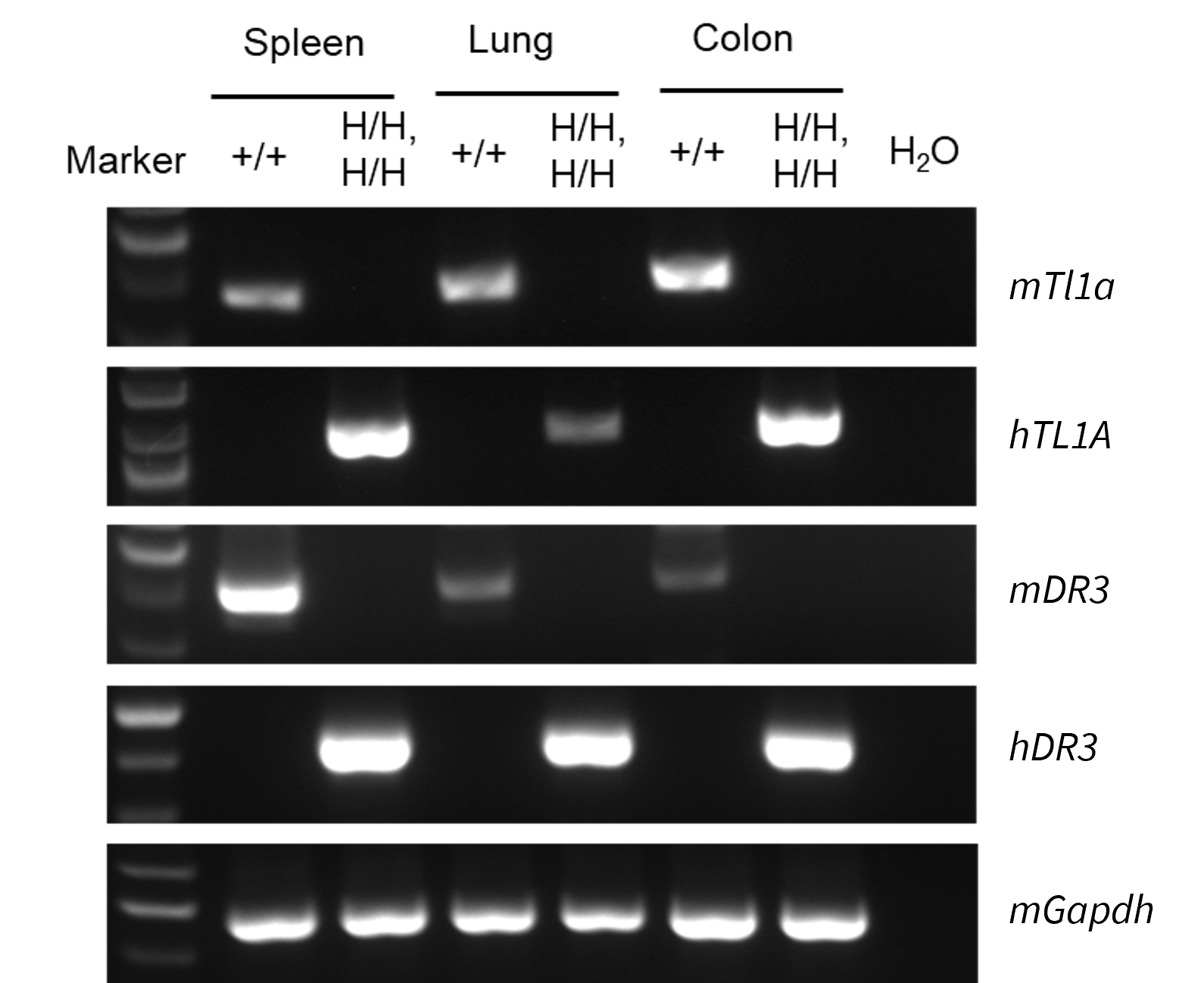
Strain specific analysis of TL1A and DR3 gene expression in wild-type C57BL/6N mice and homozygous TL1A/DR3 humanized mice by RT-PCR. Lung and colon tissues were collected from wild-type C57BL/6N mice (+/+) and homozygous TL1A/DR3 humanized mice (H/H;H/H). Mouse Tl1a and DR3 mRNA were detectable in wild-type C57BL/6N mice. Human TL1A and DR3 mRNA were exclusively detectable in homozygous B-hTL1A/hDR3 mice, but not in wild-type C57BL/6N mice.

Soluble TL1A expression analysis in TL1A/DR3 humanized mice by ELISA. Bone marrow derived dendritic cells (BMDCs) were produced by culturing the bone marrow cells isolated from wild-type C57BL/6N mice (+/+) and homozygous TL1A/DR3 humanized mice (H/H;H/H), which were stimulated with LPS in vitro. After stimulation, the supernatants were collected and the levels of soluble TL1A were measured using the species-specific human TL1A ELISA kit. Soluble human TL1A was exclusively detectable in homozygous TL1A/DR3 humanized mice but not wild-type C57BL/6N mice. Values are expressed as mean ± SEM. ND: not detectable.
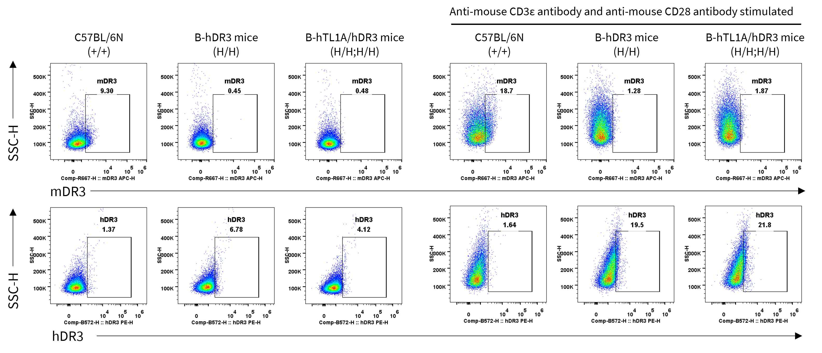
Strain specific DR3 expression analysis in wild-type C57BL/6N mice, homozygous DR3 humanized mice and TL1A/DR3 humanized mice by flow cytometry. Splenocytes were collected from wild-type C57BL/6N mice (+/+), homozygous DR3 humanized mice (H/H) and TL1A/DR3 humanized mice (H/H;H/H) stimulated with anti-mouse CD3ε antibody and anti-mouse CD28 antibody in vivo for 24 h, protein expression was analyzed with anti-mouse DR3 antibody (Biolegend, 144407) and anti-human DR3 antibody (Biolegend, 307105) by flow cytometry. Mouse DR3 was detectable in CD4+ T cells of wild-type C57BL/6N mice, human DR3 was detectable in CD4+ T cells of homozygous DR3 humanized mice and TL1A/DR3 humanized mice.
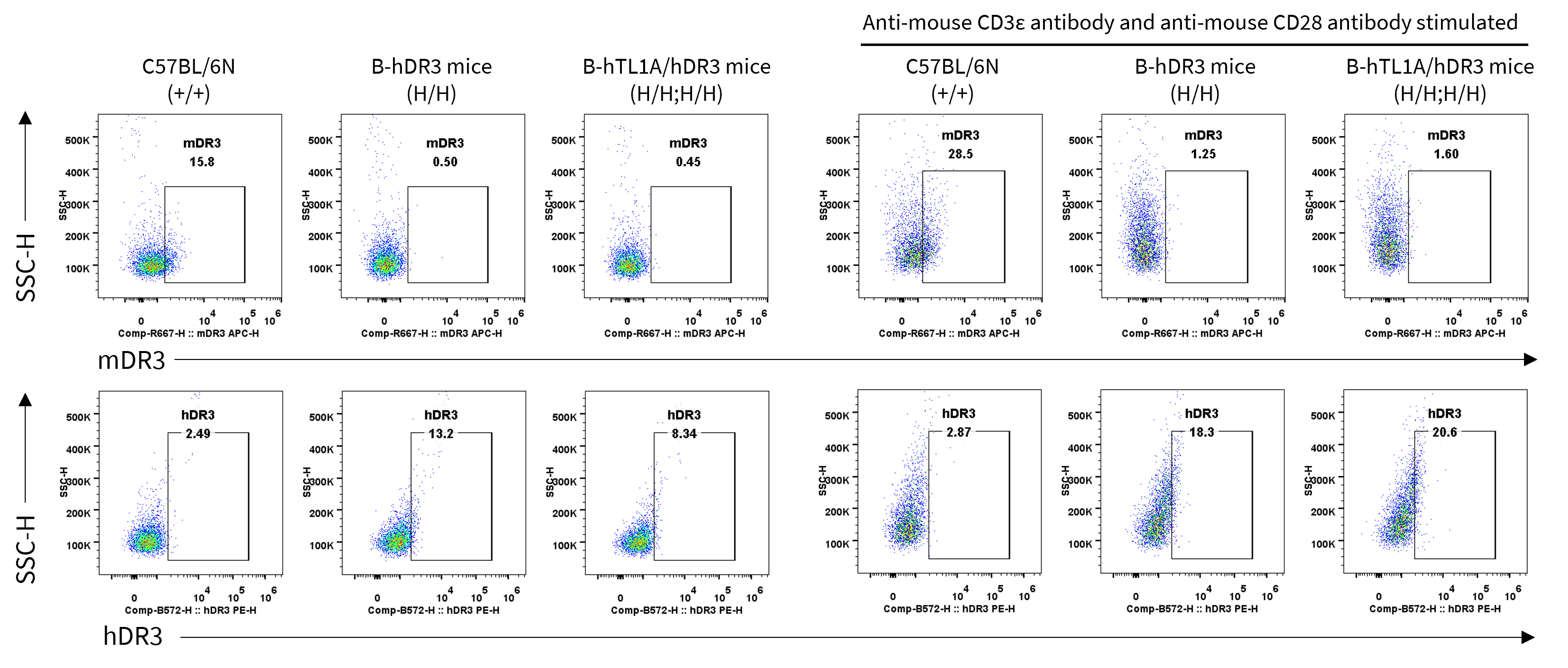
Strain specific DR3 expression analysis in wild-type C57BL/6N mice, homozygous DR3 humanized mice and TL1A/DR3 humanized mice by flow cytometry. Splenocytes were collected from wild-type C57BL/6N mice (+/+), homozygous DR3 humanized mice (H/H) and TL1A/DR3 humanized mice (H/H;H/H) stimulated with anti-mouse CD3ε antibody and anti-mouse CD28 antibody in vivo for 24 h, protein expression was analyzed with anti-mouse DR3 antibody (Biolegend, 144407) and anti-human DR3 antibody (Biolegend, 307105) by flow cytometry. Mouse DR3 was detectable in Treg cells of wild-type C57BL/6N mice, human DR3 was detectable in Treg cells of homozygous DR3 humanized mice and TL1A/DR3 humanized mice.
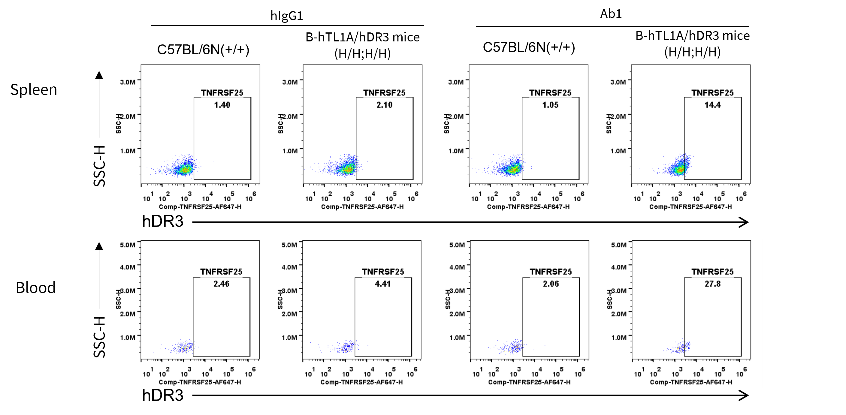
Anti-human DR3 antibody binding assessment in homozygous TL1A/DR3 humanized mice by flow cytometry. Splenocytes and blood were collected from wild-type C57BL/6N mice(+/+) and homozygous L1A/DR3 humanized mice (H/H;H/H), and analyzed by flow cytometry with anti-human DR3 antibody (Ab1), which is offered by the client. Ab1 can bind to Treg cells of homozygous TL1A/DR3 humanized mice.
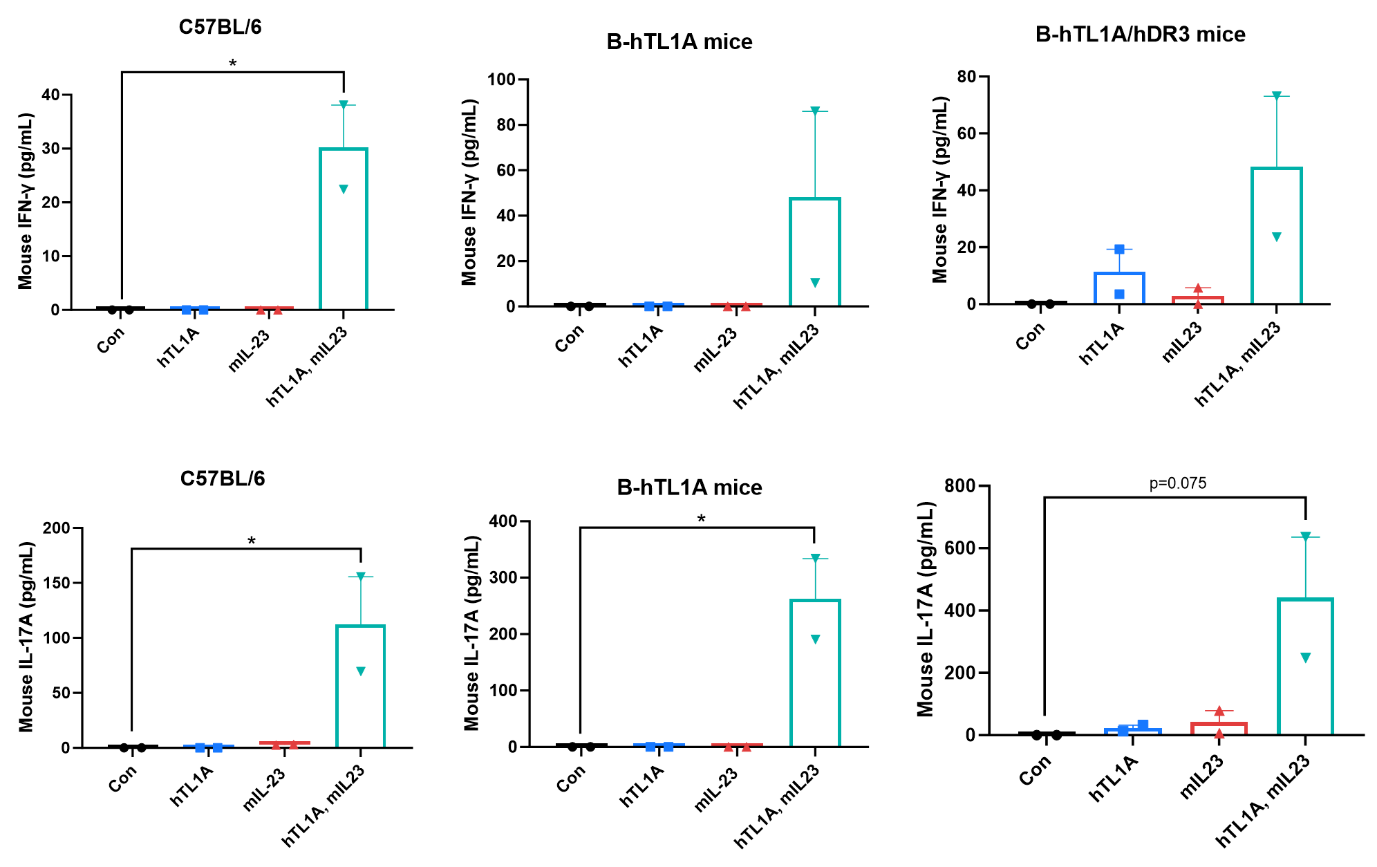
Ex vivo functional analysis in TL1A humanized and TL1A/DR3 humanized mice. Splenocytes were collected from wild-type C57BL/6 mice (+/+), homozygous TL1A humanized mice (H/H), and homozygous TL1A/DR3 humanized mice (H/H; H/H), then the production of mouse IFN-γ and mouse IL-17A in supernatants was assessed by ELISA after 72 h of incubation with mIL23 (10 ng/mL) and hTL1A (300 ng/mL) in vitro.
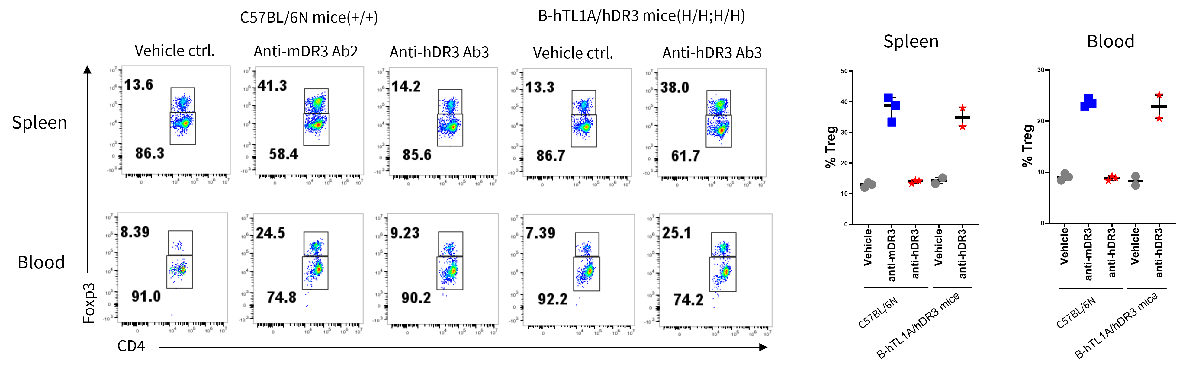
Activation of DR3 signaling pathway increased the expansion of Treg cells in wild-type C57BL/6N mice and homozygous TL1A/DR3 humanized mice. Wild-type C57BL/6N mice(+/+) and homozygous TL1A/DR3 humanized mice(H/H;H/H) were given a single intraperitoneal injection of DPBS, anti-mDR3 and anti-hDR3 antibodies on day 0. Splenocytes and blood cells were collected on day 4 for flow cytometry analysis. The agonistic anti-mouse DR3 antibody Ab2 expanded Treg cell populations in wild-type C57BL/6N mice, and the agonistic anti-human DR3 antibody Ab3 increased Treg cells in homozygous TL1A/DR3 humanized mice. These results contribute to a deeper understanding of DR3 activation in Treg cells modulation.
Note: This experiment was conducted by the client using TL1A/DR3 humanized mice.
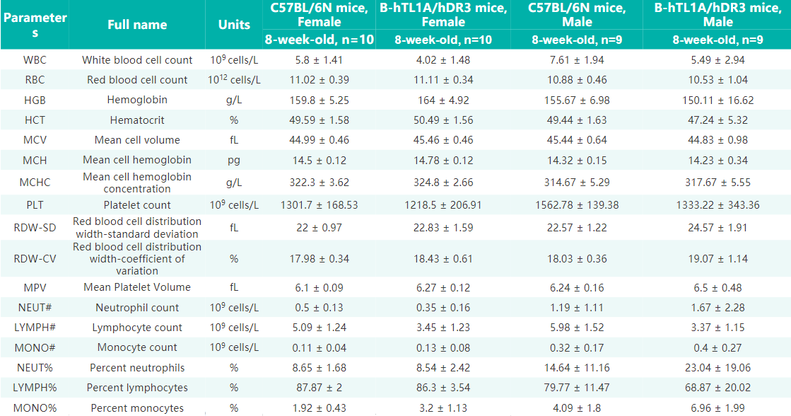
Complete blood count (CBC) of TL1A/DR3 hHumanized mice. Values are expressed as mean ± SD.
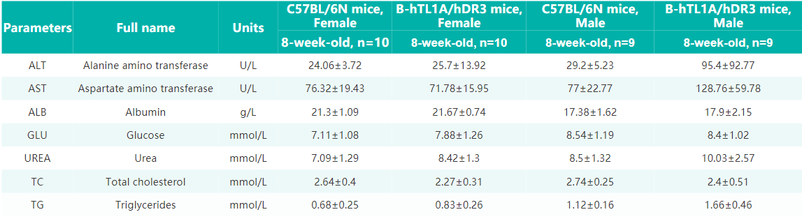
Blood biochemical parameters of TL1A/DR3 humanized mice are shown. Values are expressed as mean ± SD.
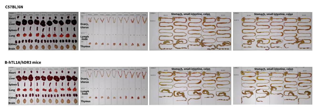
Organs of female C57BL/6N (8 week old, n=10) and TL1A/DR3 humanized mice (8 week old, n=10).
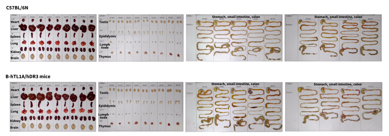
Organs of male C57BL/6N (8 week old, n=9) and TL1A/DR3 humanized mice (8 week old, n=9).
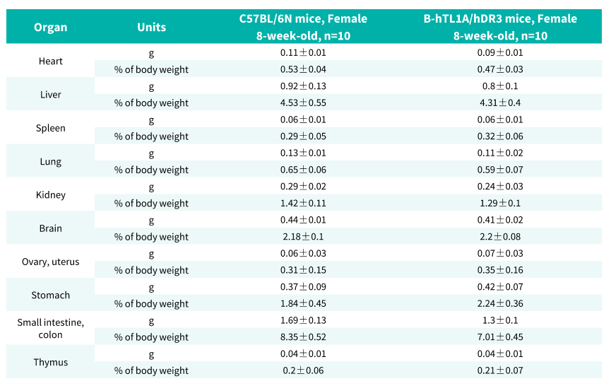
Average weight of the main organs of female C57BL/6N and TL1A/DR3 humanized mice.
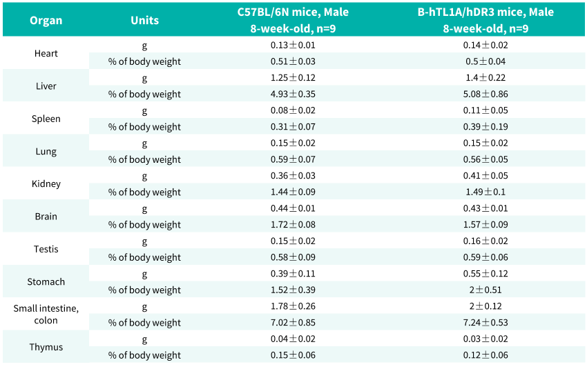
Average weight of the main organs of male C57BL/6N and TL1A/DR3 humanized mice.
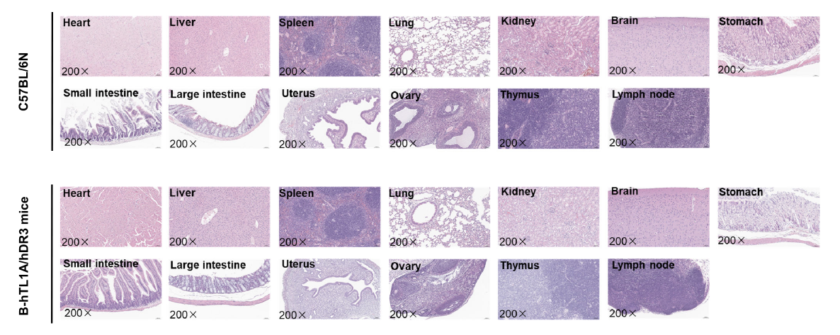
Major organs from C57BL/6N and TL1A/DR3 humanized mice were collected at 8 weeks of age and analyzed by H&E staining (female, n = 3).
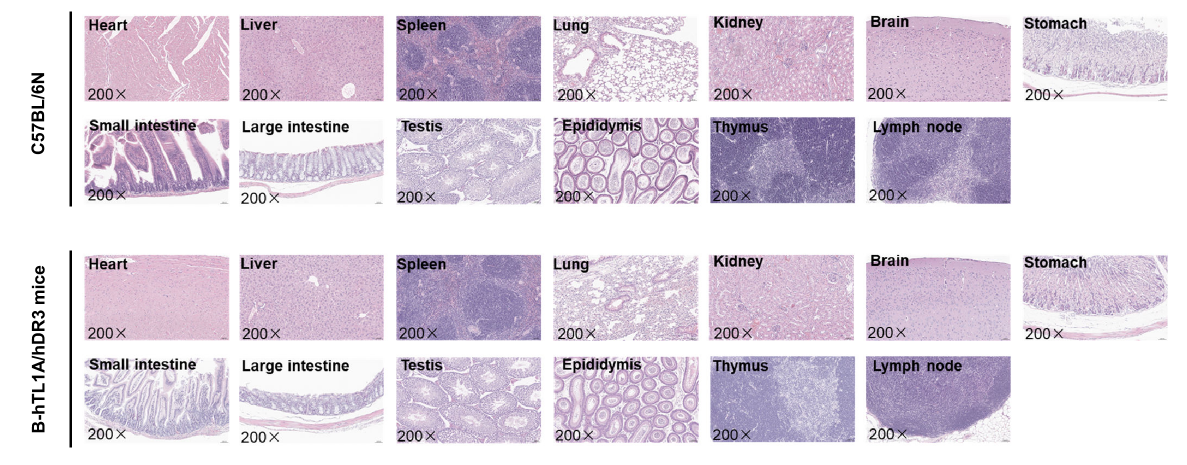
Major organs from C57BL/6N and TL1A/DR3 humanized mice were collected at 8 weeks of age and analyzed by H&E staining (male, n = 3).

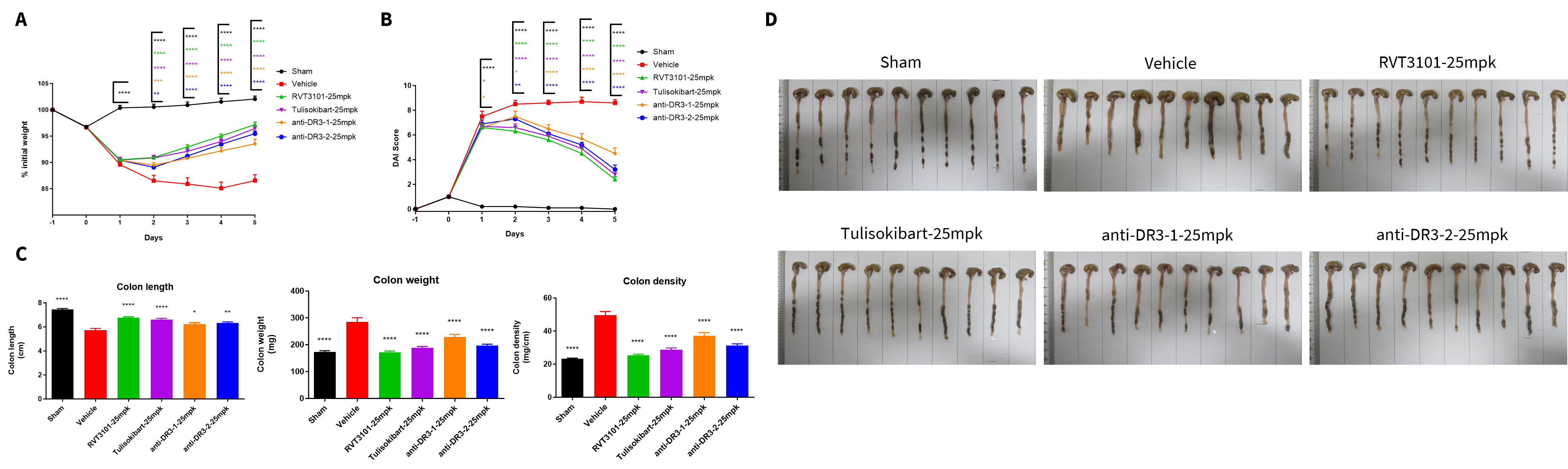
Therapeutic efficacy of anti-human TL1A antibodies and anti-human DR3 antibodies on the TNBS-induced acute colitis model in TL1A/DR3 humanized mice. TNBS solution was instilled into the colon lumen of TL1A/DR3 humanized mice (female, 8-10 weeks-old, n=8). The control group (Sham) received intrarectal injections of 50% ethanol. The treatment groups received anti-human TL1A antibodies RVT-3101 and Tulisokibart (25 mpk, provided by WuXi AppTec), anti-human DR3 antibodies anti-DR3-1 and anti-DR3-2 (25 mpk, provided by the client). (A) Body weight change. (B) DAI score. (C) Colon Index. (D) Representative colon images. A TNBS-induced acute colitis model was established inTL1A/DR3 humanized mice. Administration of anti-TL1A antibodies and anti-human DR3 antibodies effectively improved TNBS-induced acute colitis. The results indicate that TL1A/DR3 humanized mice are a powerful tool for evaluating in vivo efficacy of anti-human TL1A antibodies and anti-human DR3 antibodies in preclinical inflammatory bowel disease (IBD) research. Values are expressed as mean ± SEM. *p<0.05, **p<0.01, ***p<0.001, ****p<0.0001, versus Vehicle, ANOVA.
Note: This experiment was conducted by WuXi AppTec using TL1A/DR3 humanized mice.
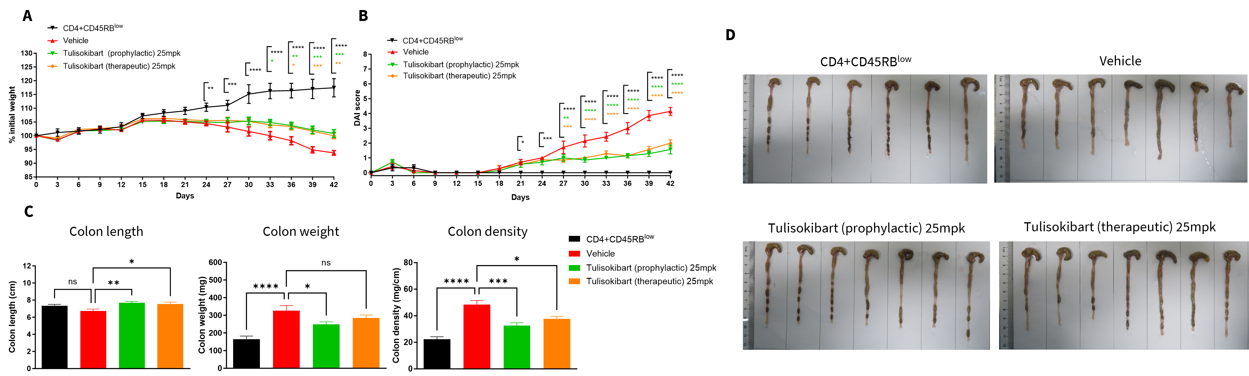
Therapeutic efficacy of anti-human TL1A antibody Tulisokibart in a T cells transfer induced colitis model in using TL1A humanized Rag2 knockout (KO) mice (B-hTL1A, Rag2 KO mice). CD4+CD45RBlow T and CD4+CD45RBhigh T cells were isolated from the spleens of TL1A/DR3 humanized mice. TL1A humanized Rag2 KO mice in group G1 were injected with CD4+CD45RBlow T cells, while TL1A humanized Rag2 KO mice in groups G2-G4 were injected with CD4+CD45RBhigh T cells. Animals in group G3 were given prophylactic administration of 25 mg/kg of anti-human TL1A antibody Tulisokibart every two days, and animals in group G4 were given therapeutic administration of 25 mg/kg of anti-human TL1A antibody Tulisokibart every two days. (A) Body weight changes. (B) Disease activity index (DAI) score. (C) Colon index. (D) Representative colon images. Administration of anti-human TL1A antibody tulisokibart effectively improved T cells transfer induced colitis. Two-way ANOVA or one-way ANOVA was used for multiple comparisons, with each group compared to Vehicle. Values are expressed as mean ± SEM. *p<0.05, **p<0.01, ***p<0.001, ****p<0.0001..
Note: This experiment was conducted by WuXi AppTec, T cell transfer induced chronic colitis in TL1A humanized Rag2 KO mice (Donor: TL1A/DR3 humanized mice).

Q1: What are TL1A/DR3 humanized mice?
A1 TL1A/DR3 humanized mice express human TL1A and DR3 in place of the corresponding murine genes, enabling translational evaluation of the human TL1A–DR3 pathway.
Q2: Why is the TL1A–DR3 pathway important?
A2 The TL1A–DR3 signaling axis plays a critical role in intestinal inflammation, fibrosis, T cell activation, and autoimmune disease progression.
Q3: Can TL1A/DR3 humanized mice be used for antibody validation?
A3 Yes. These mice are highly suitable for in vivo validation of anti-human TL1A therapeutic antibodies.
Q4: Do TL1A/DR3 humanized mice maintain normal immune homeostasis?
A4 Yes. Immune cell distributions in spleen, blood, and lymphoid tissues remain comparable to wild-type controls.
Q5: What disease areas can be studied using this model?
A5 This model supports research in IBD, Crohn’s disease, ulcerative colitis, fibrosis, autoimmune disease, and inflammatory signaling.
