

C57BL/6-Ighg2ctm1(IGHG1)Bcgen Fcgrttm1(FCGRT)Bcgen/Bcgen • 111952
on this page

B-hFcRn mice (110001): The CDS that encodes full-length human FCGRT, followed by the mouse 3’UTR-STOP, is inserted right after the mouse Fcgrt ATG to replace the exons 2-4 of the mouse Fcgrt gene. The FcRn protein expression will be driven by the endogenous mouse Fcgrt promoter, while the mouse Fcgrt gene transcription and translation will be disrupted.
B-hIGHG1/hFcRn mice were generated through secondary targeting based on B-hFcRn mice. The exons 2-4 of mouse Ighg2c gene that encode the full-length protein of the heavy chain Fc segment were replaced by human IGHG1 exon 2-4 in B-hIGHG1/hFcRn mice. The human full-length FCGRT cDNA sequence was inserted into the Fcgrt exon 2 of B-hIGHG1/hFcRn mice.

Gene targeting strategy for B-hIGHG1/hFcRn mice.
B-hFcRn mice (110001): The CDS that encodes full-length human FCGRT, followed by the mouse 3’UTR-STOP, is inserted right after the mouse Fcgrt ATG to replace the exons 2-4 of the mouse Fcgrt gene. The FcRn protein expression will be driven by the endogenous mouse Fcgrt promoter, while the mouse Fcgrt gene transcription and translation will be disrupted.
B-hIGHG1/hFcRn mice were generated through secondary targeting based on B-hFcRn mice. The exons 2-4 of mouse Ighg2c gene that encode the full-length protein of the heavy chain Fc segment were replaced by human IGHG1 exon 2-4 in B-hIGHG1/hFcRn mice. The human full-length FCGRT cDNA sequence was inserted into the Fcgrt exon 2 of B-hIGHG1/hFcRn mice.

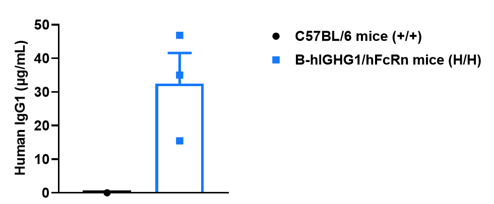
Strain specific human IgG1 expression analysis in homozygous B-hIGHG1/hFcRn mice by ELISA. Serum was collected from wild-type C57BL/6 mice (+/+) and homozygous B-hIGHG1/hFcRn mice (H/H), and analyzed by ELISA. Human IgG1 was exclusively detectable in homozygous B-hIGHG1/hFcRn mice but not in wild-type mice.
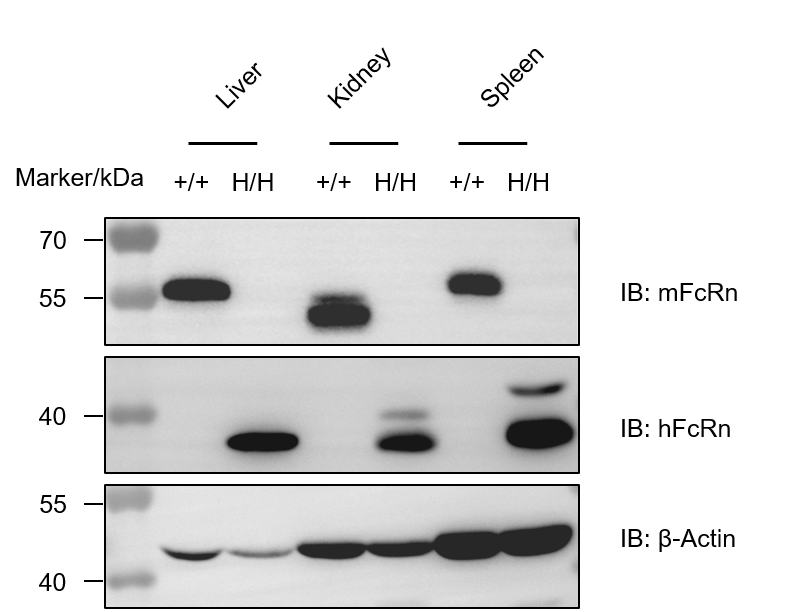
Strain specific FcRn expression analysis in homozygous B-hIGHG1/hFcRn mice by western blot. Various tissue lysates were collected from wild-type C57BL/6 mice (+/+) and homozygous B-hIGHG1/hFcRn mice (H/H), and analyzed by western blot with anti-FcRn antibody (Mouse FCRN Antibody, R&D, AF6775; FCRN/FCGRT Antibody, Novus Biologicals, AF6775). Mouse FcRn was detectable in wild type mice. Human FcRn was exclusively detectable in homozygous B-hIGHG1/hFcRn mice but not in wild-type C57BL/6 mice.

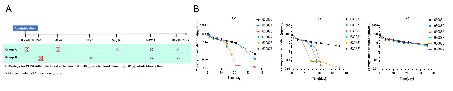
Pharmacokinetics of Yervoy-analog in B-hIGHG1/hFcRn mice. C57BL/6JNifdc, B-hFcRn mice and B-hIGHG1/hFcRn mice were intravenously injected with Yervoy-analog (in-house), and serum was collected for pharmacokinetic (PK) analysis (n=6). A. Design of Blood Collection Timepoints. B. The PK curve of single mice in each group.
ADA was observed in 4–5 mice in both G1 and G2 (ADA was not tested but inferred based on the pharmacokinetic curves), while no ADA was observed in G3. This is likely because B-hIGHG1/hFcRn mice express the human IgG1 Fc region, which may help reduce ADA to some extent.
ADA: anti-drug-antibody.
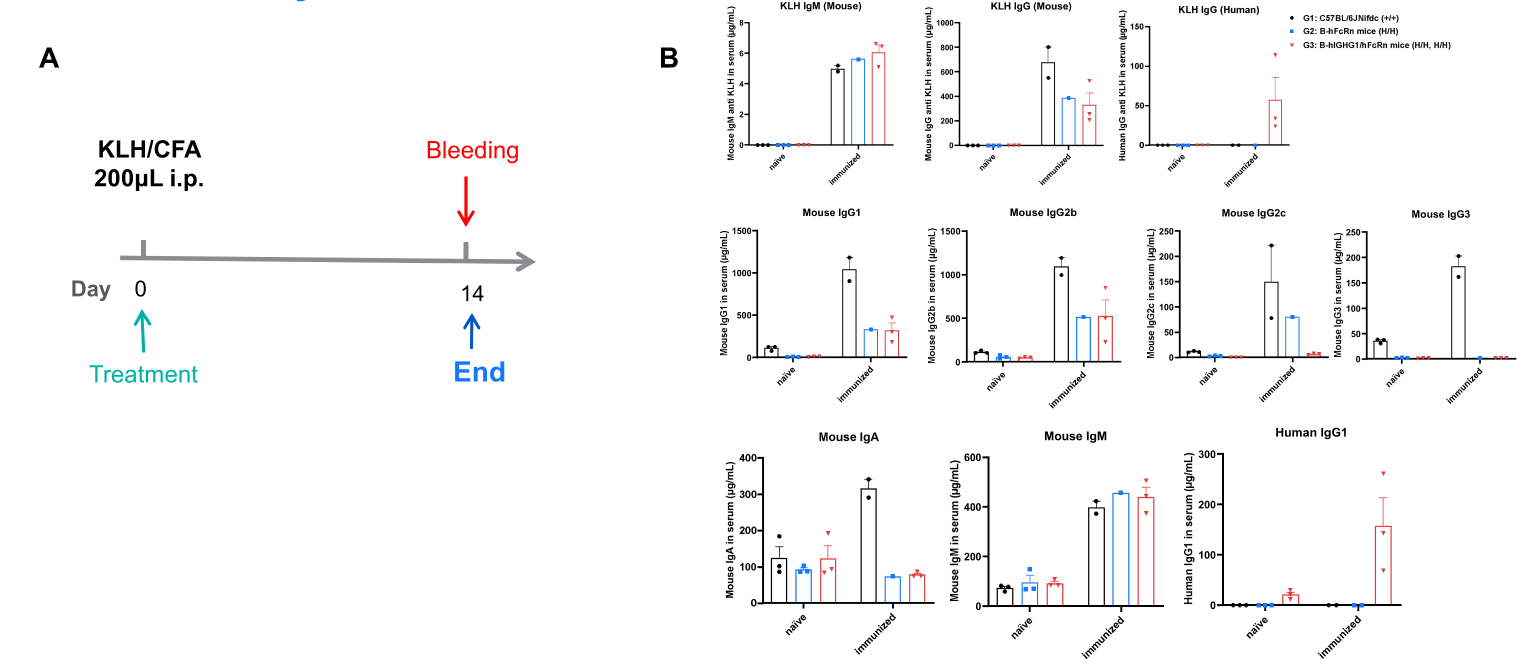
Immunoglobulin expression analysis in wild-type C57BL/6JNifdc mice, homozygous B-hFcRn mice, and B-hIGHG1/hFcRn mice by ELISA.
(A) Schematic diagram of KLH/CFA intraperitoneal (i.p.) immunization and serum collection schedule (10-week-old male mice, immunized on Day 0, bled on Day 14). (B) ELISA quantification of antigen-specific antibodies (mouse anti-KLH IgM, mouse anti-KLH IgG, human anti-KLH IgG; Abnova: KA2461, KA2460, KA6737) and total immunoglobulins (mouse IgG1, IgG2b, IgG2c, IgG3, IgA, IgM, and human IgG1; SouthernBiotech SBA Clonotyping System-C57BL/6-HRP: SouthernBiotech, 5300-05B) in naive and immunized WT (C57BL/6JNifdc, n=2–3), B-hFcRn (n=1–3), and B-hIGHG1/hFcRn (n=3) mice. Values are expressed as mean ± SEM.
B-hIGHG1/hFcRn mice selectively produced high levels of human anti-KLH IgG and serum human IgG1 post-immunization, confirming functional expression of the humanized loci and intact human-type humoral immunity.
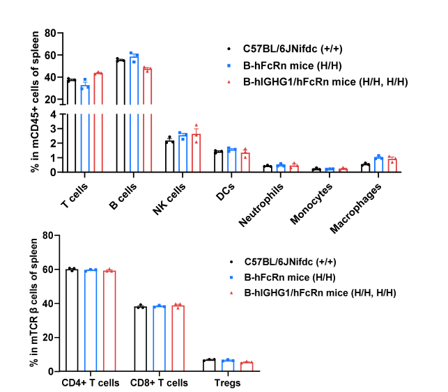
Analysis of leukocyte subpopulations by flow cytometry in immune organs. Splenocytes were isolated from C57BL/6, B-hFcRn and B-hIGHG1/hFcRn mice (female, 8-week-old, n = 3). Single live cells were gated on the CD45⁺ population and analyzed by flow cytometry as indicated. Values are expressed as mean ± SEM.
