

C57BL/6JNifdc-Msh3tm1(MSH3)Bcgen/Bcgen • 114352

MSH3: Biological Roles and Therapeutics Strategies

MSH3

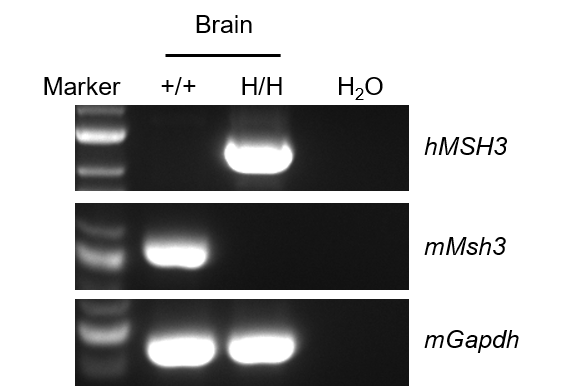
Strain specific analysis of MSH3 mRNA expression in wild-type C57BL/6JNifdc mice and homozygous B-hMSH3 mice by RT-PCR. Brain RNA were isolated from wild-type C57BL/6JNifdc mice (+/+, female) and homozygous B-hMSH3 mice (H/H, female), then cDNA libraries were synthesized by reverse transcription, followed by PCR with mouse or human MSH3 primers. Mouse Msh3 mRNA was detectable in wild-type mice. Human MSH3 mRNA was only detectable in homozygous B-hMSH3 mice but not in wild-type mice. And human sequences were confirmed via Sanger sequencing.
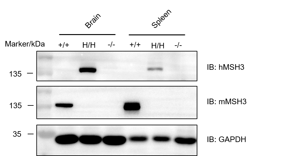
Western blot analysis of MSH3 protein expression in homozygous B-hMSH3 mice. Brain and spleen lysates were collected from wild-type C57BL/6JNifdc mice (+/+, female), homozygous B-hMSH3 mice (H/H, female) and homozygous B-Msh3 KO mice (-/-), and then analyzed by western blot with species-specific anti-MSH3 antibodies. 40 μg total protein was loaded for western blot analysis. Mouse MSH3 was absent in B-Msh3 KO mice and B-hMSH3 mice, but present in wild-type mice for mouse-specific antibody results. Human MSH3 protein was only detectable in brain and spleen from homozygous B-hMSH3 mice.

Strain specific analysis of MSH3 mRNA expression in wild-type C57BL/6JNifdc mice and homozygous B-hMSH3 mice by RT-qPCR. Brain, spleen and liver RNA were isolated from wild-type C57BL/6JNifdc mice (+/+, male and female, n=3, 9-week-old) and homozygous B-hMSH3 mice (H/H, male and female, n=3, 9-week-old), then cDNA libraries were synthesized by reverse transcription, followed by RT-qPCR with mouse or human MSH3 primers. Mouse Gapdh served as an internal reference gene. Human MSH3 mRNA was detectable only in homozygous B-hMSH3 mice but not in wild-type mice, and MSH3 expression in other tissues was normalized to that in the brains from female mice both from wild-type and B-hMSH3 mice. Values are expressed as mean ± SEM.

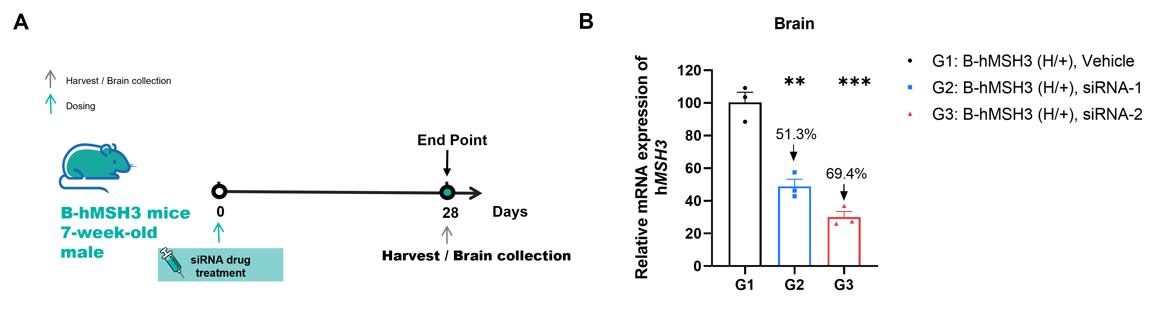
The inhibitory efficiency of the MSH3-targeted small nucleic acid drug in heterozygous B-hMSH3 mice. B-hMSH3 mice were randomly divided into 3 groups (n=3, 7-week-old, female). The human MSH3-targeted nucleic acid drug were administered to the mice individually. The mice were sacrificed on day 28, and the brains were collected to detect the human MSH3 expression by qRT-PCR. (A) The schematic diagram of experimental processing. (B) The expression of human MSH3 mRNA in brain from different groups. The human MSH3 mRNA in the treatment group was significantly reduced compared to the control group. Values are expressed as mean ± SEM. Significance was determined by unpaired t test. *P < 0.05, **P < 0.01, ***P < 0.001.
Note: Data are shared upon the client's approval.
