

C57BL/6JNifdc-Opgtm1Bcgen/Bcgen • 114526
on this page

The RANKL/RANK/OPG axis is the major determinant of bone remodeling.

OPG

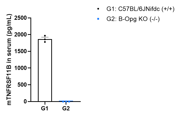
Mouse-specific OPG expression analysis in homozygous B-Opg KO mice by ELISA. Serum were collected from wild-type mice C57BL/6JNifdc mice (+/+) (female, 16-week-old, n=3) and homozygous B-Opg KO mice (-/-) (female, 16-week-old,n=3), then analyzed by ELISA with mouse-specific OPG ELISA kit (Mouse Osteoprotegerin/TNFRSF11B Immunoassay: R&D, MOP00). Mouse OPG was exclusively detectable in C57BL/6JNifdc mice but not in homozygous B-Opg KO mice. Values are expressed as mean ± SEM.
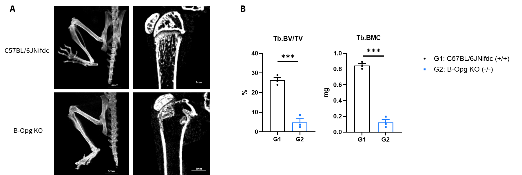
Micro-CT images of the bones of wild-type C57BL/6JNifdc and homozygous B-Opg KO mice. (A) Representative whole-hindlimb radiographs and magnified radiographs of the distal femur. The distal portion of the femur of the homozygous B-Opg KO mice was highly translucent compared with that of wild-type mice. The distal femur of B-Opg KO mice exhibited marked radiolucency, with severe trabecular bone loss (asterisk) and disrupted growth plate (arrow); wild-type mice showed intact bone architecture without lesions. (B) Quantitative micro-CT analysis of trabecular bone parameters (bone volume/tissue volume (BV/TV) and bone mineral content (BMC)) further confirmed profound bone loss in B-Opg KO mice. Values are expressed as mean ± SEM. Significance was determined by unpaired t test. *P < 0.05, **P < 0.01, ***P < 0.001.

Q: Is there any difference in bone mineral density (BMD) between wild-type mice and B-Opg KO mice(#114526)?
A: Micro-CT analysis of femurs from homozygous B-Opg KO mice revealed severe trabecular bone and growth plate loss, whereas trabecular BMD exhibited no obvious decline. Trabecular BMD was calculated using the formula TMC/BV. Original data showed that both TMC and BV were significantly reduced in B-Opg KO mice, and the quotient of the two parameters ended up at a similar level to that of wild-type mice, which could be verified from the raw data in the table.

