on this page

Key Advantages
Validation
Application

TNFA
TNFR2
TNFR1

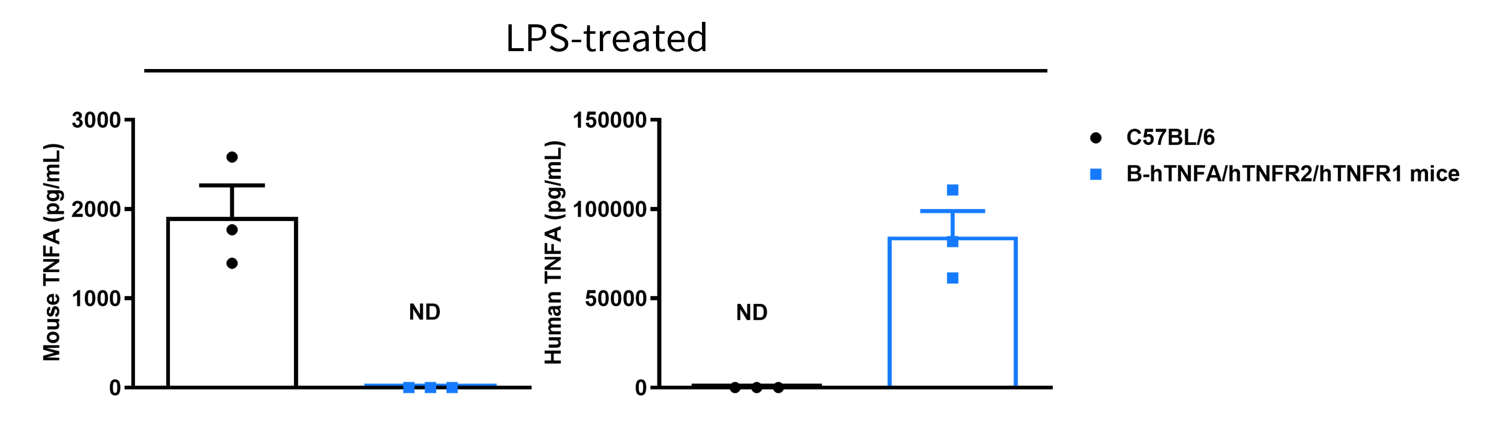
Strain-specific analysis of mouse and human TNFA protein expression in wild-type C57BL/6 mice and TNFA/TNFR2/TNFR1 humanized mice by ELISA. Wild-type C57BL/6 mice (+/+) and homozygous TNFA/TNFR2/TNFR1 humanized mice (H/H; H/H; H/H) were stimulated in vivo with lipopolysaccharide (LPS). Serum samples were subsequently collected, and mouse and human TNFA protein levels were measured by ELISA using species-specific TNFA ELISA kits. Mouse TNFA was detected exclusively in wild-type C57BL/6 mice, whereas human TNFA was detected exclusively in homozygous TNFA/TNFR2/TNFR1 humanized mice and was not detected in wild-type C57BL/6 mice. Values are expressed as mean ± SEM. ND: not detectable.
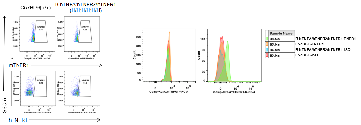
Strain-specific analysis of mouse and human TNFR1 protein expression in wild-type C57BL/6 mice and TNFA/TNFR2/TNFR1 humanized mice by flow cytometry. Splenocytes were collected from wild-type C57BL/6 mice (+/+) and homozygous TNFA/TNFR2/TNFR1 humanized mice (H/H; H/H; H/H). Mouse and human TNFR1 protein expression was analyzed by flow cytometry using species-specific anti-TNFR1 antibodies. Mouse TNFR1 was detected in wild-type C57BL/6 mice, whereas human TNFR1 was detected exclusively in homozygous TNFA/TNFR2/TNFR1 humanized mice and was not detected in wild-type C57BL/6 mice.
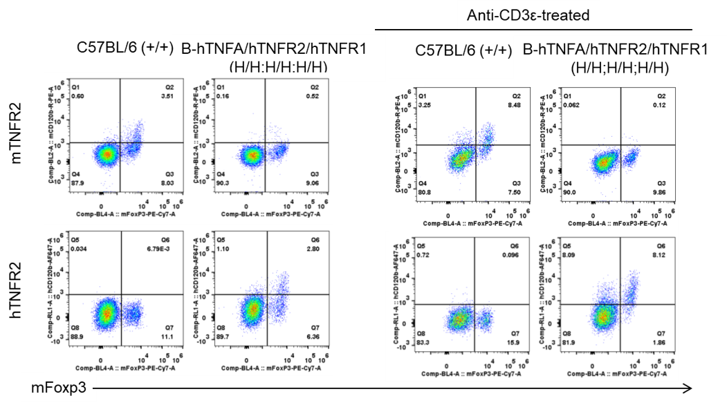
Strain-specific analysis of mouse and human TNFR2 protein expression in wild-type C57BL/6 mice and TNFA/TNFR2/TNFR1 humanized mice by flow cytometry. Wild-type C57BL/6 mice (+/+) and homozygous TNFA/TNFR2/TNFR1 humanized mice (H/H; H/H; H/H) were stimulated in vivo with anti-CD3ε antibody. Splenocytes were subsequently collected, and mouse and human TNFR2 protein expression was analyzed by flow cytometry using species-specific anti-TNFR2 antibodies. Mouse TNFR2 was detected in wild-type C57BL/6 mice, whereas human TNFR2 was detected exclusively in homozygous TNFA/TNFR2/TNFR1 humanized mice and was not detected in wild-type C57BL/6 mice.
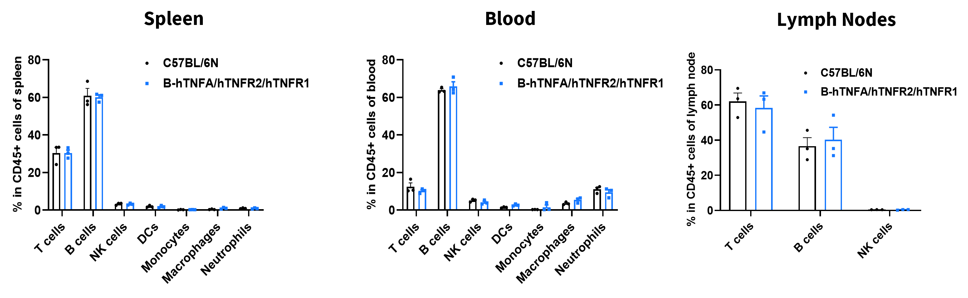
Flow cytometry analysis of leukocyte subpopulations in immune organs and peripheral blood of TNFA/TNFR2/TNFR1 humanized mice. Splenocytes, peripheral blood, and lymph nodes were collected from female wild-type C57BL/6N mice and TNFA/TNFR2/TNFR1 humanized mice (9 weeks old, n = 3). Single live cells were gated on the CD45⁺ population and analyzed by flow cytometry as indicated. Values are expressed as mean ± SEM.

Flow cytometry analysis of T-cell subpopulations in immune organs and peripheral blood of TNFA/TNFR2/TNFR1 humanized mice. Splenocytes, peripheral blood, and lymph nodes were collected from female wild-type C57BL/6N mice and TNFA/TNFR2/TNFR1 humanized mice (9 weeks old, n = 3). Single live cells were gated on the CD3⁺ T-cell population and analyzed by flow cytometry as indicated. Values are expressed as mean ± SEM.
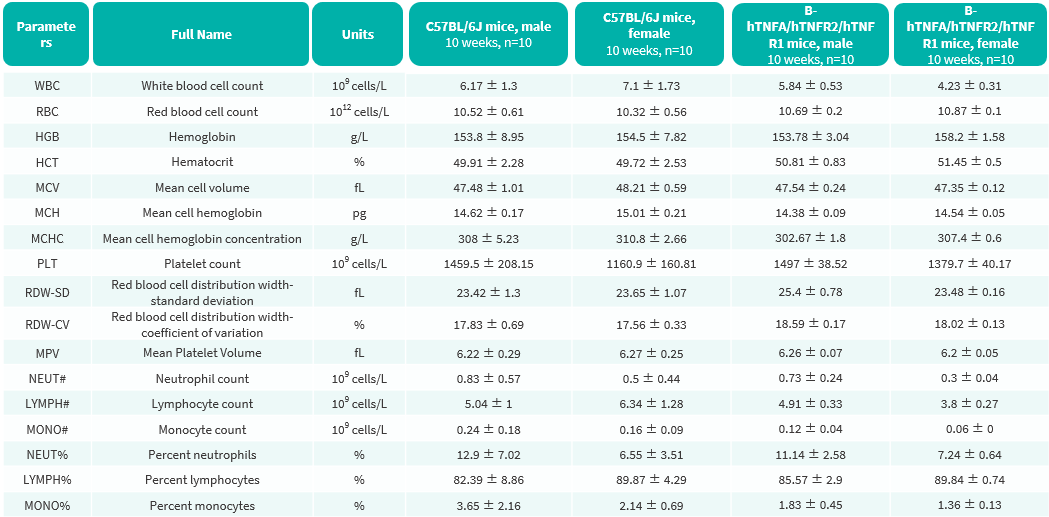
Complete blood count (CBC) of TNFA/TNFR2/TNFR1 humanized mice. Values are expressed as mean ± SD.
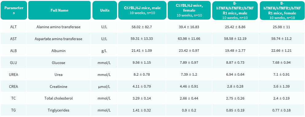
Blood biochemical parameters of TNFA/TNFR2/TNFR1 humanized mice are shown. Values are expressed as mean ± SD.

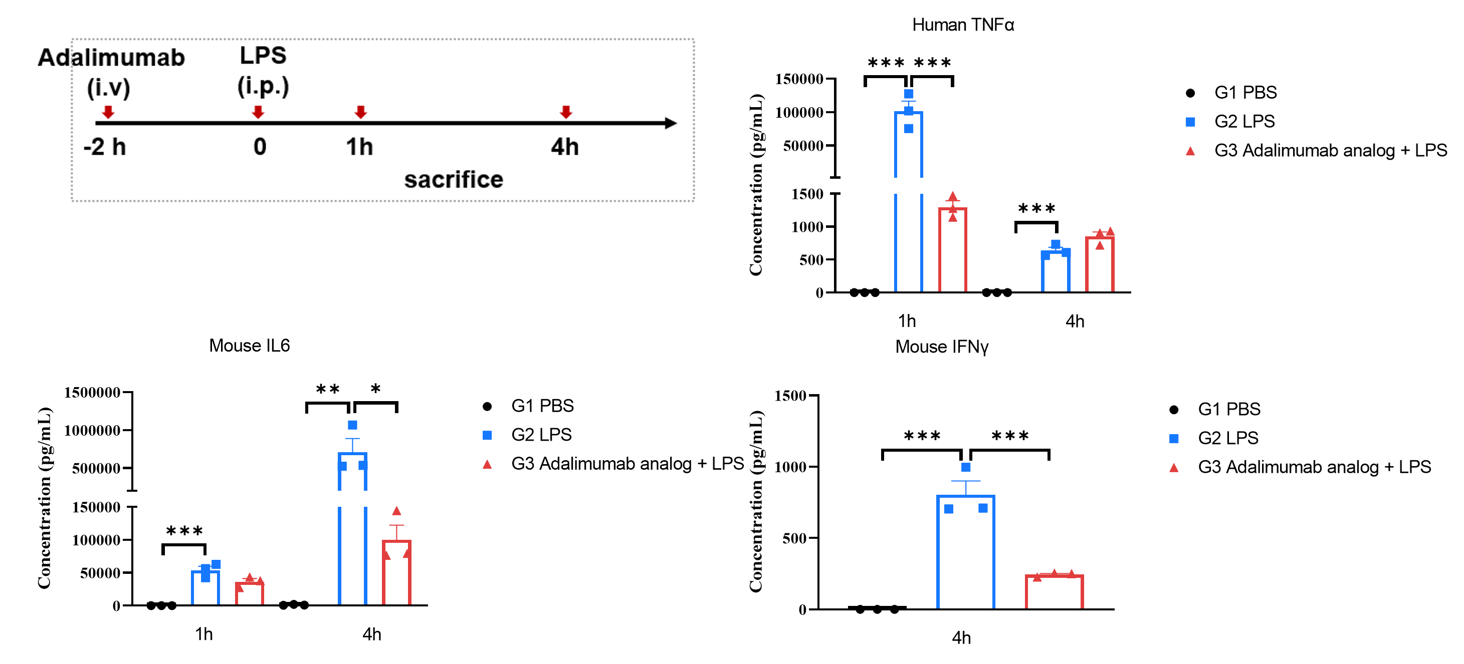
ELISA analysis of human TNFA, mouse IL-6, and mouse IFN-γ levels following LPS stimulation and adalimumab analog treatment in TNFA/TNFR2/TNFR1 humanized mice. Homozygous TNFA/TNFR2/TNFR1 humanized mice (H/H; H/H; H/H) received an intraperitoneal (i.p.) injection of lipopolysaccharide (LPS) 2 hours after treatment with PBS or an in-house adalimumab analog administered intravenously (i.v.). Serum samples were collected 1 and 4 hours after LPS injection. Human TNFA, mouse IL-6, and mouse IFN-γ levels were measured by ELISA using species-specific kits (ELISA MAX™ Deluxe Set Human TNF-α, BioLegend, 430204; ELISA MAX™ Deluxe Set Mouse IL-6, BioLegend, 430304; ELISA MAX™ Deluxe Set Mouse IFN-γ, BioLegend, 430804). Following LPS stimulation, human TNFA and mouse IL-6 levels were significantly increased at both 1 and 4 hours, while mouse IFN-γ levels were significantly increased at 4 hours. Pretreatment with the adalimumab analog significantly reduced human TNFA levels at 1 hour and mouse IL-6 and IFN-γ levels at 4 hours after LPS injection, indicating effective inhibition of downstream inflammatory responses mediated by human TNFA. Values are expressed as mean ± SEM. Statistical significance was determined using one-way ANOVA. *p < 0.05, **p < 0.01, ***p < 0.001.
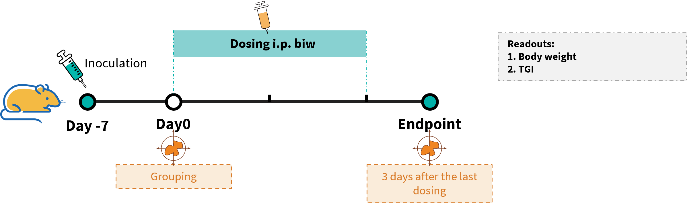
Establishment of a subcutaneous MC38 tumor model and in vivo efficacy evaluation of an anti-human TNFR2 antibody in TNFA/TNFR2/TNFR1 humanized mice. MC38 mouse colon carcinoma cells were subcutaneously implanted into homozygous TNFA/TNFR2/TNFR1 humanized mice (female, 7 weeks old, n = 6). When the average tumor volume reached approximately 110 mm³, mice were randomized into treatment groups and subsequently administered an anti-human TNFR2-specific antibody via intraperitoneal injection.
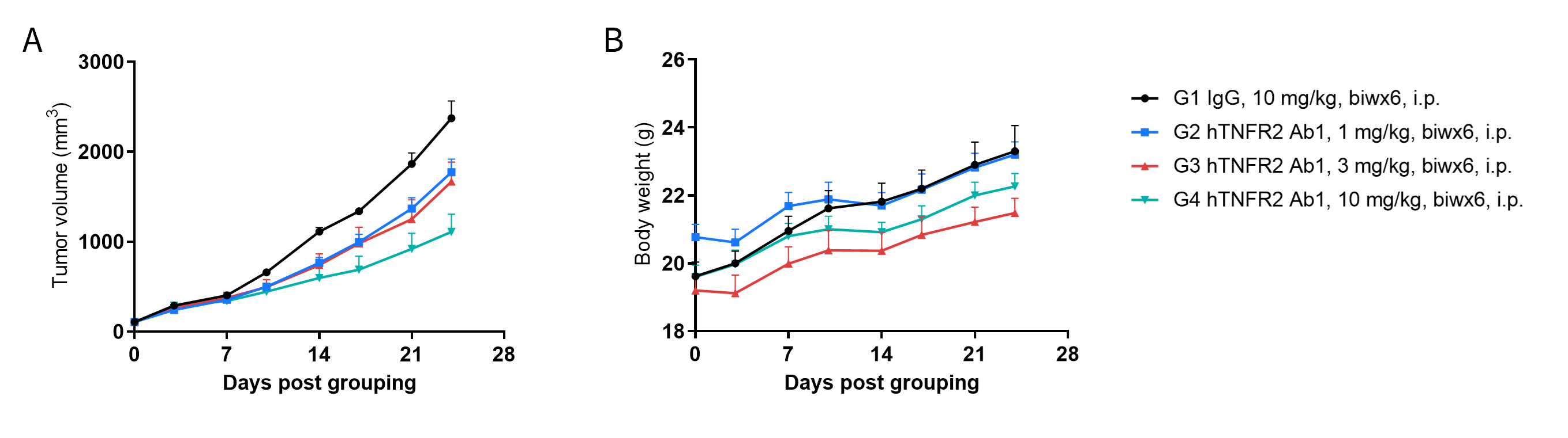
In vivo antitumor efficacy of anti-human TNFR2 antibodies in a subcutaneous MC38 tumor model using TNFA/TNFR2/TNFR1 humanized mice. (A) Tumor growth curves. Murine colon carcinoma MC38 cells were subcutaneously implanted into homozygous TNFA/TNFR2/TNFR1 humanized mice (female, 8 weeks old, n = 6). Mice were assigned to treatment groups based on body weight differences and subsequently administered the client-provided anti-human TNFR2 antibody (Ab1) at the doses and schedules indicated in panel A. (B) Body weight changes during treatment. As shown in panel A, treatment with the anti-human TNFR2 antibody inhibited MC38 tumor growth in TNFA/TNFR2/TNFR1 humanized mice in a dose-dependent manner. These results demonstrate the utility of TNFA/TNFR2/TNFR1 humanized mice as a preclinical model for evaluating the in vivo antitumor efficacy of anti-human TNFR2 antibodies. Values are expressed as mean ± SEM.

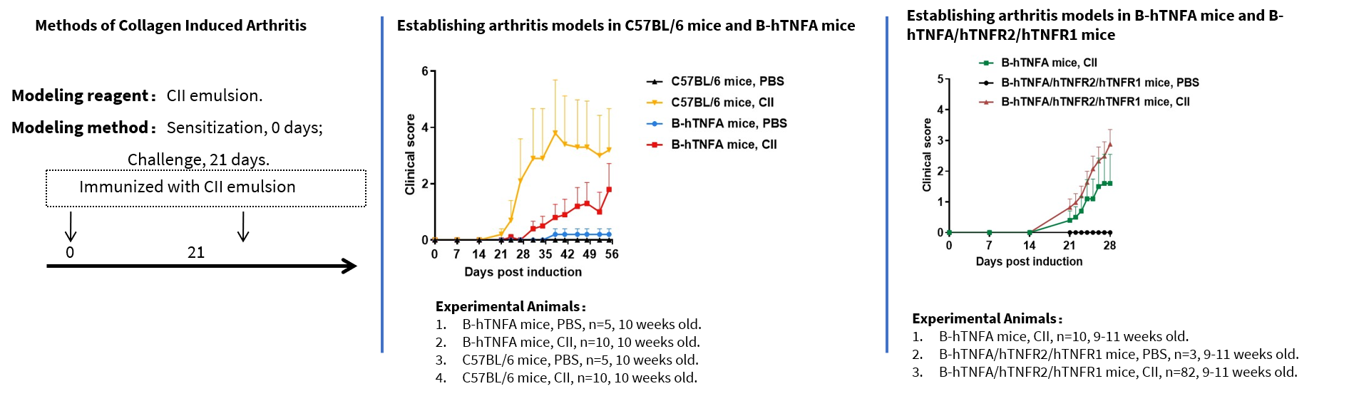
Establishment and clinical evaluation of a collagen-induced arthritis (CIA) model in TNFA/TNFR2/TNFR1 humanized mice. Arthritis was induced in TNFA/TNFR2/TNFR1 humanized mice and wild-type C57BL/6 mice using type II collagen (CII). (A) Body weight changes. (B) Clinical arthritis scores. The results showed significantly increased clinical arthritis scores in TNFA/TNFR2/TNFR1 humanized mice, indicating successful establishment of the collagen-induced arthritis model.
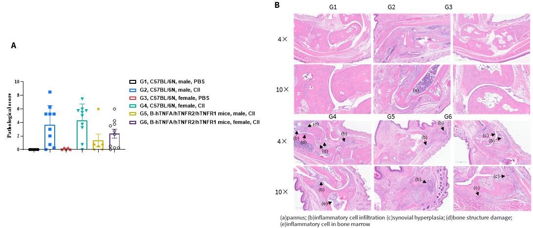
Histopathological analysis of collagen-induced arthritis (CIA) in TNFA/TNFR2/TNFR1 humanized mice and wild-type C57BL/6 mice. (A) Histopathological scores. (B) Hematoxylin and eosin (H&E) staining of joint tissue sections. In the arthritis model groups, histopathological examination revealed subcutaneous mixed inflammatory cell infiltration, periarticular joint space narrowing, articular cartilage destruction, bone tissue damage, and other arthritis-associated lesions in some or all limb joints. These findings indicate the successful establishment of the collagen-induced arthritis model in TNFA/TNFR2/TNFR1 humanized mice and wild-type C57BL/6 mice.

In vivo efficacy of anti-human TNFA antibody adalimumab analog in a collagen-induced arthritis (CIA) model using TNFA/TNFR2/TNFR1 humanized mice. Collagen-induced arthritis was established in female TNFA/TNFR2/TNFR1 humanized mice (n = 9–10 per group) by subcutaneous injection of type II collagen (CII) emulsion on days 0 and 21. Arthritis development was monitored, and clinical arthritis scores were assessed daily. At the onset of inflammation (defined as a clinical score > 1 or a persistent score of 1), mice were assigned to treatment groups, with the treatment initiation day designated as day 0. Mice received intraperitoneal injections of an in-house anti-human TNFA antibody (adalimumab analog) at different doses. (A) Body weight changes. (B) Clinical arthritis scores. Body weight and clinical arthritis scores were monitored daily during treatment. Mice were euthanized 2 days after the final treatment, and paws were collected for histopathological evaluation using decalcified hematoxylin and eosin (H&E)-stained joint sections. No significant changes in body weight were observed, while clinical arthritis scores increased in all groups except the control group, confirming successful establishment of the CIA model. Treatment with the adalimumab analog reduced clinical arthritis scores in a dose-dependent manner. These results demonstrate the utility of TNFA/TNFR2/TNFR1 humanized mice as a preclinical CIA model for evaluating the in vivo therapeutic efficacy of anti-human TNFA antibodies.
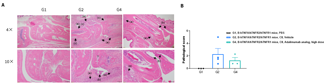
Histopathological evaluation of anti-human TNFA antibody efficacy in a collagen-induced arthritis (CIA) model using TNFA/TNFR2/TNFR1 humanized mice. Histopathological examination of limb joints was performed at the study endpoint. (A) Hematoxylin and eosin (H&E) staining of joint tissue sections showing inflammatory cell infiltration (a), synovial hyperplasia (b), and bone structural damage (c). (B) Histopathological scores. The histopathological score was higher in group G2 than in group G1, confirming successful establishment of the CIA model. The score was also higher in group G2 than in group G4, indicating the therapeutic efficacy of anti-human TNFA antibody treatment in reducing arthritis-associated joint pathology. These results demonstrate the utility of TNFA/TNFR2/TNFR1 humanized mice as a preclinical CIA model for evaluating the in vivo therapeutic efficacy of anti-human TNFA antibodies.

Q: Do TNFA humanized mice (B-hTNFA) have efficacy data from autoimmune disease models, such as arthritis and psoriasis?
A: TNFA humanized mice (B-hTNFA) only have modeling data for collagen induced arthritis (CIA), and no data for other autoimmune disease models.
In two collagen-induced arthritis experiments, the clinical scores of TNFA humanized mice (B-hTNFA) were lower than those of C57BL/6 and TNFA/TNFR2/TNFR1 humanized mice (B-hTNFA/hTNFR2/hTNFR1), respectively. Therefore, we recommend using TNFA/TNFR2/TNFR1 humanized mice (B-hTNFA/hTNFR2/hTNFR1) for modeling collagen-induced arthritis.