Description
HER3: HER3 is an ERBB family receptor that promotes tumor growth, survival, and therapeutic resistance through heterodimerization with other ERBB receptors.
- Gene Information: HER3 (ERBB3) is a member of the ERBB receptor tyrosine kinase family encoded on human chromosome 12. Although it has limited kinase activity, HER3 functions as a potent signaling partner through heterodimerization.
- Protein Expression: HER3 is expressed in a variety of normal epithelial tissues and is frequently overexpressed in breast, lung, colorectal, ovarian, and other solid tumors. Increased HER3 expression is associated with tumor progression and drug resistance.
- Signaling Pathway: HER3 forms heterodimers with EGFR or HER2 to activate PI3K/AKT and MAPK signaling pathways. These pathways promote cell proliferation, survival, metastasis, and resistance to targeted therapies.
- Therapeutic Inhibition: HER3-targeted antibodies, ADCs, and bispecific antibodies inhibit HER3 signaling and eliminate HER3-positive tumor cells. These therapies are being actively developed for multiple solid tumors, particularly in combination with EGFR- or HER2-targeted agents.
Targeting strategy
HER3
- The exons 1-17 of mouse Her3 gene that encode the extracellular domain and transmembrane domain were replaced by human HER3 exons 1-17 in B-hHER3 mice.
- The genomic region of mouse Her3 gene that encodes signal peptide, transmembrane domain and cytoplasmic portion is retained. The promoter, 5’UTR and 3’UTR region of the mouse gene are also retained. The chimeric HER3 expression is driven by endogenous mouse Her3 promoter, while mouse Her3 gene transcription and translation will be disrupted.
HER3 mRNA expression analysis
Strain specific analysis of HER3 mRNA expression in wild-type C57BL/6 mice and homozygous B-hHER3 mice by RT-PCR. Liver RNA were isolated from wild-type C57BL/6 mice (+/+) and homozygous B-hHER3 mice (H/H), then cDNA libraries were synthesized by reverse transcription, followed by PCR with mouse or human HER3 primers. Sequencing of the short-form PCR products confirmed that the amplified sequences were consistent with database reference sequences.
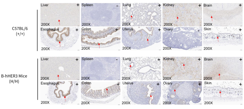
Immunohistochemical Analysis of HER3 Expression
Immunohistochemical (IHC) analysis of HER3 expression in homozygous B-hHER3 mice. The liver and colon were collected from WT and homozygous B-hHER3 mice (H/H) and analyzed by IHC with anti-HER3 antibody. HER3 was detectable in WT mice and homozygous B-hHER3 mice due to the cross-reactivity of the antibody. The arrow indicates tissue cells with positive HER3 staining (brown).
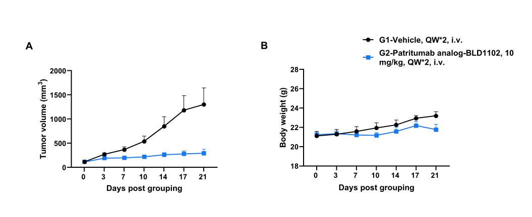
In vivo Efficacy of Anti-human HER3 Antibody-Drug Conjugate (ADC)
Establishment of a MC38 model in B-hHER3 mice and in vivo efficacy study of anti-HER3 ADC (Patritumab analog-BLD1102, in-house). Murine colon cancer B-hHER3 MC38 (Cat# 322045) were subcutaneously implanted into homozygous B-hHER3 mice (female, 9-week-old, n=6). Mice were grouped when tumor volume reached approximately 100 mm3, at which time they were intravenously injected with Patritumab analog-BLD1102 (ADC) indicated in panel. Values are expressed as mean ± SEM.
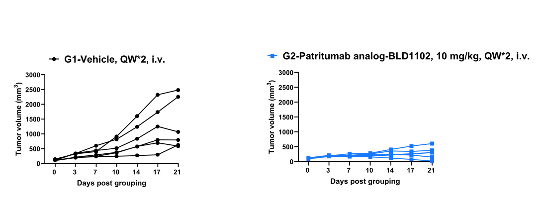
In vivo efficacy of anti-human HER3 ADC-individualtumor growth curves.
* When publishing results obtained using this animal model, please acknowledge the source as follows: The animal model [B-hHER3 mice] (Cat# 110791) was purchased from Biocytogen.