
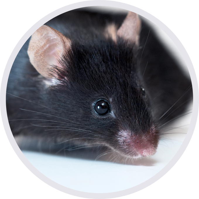

HSD17B13: A key regulator in lipid metabolism and liver homeostasis
Gene Information: HSD17B13 is a direct target gene of SREBP-1c. Its transcription is upregulated by the expression and maturation of SREBP-1c, which can be induced by insulin, saturated fatty acids (FA), and LXR agonists like T0901317.
Signaling Pathway: HSD17B13 plays a key role in maintaining liver lipid droplet (LD) homeostasis and contributes to de novo lipogenesis. Its upregulation creates a positive feedback loop that further enhances lipogenesis.
Pathological Impact: An increase in HSD17B13 levels leads to excessive lipid synthesis and LD enlargement, which can potentially cause liver steatosis.
Knockout Phenotype: If HSD17B13 is knocked out, the long-term accumulation of its substrates or lack of downstream metabolites may disrupt lipid homeostasis, ultimately leading to age-related steatosis and inflammation.


Strain-specific analysis of HSD17B13 mRNA expression in wild-type C57BL/6JNifdc mice and HSD17B13 humanized mice by RT-PCR and sequencing. Liver RNA were isolated from wild-type C57BL/6JNifdc mice (+/+) and homozygous B-hHSD17B13 mice plus (H/H), then cDNA libraries were synthesized by reverse transcription, followed by PCR with mouse or human HSD17B13 primers.

Strain-specific HSD17B13 expression was evaluated by Western blot in wild-type C57BL/6 mice and HSD17B13 humanized mice. Various tissue lysates were collected from wild-type C57BL/6 mice (+/+) and homozygous B-hHSD17B13 mice plus (H/H), and then analyzed by western blot with anti-HSD17B13 antibody (abcam, ab316153). 40 μg total proteins were loaded for western blotting analysis.

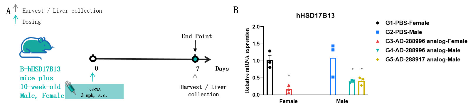
The inhibitory efficiency of the nucleic acid drugs against human HSD17B13 in HSD17B13 humanized mice. B-hHSD17B13 mice plus were randomly divided into five groups (10 weeks old). The human HSD17B13 targeted nucleic acid drugs (synthesized according to patents) and PBS were administered to the mice individually. The nucleic acid drugs were administered in the form of a PBS aqueous solution. The mice were sacrificed on day 7, and the liver tissue was collected to detect the expression level of human HSD17B13 mRNA by qPCR. (A) The schematic diagram of experimental processing. (B) The expression of human HSD17B13 mRNA in the liver after treatment. Significance was determined by a one-way ANOVA, *p<0.05. Values are expressed as mean ± SEM.
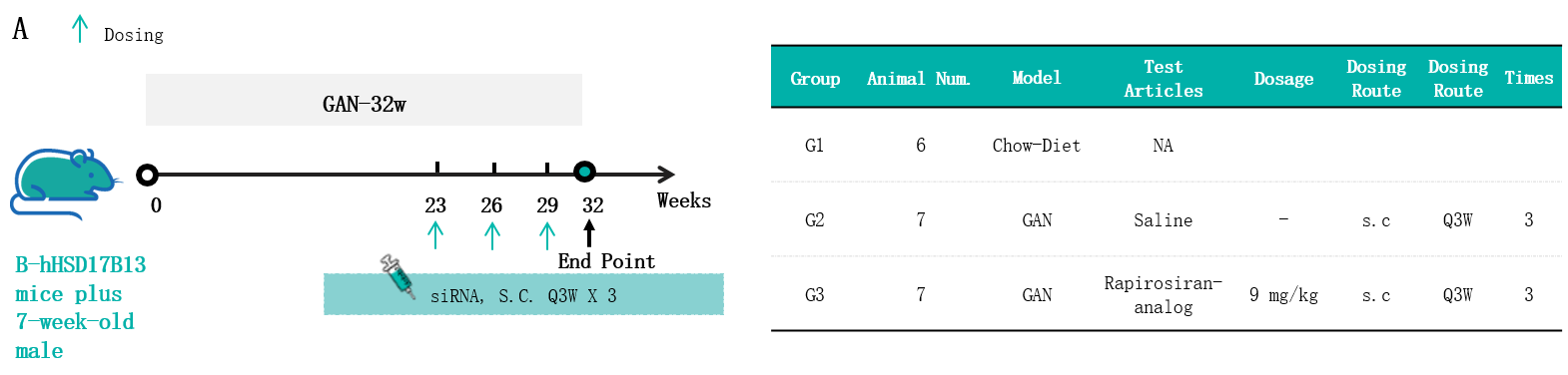
In vivo efficacy of the GAN-induced model in HSD17B13 humanized mice. (A) The schematic diagram of experimental processing. Male B-hHSD17B13 mice plus (7 weeks old) were fed either a standard chow diet (G1, n = 6) or a GAN diet (n = 20). Following a 23-week induction period, 14 mice from the GAN-fed cohort were evenly allocated into two groups (G2-G3) based on their body weight and total cholesterol (TC) levels. The mice in the siRNA treatment groups were injected subcutaneously with the saline or Rapirosiran-analog (MCE) from week 23 once every 3 weeks for 9 weeks.
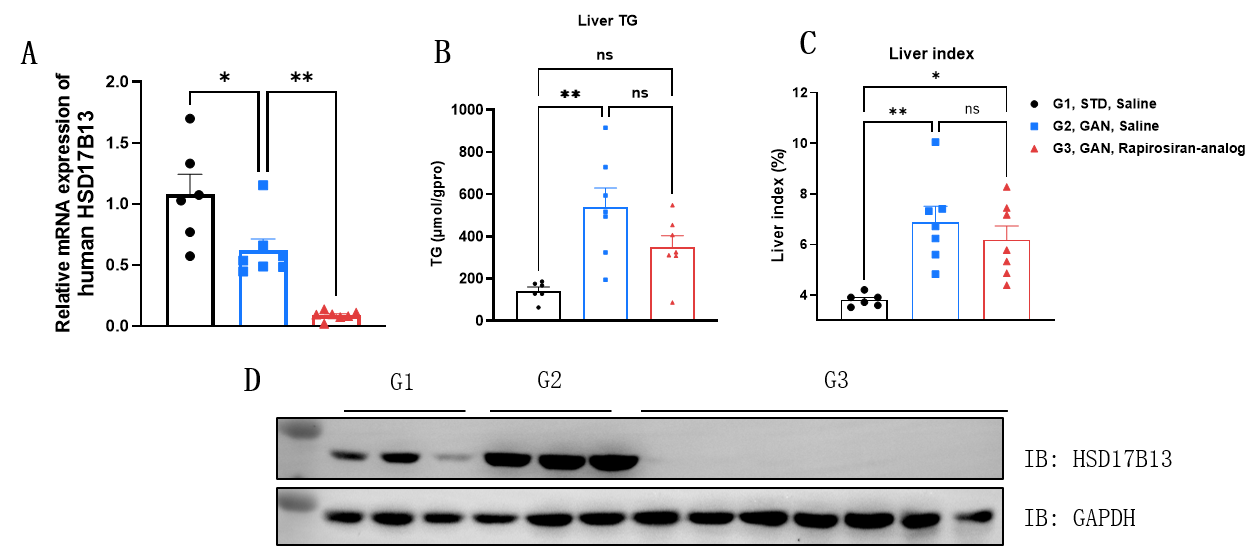
In vivo efficacy of the GAN-induced model in HSD17B13 humanized mice. (A) The expression of human HSD17B13 mRNA in the liver after treatment was measured by qPCR. (B) Liver TG. (C) Liver index was calculated by the following formula: Liver index (%) = weight of liver/weight of body x 100%. (D) The expression of human HSD17B13 protein in the liver after treatment was measured by Western blot with anti-HSD17B13 antibody (abcam, ab316153). Data are shown as mean ± SEM, analyzed by one-way ANOVA (*p<0.05, **p<0.01).
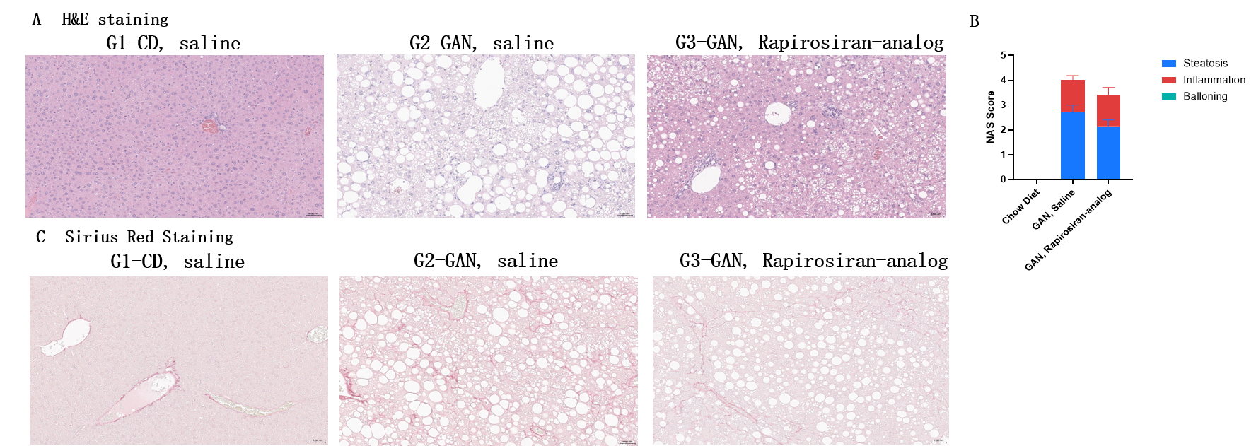
In vivo efficacy of the GAN-induced model in HSD17B13 humanized mice. (A) Representative pictures of H&E staining showing the degree of NASH. (B) NAFLD activity score (NAS) in different treatment groups. (C) Representative pictures of Sirius Red Staining showing the degree of liver fibrosis. Scale bar:50μm.