

Non-disclosure • 112744

Thymic stromal lymphopoietin (TSLP) is an epithelial-cell–derived cytokine, while TSLP receptor (TSLPR, also known as CRLF2) mediates TSLP signaling in immune cells. Dysregulated TSLP–TSLPR signaling has been implicated in allergic asthma, atopic dermatitis, inflammatory bowel disease (IBD), and autoimmune disorders.
The B-hTSLP/hTSLPR humanized mouse model was generated by replacing murine Tslp and Tslpr genes with their human orthologs. These mice express functional human TSLP and TSLPR, enabling the study of human-specific cytokine signaling and therapeutic interventions. This double humanized system provides a robust platform for preclinical efficacy evaluation, antibody validation in vivo, and autoimmune/inflammatory disease modeling.
1.Complete humanized axis: Both TSLP and TSLPR genes replaced with human sequences.
2.Validated expression: Physiological expression in epithelial and immune compartments.
3.Immune system stability: Comparable immune cell distribution and cytokine profiles to wild-type mice.
4.Preclinical disease models: Effective in colitis (DSS/TNBS) and allergic asthma studies.
5.In vivo antibody validation: Suitable for testing human-specific TSLP/TSLPR antibodies.
6.Competitive differentiation: Offers dual-target humanization advantage over single-target models from Charles River or Cyagen.
1.Molecular Validation: Human TSLP and TSLPR mRNA and protein expression confirmed in relevant tissues.
2.Functional Validation: Human TSLP binding to humanized TSLPR in vivo results in downstream STAT5 phosphorylation and T-cell activation.
3.Pathology Correlation: Enhanced disease phenotype in colitis and asthma models, responsive to anti-human TSLP antibody treatment.
4.Antibody Validation in vivo: Demonstrated binding and efficacy of clinical-stage anti-TSLP/TSLPR antibodies in B-hTSLP/hTSLPR humanized mice.
1.Autoimmune & Inflammatory Disease Research: Crohn's disease, ulcerative colitis, rheumatoid arthritis.
2.Allergic Disease Modeling: Atopic dermatitis, asthma.
3.Therapeutic Antibody Development: In vivo validation of anti-TSLP/TSLPR antibodies.
4.Mechanistic Studies: Dissection of TSLP–TSLPR signaling in epithelial–immune cell interactions.
5.Drug Safety Assessment: Preclinical toxicology of cytokine-targeting biologics.

TSLP
TSLPR
IL7R

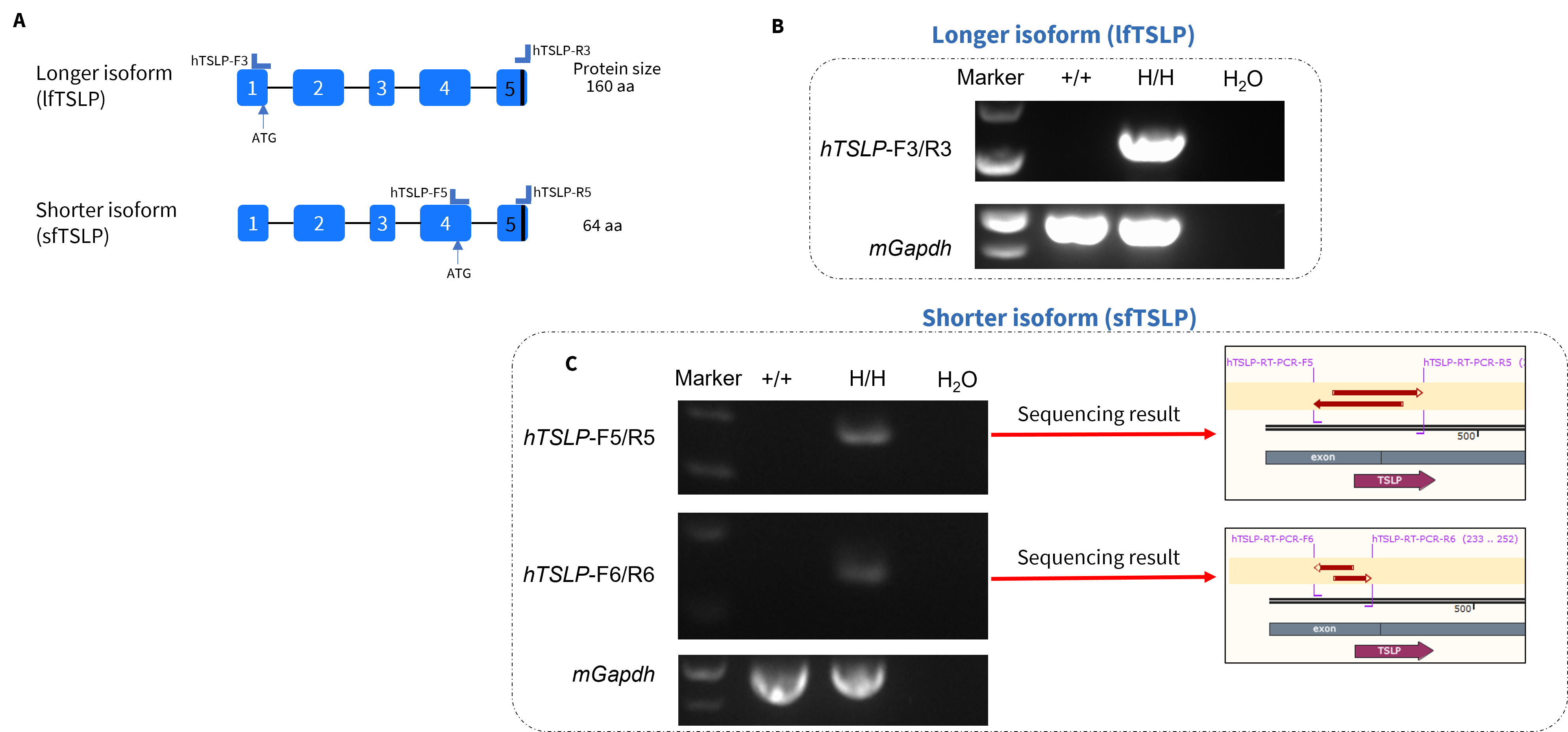
Long and short isoforms of human TSLP were assayed by RT-PCR and Sanger sequencing. Ear tissues were collected from wild-type C57BL/6 (+/+) and homozygous TSLP/TSLPR humanized mice plus (H/H). (A) Isoform-specific primers were designed to detect long (lfTSLP) and short (sfTSLP) human TSLP transcripts. (B) lfTSLP mRNA was detectable in TSLP/TSLPR humanized mice plus, but not in wild-type mice. (C) sfTSLP mRNA was detectable in TSLP/TSLPR humanized mice plus, but not in wild-type mice. Sequencing of sfTSLP PCR products confirmed identity to database reference sequences.
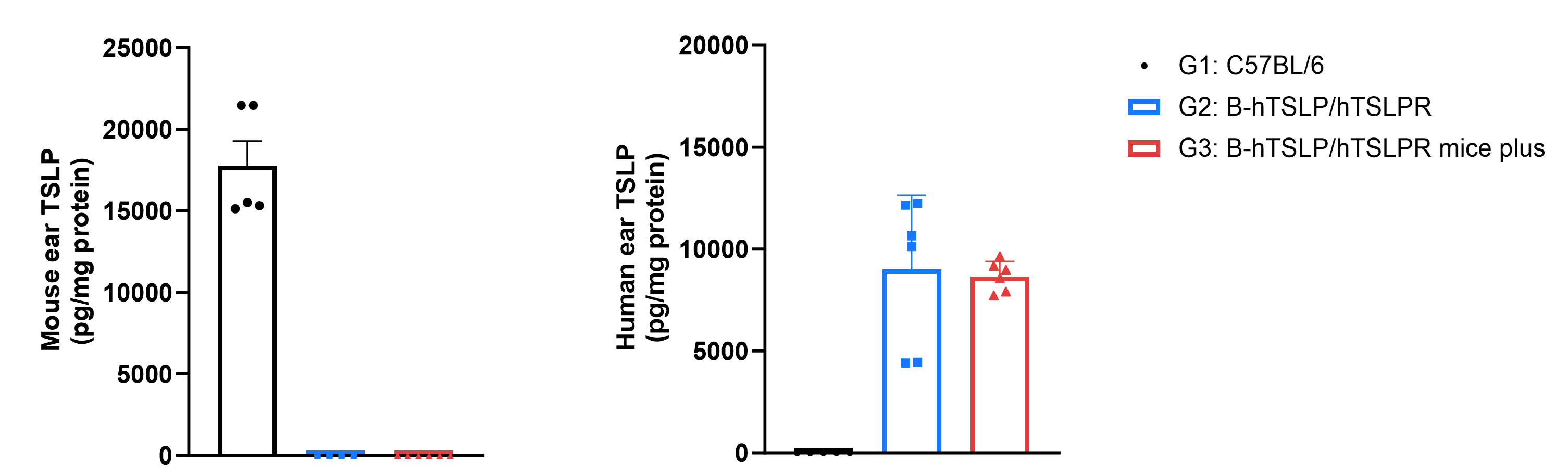
Strain-specific TSLP expression was analyzed in wild-type C57BL/6, homozygous TSLP/TSLPR humanized mice, and TSLP/TSLPR humanized mice plus by ELISA. Calcipotriol (MC903) was dissolved in ethanol and topically applied to ears for 7 days (male, 8-week-old, n=3 per strain). Mouse and human TSLP in ear-grinding supernatants were quantified by ELISA. Mouse TSLP was detectable only in wild-type mice. Human TSLP was detectable in homozygous TSLP/TSLPR humanized mice and TSLP/TSLPR humanized mice plus, but not in wild-type mice. Values are mean ± SEM.
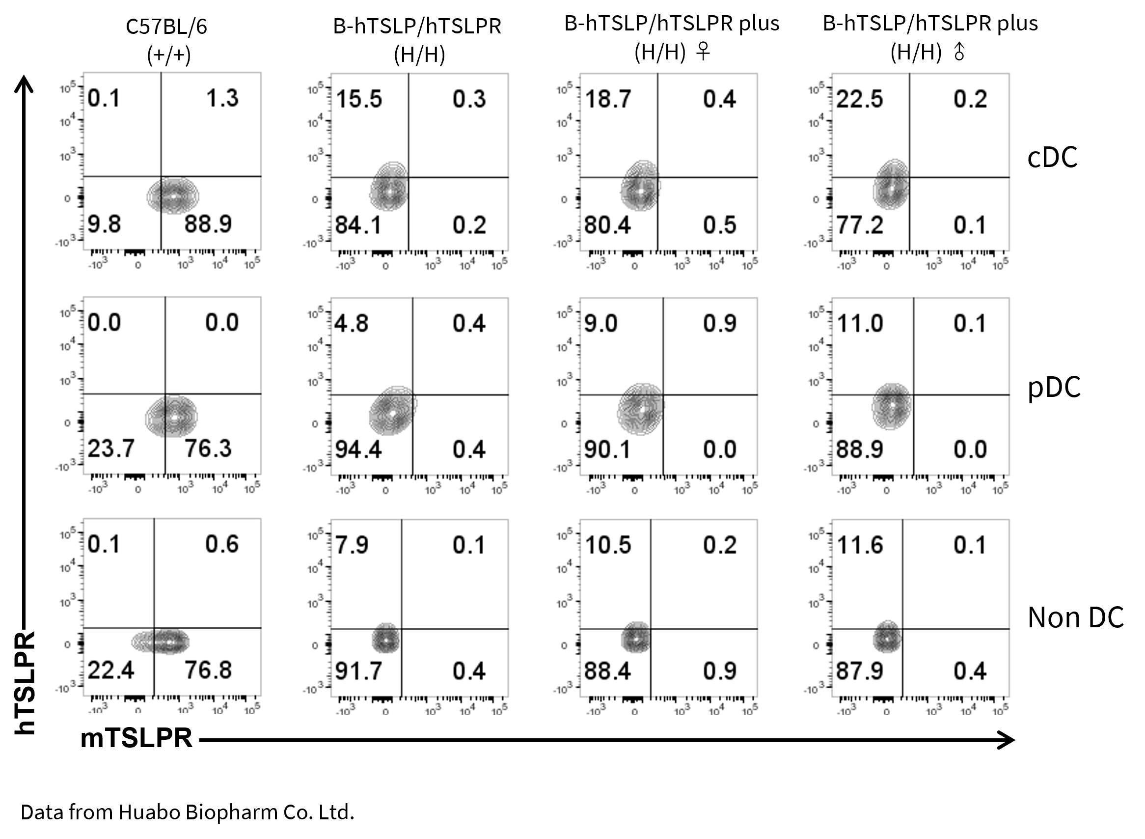
Mouse and human TSLPR protein expression analysis in splenocytes. Splenocytes from wild-type C57BL/6, homozygous TSLP/TSLPR humanized mice, and TSLP/TSLPR humanized mice plus were stained with species-specific anti-TSLPR antibodies. Mouse TSLPR was detected on cDC, pDC, and non-DCs in wild-type mice, but not in TSLPR-humanized strains. Human TSLPR was detected on cDC, pDC, and non-DCs in TSLP/TSLPR humanized mice and TSLP/TSLPR humanized mice plus, but not in wild-type mice. Values are mean ± SEM.
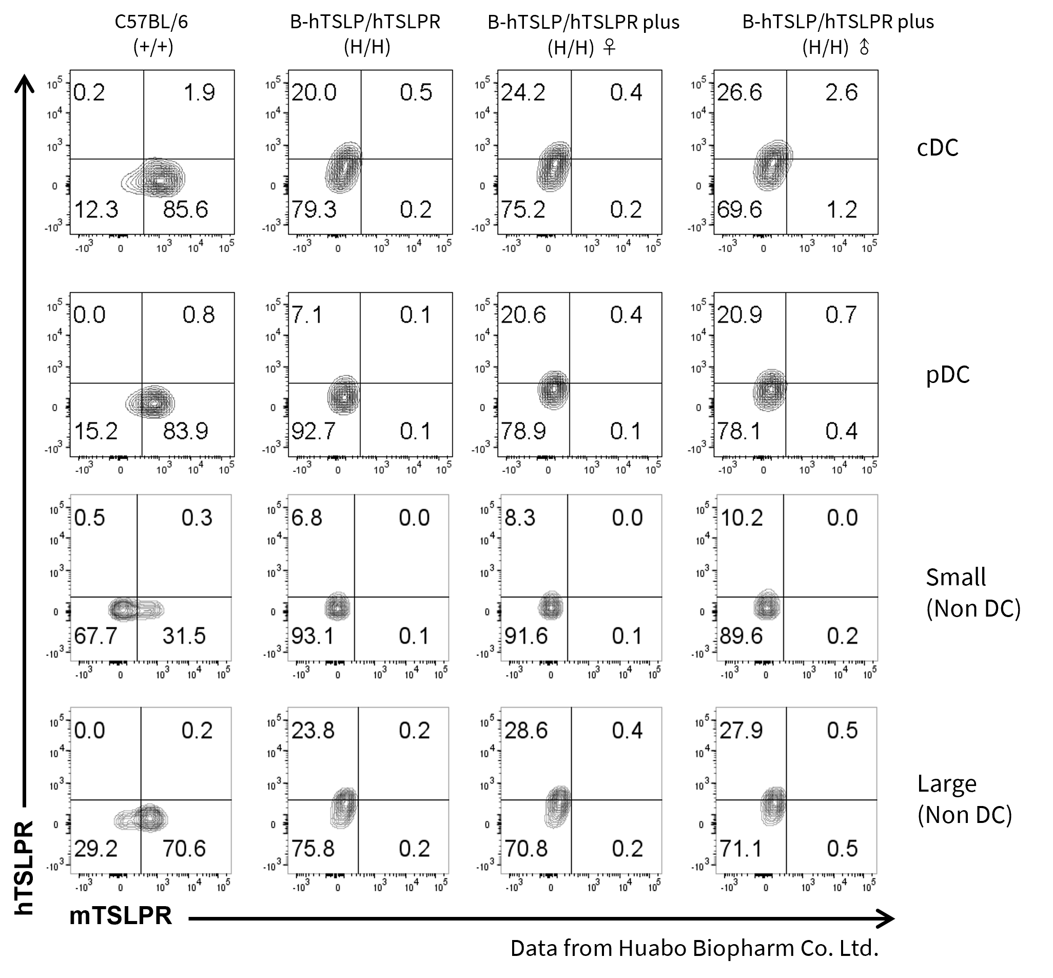
Mouse and human TSLPR protein expression analysis in bone marrow. Bone marrow cells from wild-type C57BL/6, homozygous TSLP/TSLPR humanized mice, and TSLP/TSLPR humanized mice plus were analyzed by flow cytometry. Mouse TSLPR was detectable on cDC, pDC, and non-DCs in wild-type mice, but absent in TSLPR-humanized strains. Human TSLPR was detectable on cDC, pDC, and non-DCs in both humanized strains, but absent in wild-type mice. Values are mean ± SEM.
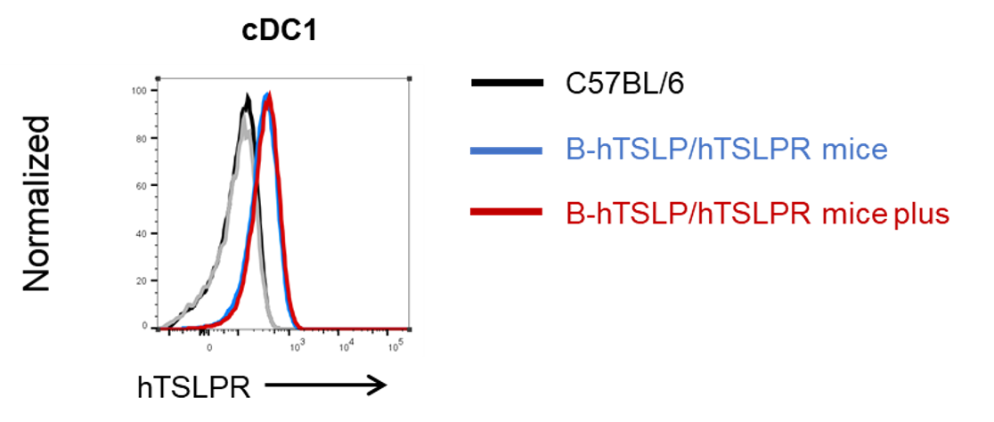
Human TSLPR expression analysis in cDC1 from bone marrow. Bone marrow from wild-type C57BL/6, homozygous TSLP/TSLPR humanized mice, and TSLP/TSLPR humanized mice plus was analyzed by flow cytometry with species-specific antibodies. Human TSLPR was highly expressed on cDC1 in both humanized strains.
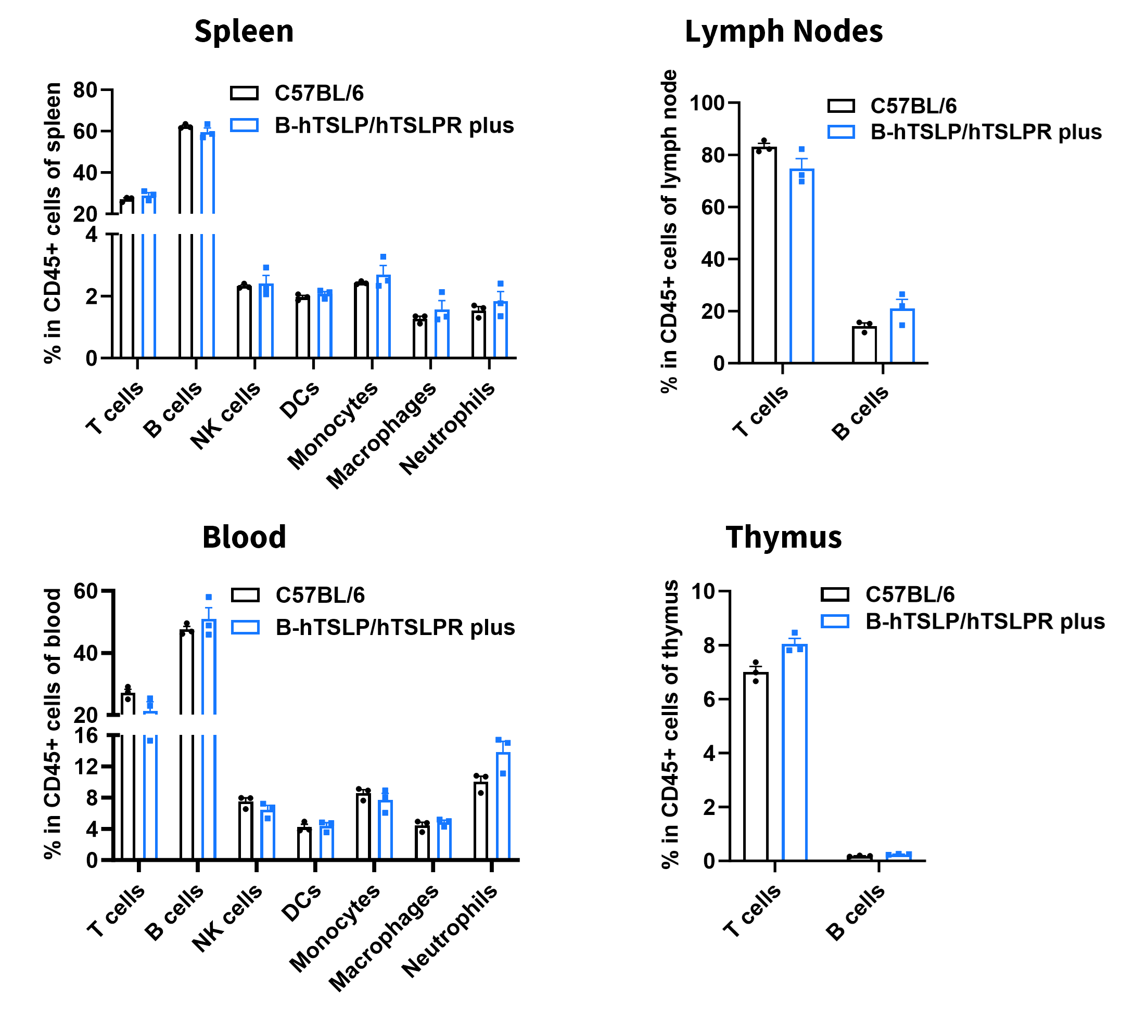
Analysis of leukocyte subpopulations by flow cytometry in immune organs and blood. Splenocytes, peripheral blood, lymph nodes, and thymus were isolated from C57BL/6 and TSLP/TSLPR humanized mice plus (female, 9-week-old, n = 3). Single live cells were gated on the CD45⁺ population and analyzed by flow cytometry as indicated. Values are expressed as mean ± SEM.
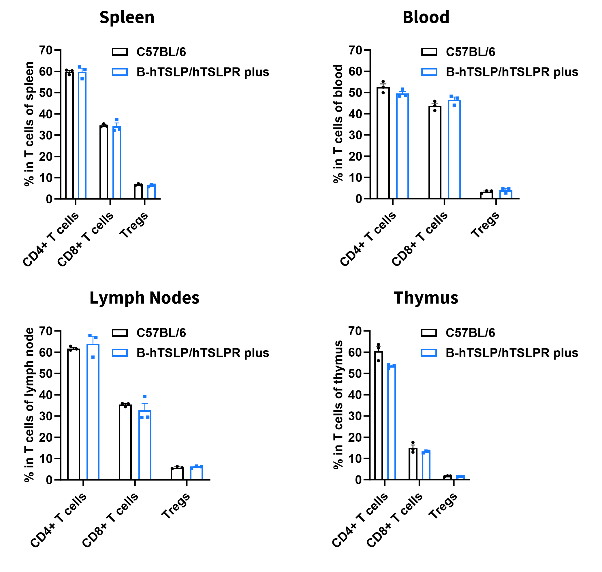
Analysis of T-cell subpopulations by flow cytometry in immune organs and blood. Splenocytes, peripheral blood, lymph nodes, and thymus were isolated from C57BL/6 and TSLP/TSLPR humanized mice plus (female, 9-week-old, n = 3). Single live cells were gated on the CD3⁺ T-cell population and analyzed by flow cytometry as indicated. Values are expressed as mean ± SEM.
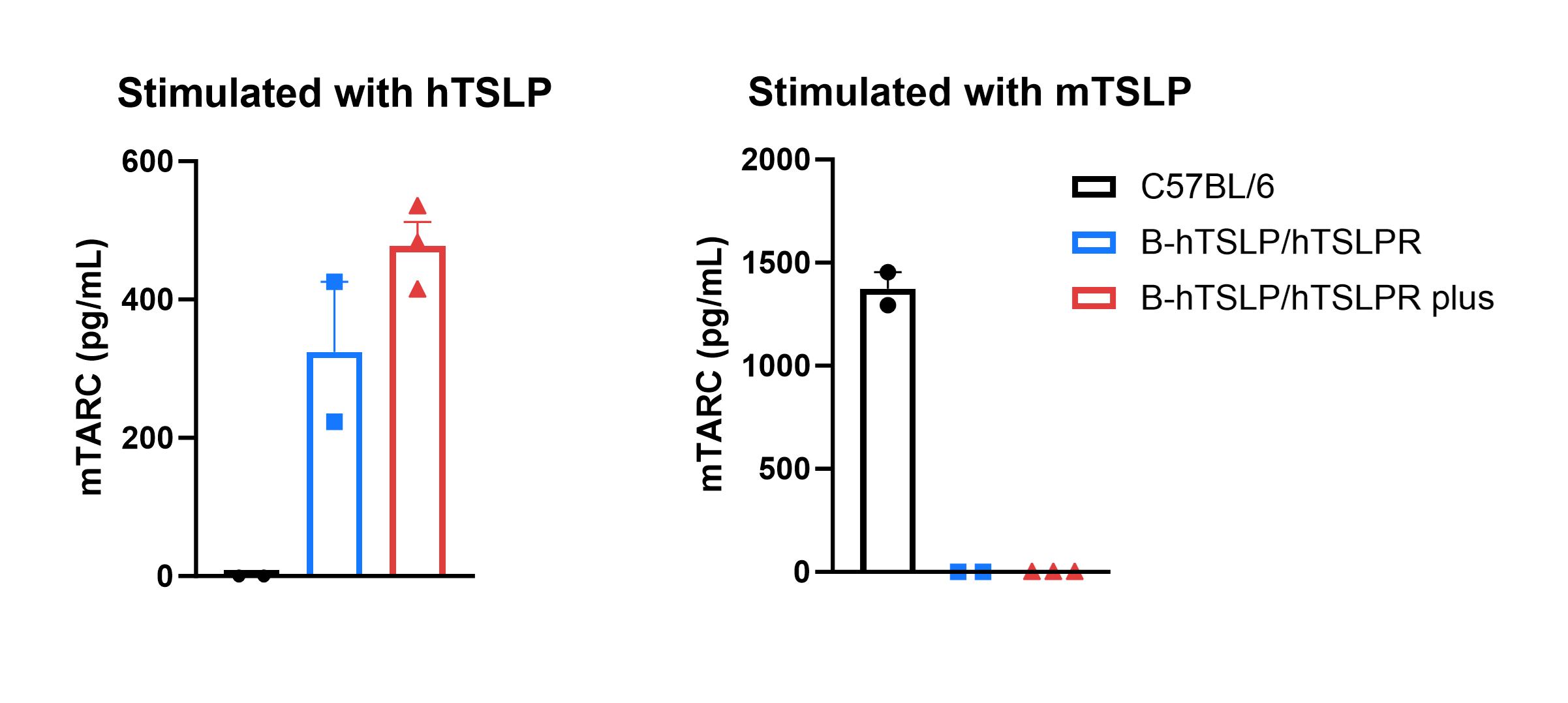
Mouse TARC production was analyzed following stimulation with human TSLP or mouse TSLP in wild-type C57BL/6 mice, homozygous TSLP/TSLPR humanized mice, and TSLP/TSLPR humanized mice plus. Dendritic cells (DCs) were generated from bone marrow using FLT3L and subsequently stimulated with human TSLP or mouse TSLP in vitro. Supernatant concentrations of mouse TARC were measured by ELISA. In homozygous TSLP/TSLPR humanized mice and TSLP/TSLPR humanized mice plus, human TSLP successfully induced mouse TARC secretion, whereas mouse TSLP did not. In contrast, in wild-type C57BL/6 mice, mouse TSLP induced TARC, while human TSLP did not. The level of mouse TARC in TSLP/TSLPR humanized mice plus was higher than in homozygous TSLP/TSLPR humanized mice, confirming stronger responsiveness. These results demonstrate that TSLP–TSLPR signaling is species-specific, with no cross-reactivity between mouse and human. Importantly, human TSLP activates dendritic cells only in TSLP/TSLPR humanized mice, validating the model for antibody efficacy testing and translational immunology research.
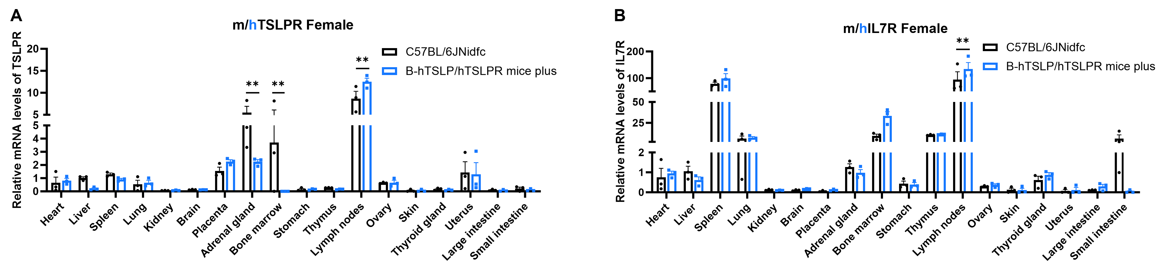
Strain specific analysis of TSLPR and IL7R expression in wild-type C57BL/6JNidfc mice and homozygous B-hTSLP/hTSLPR mice plus by RT-qPCR. RNA was isolated from multiple organs of wild-type C57BL/6JNidfc mice (+/+) (female, 8-week-old, n = 3) and homozygous B-hTSLP/hTSLPR mice plus (female, 8-week-old, n = 3), followed by cDNA synthesis via reverse transcription and quantitative real-time PCR using TSLPR- and IL7R-specific primers. Values are expressed as mean ± SEM. Statistical significance was determined by two-way ANOVA. *P < 0.05, **P < 0.01, ***p < 0.001. Data were normalized to C57BL/6JNidfc liver.
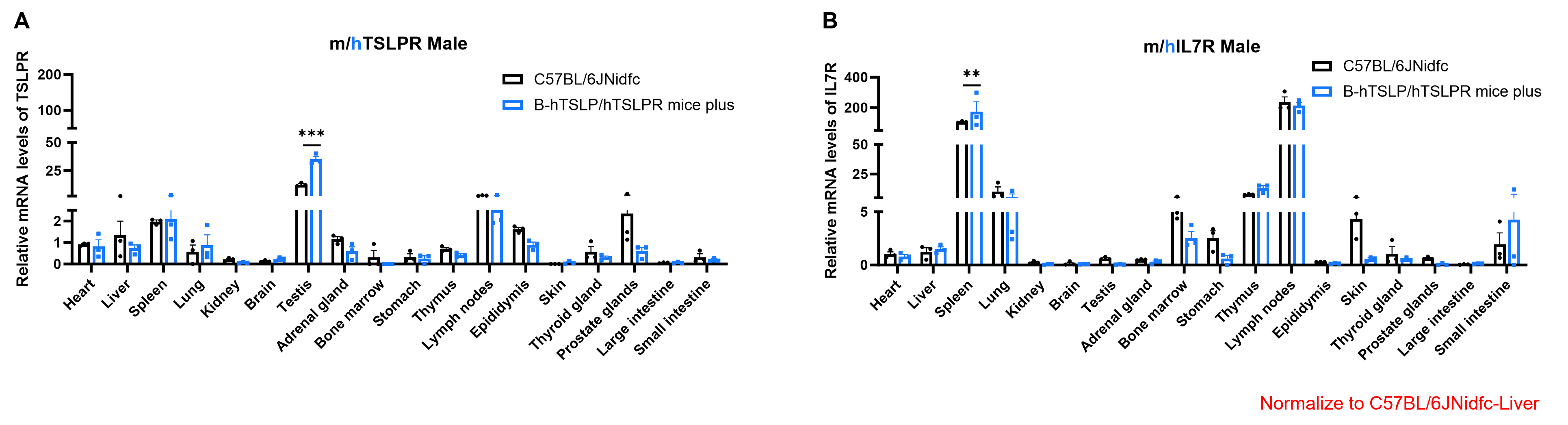
Strain-specific analysis of TSLPR and IL7R expression in wild-type C57BL/6JNidfc mice and homozygous B-hTSLP/hTSLPR mice plus by RT–qPCR. RNA was isolated from multiple organs of wild-type C57BL/6JNidfc mice (+/+) (male, 8-week-old, n = 3) and homozygous B-hTSLP/hTSLPR mice plus (male, 8-week-old, n = 3), followed by cDNA synthesis by reverse transcription and quantitative real-time PCR using TSLPR- and IL7R-specific primers. Values are expressed as mean ± SEM. Statistical significance was determined by two-way ANOVA. *P < 0.05, **P < 0.01, ***P < 0.001. Data were normalized to C57BL/6JNidfc liver.
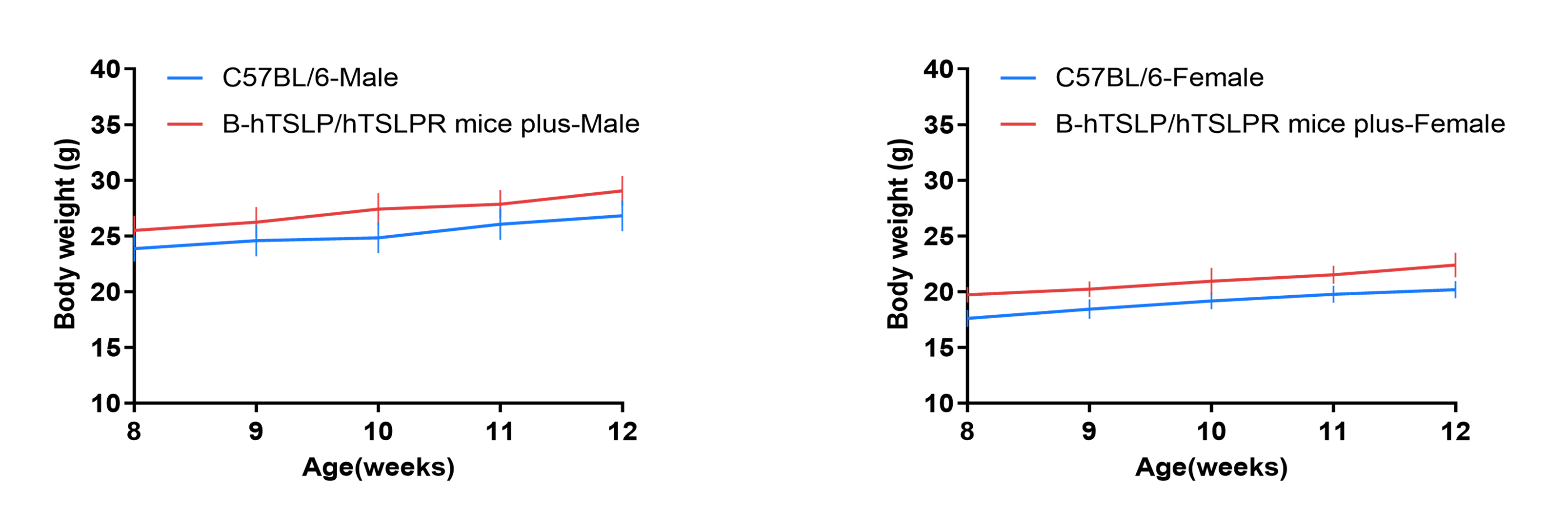
Eight-week-old TSLP/TSLPR humanized mice plus were grouped by sex (10 males and 10 females, respectively). Body weight was measured weekly on the same day for 12 consecutive weeks. The lowest and highest values of body weight in the table were calculated from mean ± SD. The growth curve conformed to a normal distribution, with the probability of random error falling within ± SD estimated at 68%.
These results confirm that TSLP/TSLPR humanized mice plus display normal growth and physiological development, supporting their suitability for long-term pharmacology, toxicology, and antibody validation studies.
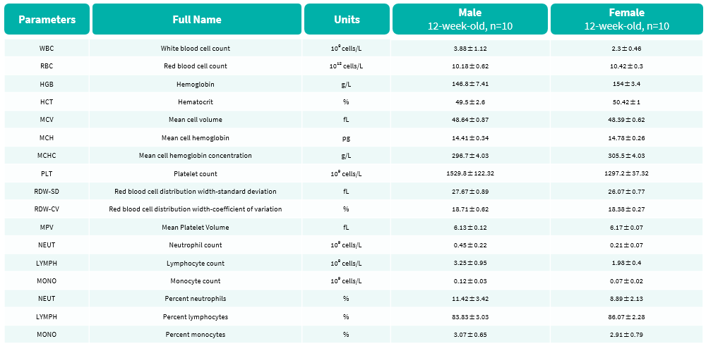
Complete blood count (CBC) was performed in TSLP/TSLPR humanized mice plus. Values are presented as mean ± SD.
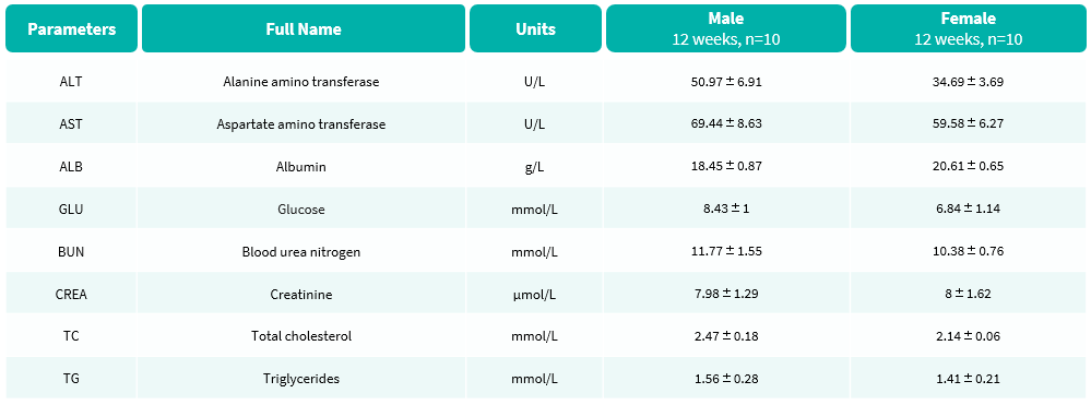
Blood biochemical tests were performed in TSLP/TSLPR humanized mice plus. Values are expressed as mean ± SD.
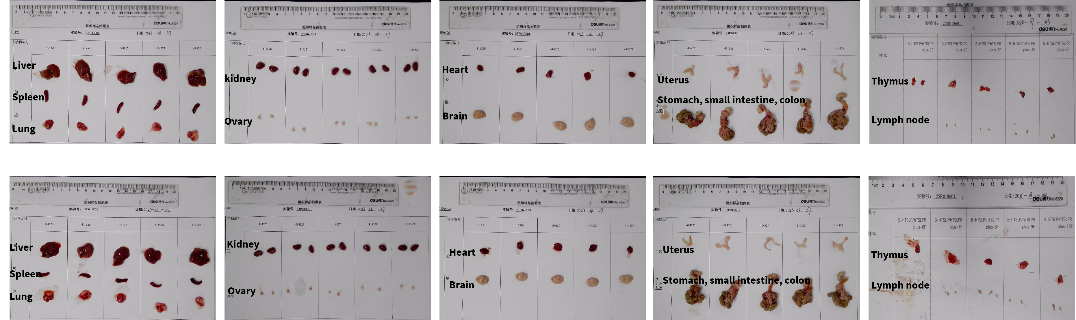
The organs of female TSLP/TSLPR humanized mice plus (12-week-old, n=10) were collected and examined.
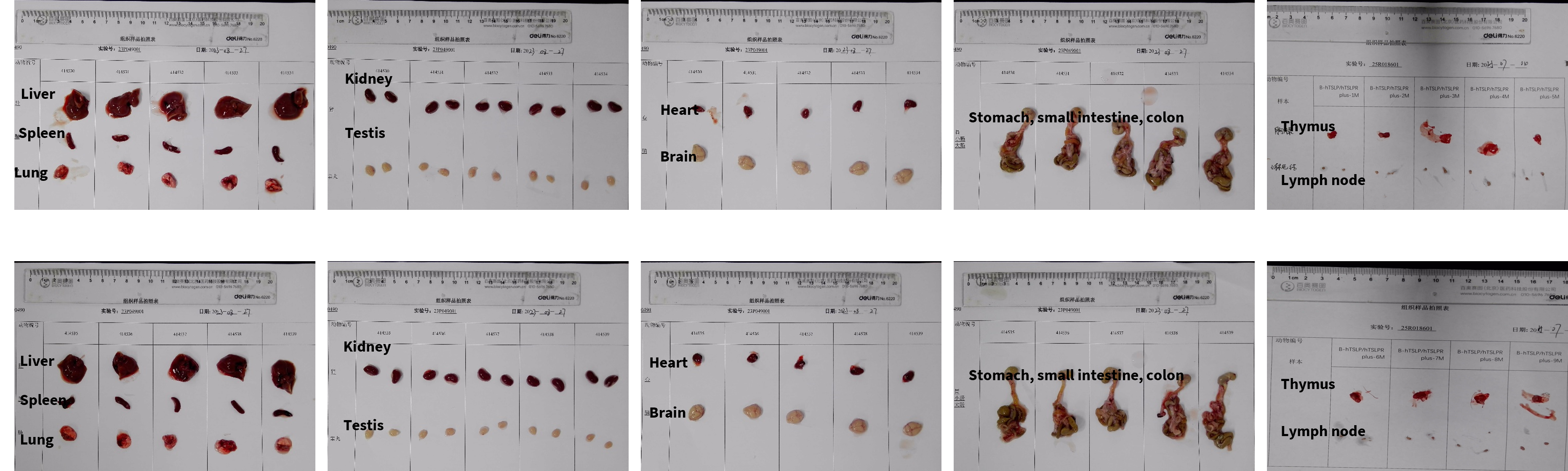
The organs of male TSLP/TSLPR humanized mice plus (12-week-old, n=10) were collected and examined.
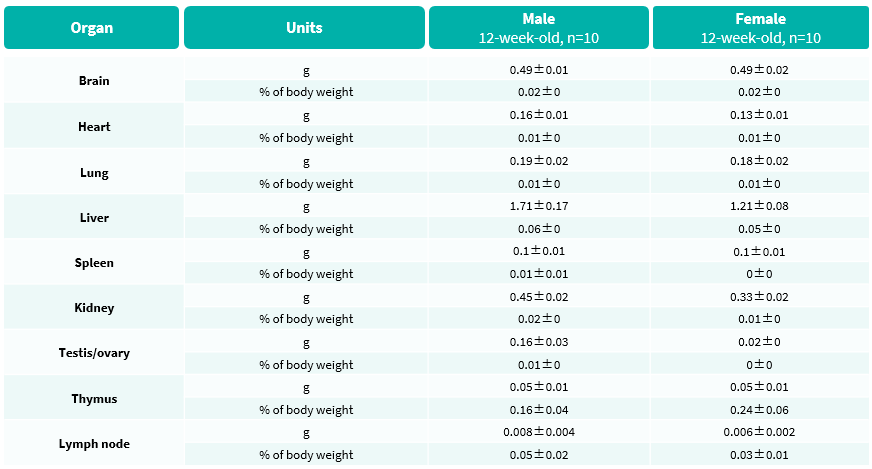
Average weight of the main organs was measured in TSLP/TSLPR humanized mice plus.
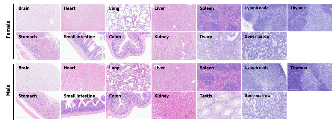
The main organs of TSLP/TSLPR humanized mice plus were collected at 12 weeks of age and analyzed by H&E staining (male, n=10; female, n=10). Results showed that no obvious abnormalities were detected in any of the examined organs, including brain, heart, lung, liver, spleen, stomach, small intestine, colon, kidney, ovary, uterus, and testis. These findings confirm that TSLP/TSLPR humanized mice maintain normal histopathological profiles, supporting their use in toxicology studies, long-term pharmacology, and preclinical antibody validation.

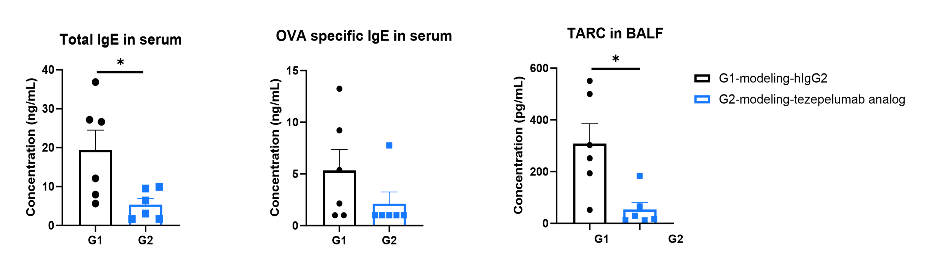
In a mouse asthma model using TSLP/TSLPR humanized mice plus, treatment with an anti-humanized TSLP antibody (tezepelumab, synthesized in-house) significantly reduced OVA-specific IgE in serum and TARC in bronchoalveolar lavage fluid (BALF). Serum was collected at the study endpoint, and both IgE and TARC levels were measured by ELISA. Results demonstrated that levels of OVA-specific IgE and TARC were lower in the antibody-treated group compared with untreated mice. Values are expressed as mean ± SEM.
TARC: thymic and activation-regulated chemokine, also known as CCL17 (C-C motif chemokine ligand 17). These findings confirm that TSLP/TSLPR humanized mice provide a robust preclinical asthma model for evaluating the efficacy of anti-TSLP biologics.
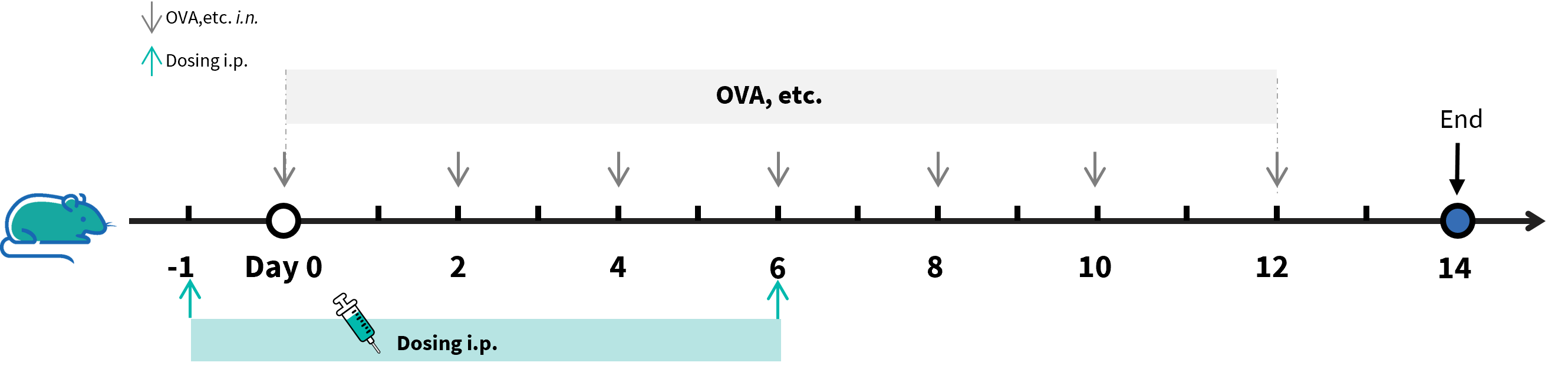
Experimental schedule for the induction of asthma model and in vivo efficacy of anti-human TSLP antibody in B-hTSLP/hTSLPR mice plus. B-hTSLP/hTSLPR mice plus (female, 7-week-old, n=6) were immunized with OVA etc. inducer to induce asthma. The anti-human TSLP antibody tezepelumab analog (in-house) was administered by intraperitoneal injection (n = 6).
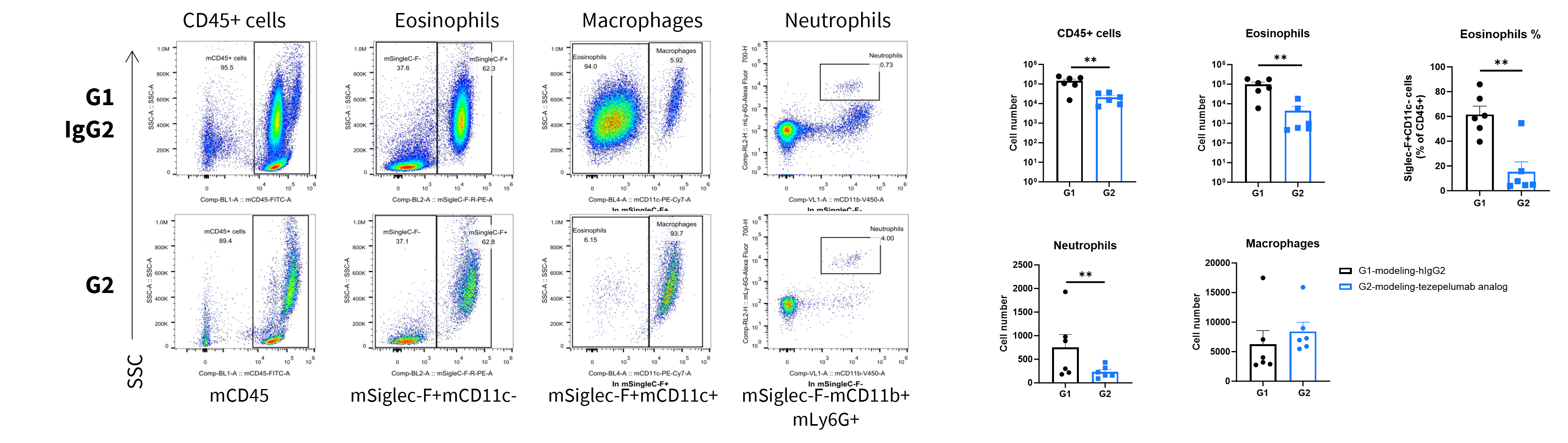
Analysis of inflammatory cells in BALF by FACS. A mouse asthma model was induced in TSLP/TSLPR humanized mice plus and treated with an anti-human TSLP antibody (Tezepelumab analog, synthesized in-house). BALF was collected at the end of the experiment to assess infiltrating inflammatory cells in lung tissue. Values are expressed as mean ± SEM. *P < 0.05, **P < 0.01, ***P < 0.001.
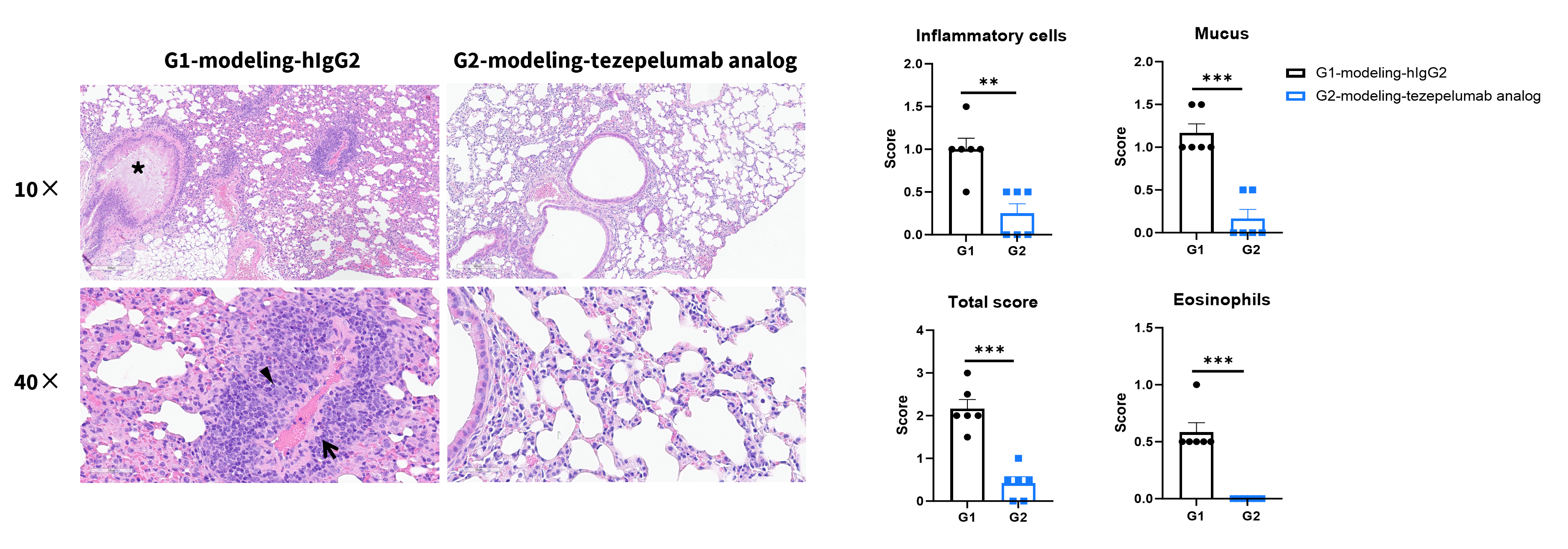
Lung tissues were collected at the study endpoint from TSLP/TSLPR humanized mice plus subjected to an asthma-like model and analyzed by H&E staining.
Compared to the untreated group (G1), mice treated with anti-humanized TSLP antibody (tezepelumab, synthesized in-house) exhibited a significant reduction in inflammatory cell infiltration and mucus secretion in lung tissue.
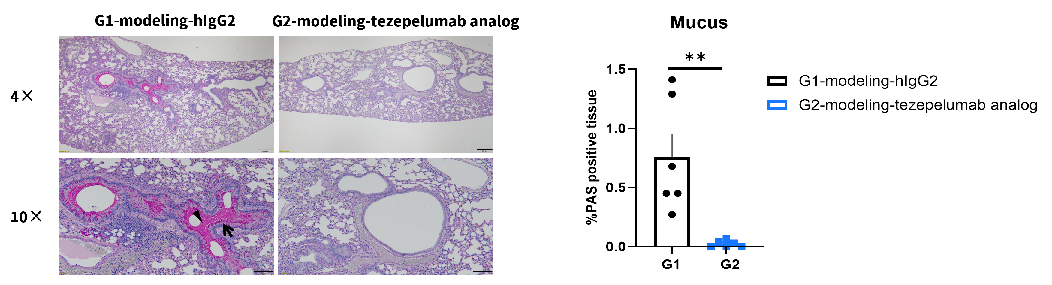
Periodic acid–Schiff (PAS) staining of an asthma model in B-hTSLP/hTSLPR mice plus. Lung tissues were collected at the study endpoint and analyzed by PAS staining. Arrows: goblet cells, triangles: mucus. Values are expressed as mean ± SEM. *P < 0.05, **P < 0.01, ***P < 0.001.
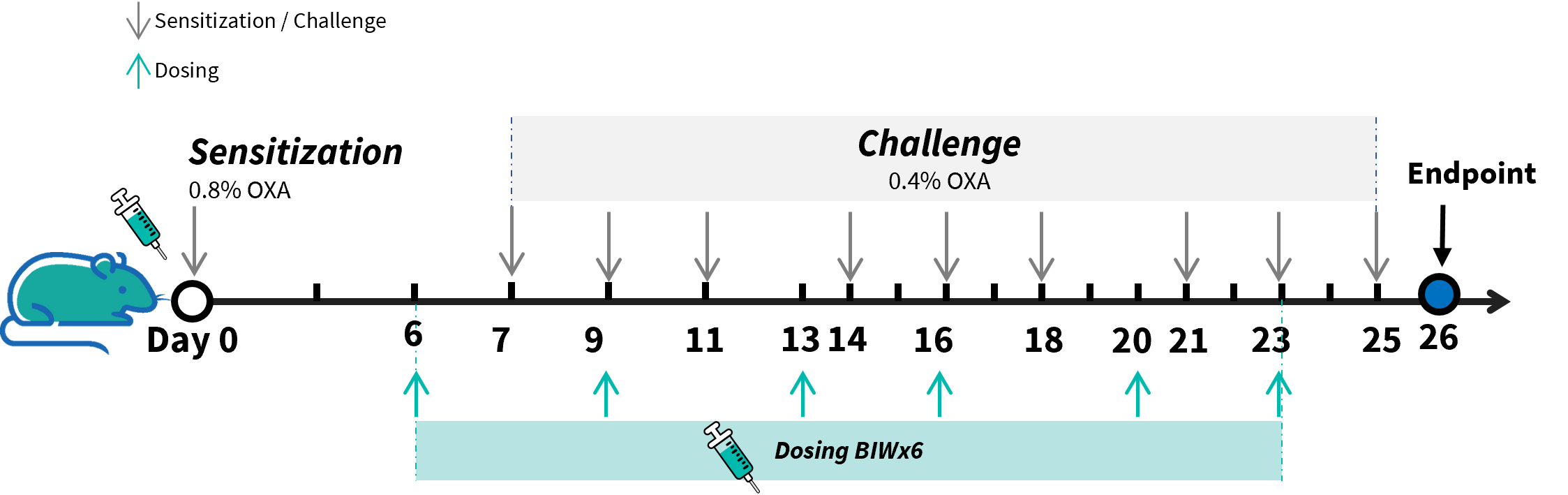
Experimental schedule for the induction of atopic dermatitis (AD) skin lesions and in vivo efficacy of anti-human TSLP antibody in B-hTSLP/hTSLPR mice plus. OXA was applied to the ear skin of mice on day 0, followed by nine challenges to the same site from days 7 to 25. The anti-human TSLP antibody tezepelumab analog (in-house) was administered by intraperitoneal injection (n = 6). OXA, oxazolone.
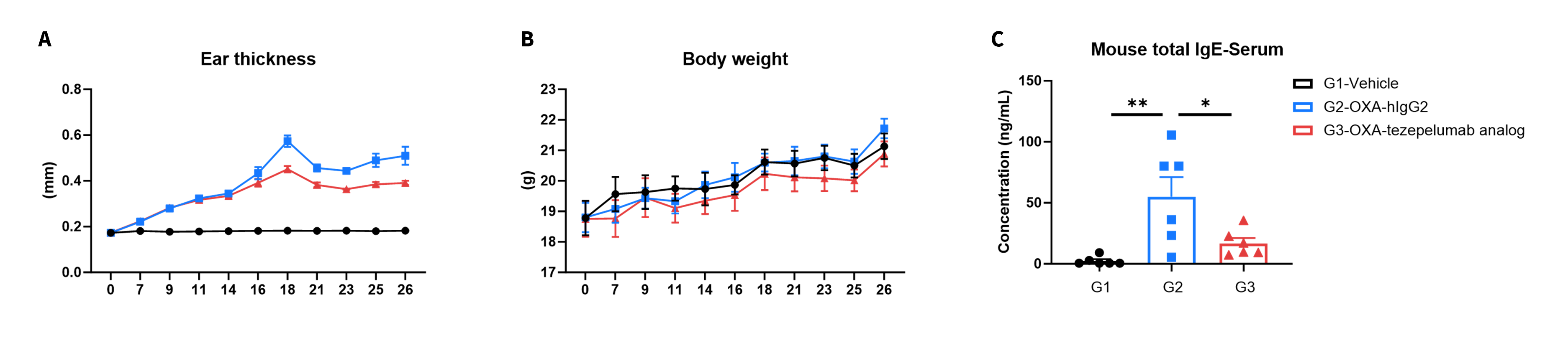
Efficacy of anti-human TSLP antibody in B-hTSLP/hTSLPR mice plus. Mice in each group were treated with the anti-hTSLP antibody tezepelumab analog (in-house). (A) Statistical analysis of ear thickness in each group. Epidermal desquamation of the ear began on day 18; therefore, ear thickness decreased from day 18, as shown in the figure. (B) Body weight changes during treatment. (C) Total serum IgE levels, measured by ELISA using serum collected on day 26 (n = 6). Values are expressed as mean ± SEM. *P < 0.05, **P < 0.01, ***P < 0.001.
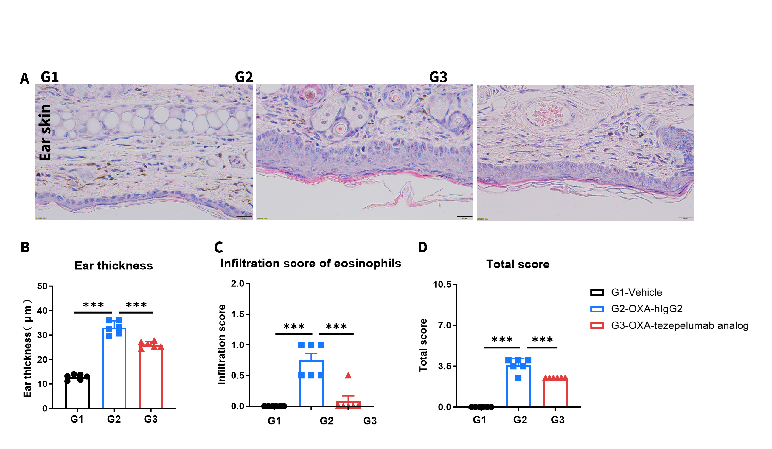
Effects of anti-human TSLP antibody on ear skin in an AD mouse model. (A) Hematoxylin and eosin (H&E) staining. (B) Ear epidermal thickness. (C) Eosinophil infiltration score in ear epidermal skin. (D) Total score of ear epidermal skin. Eosinophil infiltration was scored as follows: 1 = slight; 2 = mild; 3 = moderate; 4 = severe. The total pathology score included epidermal hyperplasia, erosion/crusting, hyperkeratosis, parakeratosis, and inflammatory cell infiltration in the dermis and subcutaneous tissue. *P < 0.05, **P < 0.01, ***P < 0.001.
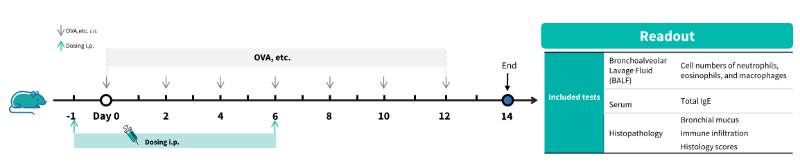
Experimental schedule for the induction of asthma model and in vivo efficacy of anti-human TSLPR antibody in B-hTSLP/hTSLPR mice plus. B-hTSLP/hTSLPR mice plus (female, 7-week-old, n=6) were immunized with OVA etc. inducer to induce asthma. The anti-human TSLPR antibody Verekitug analog (in-house) was administered by intraperitoneal injection (n = 6).
CD45⁺ cells, eosinophils were significantly reduced in the anti–human TSLPR antibody–treated group (G3 G4) compared with the isotype control group (G2).
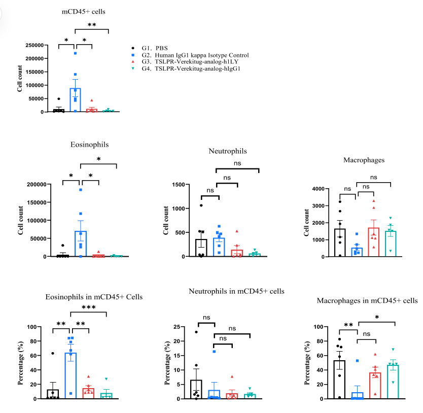
Analysis of inflammatory cells in BALF by FACS. A mouse asthma model wasinduced in B-hTSLP/hTSLPR mice plus and treated with an anti-human TSLPRantibody (Verekitug analog, synthesized in-house). BALF was collected at the end ofthe experiment to assess infiltrating inflammatory cells in lung tissue. Values areexpressed as mean ± SEM. *P < 0.05, **P < 0.01, ***P < 0.001.
Verekitug analog treatment reduced IgE levels compared with untreated controls.
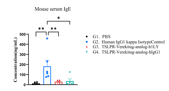
IgE in serum was significantly reduced in the mouse asthma model treated with anti-TSLPR antibody. Serum was collected at the study endpoint, and IgElevels were analyzed by ELISA. Values are expressed as mean ± SEM. *P < 0.05, **P < 0.01.
• Verekitug analog significantly reduced inflammatory infiltration and mucus secretion in lung tissue compared with untreated controls (G2).
• B-hTSLP/hTSLPR mice plus provide a robust in vivo preclinical model for evaluating anti human TSLPR antibodies.
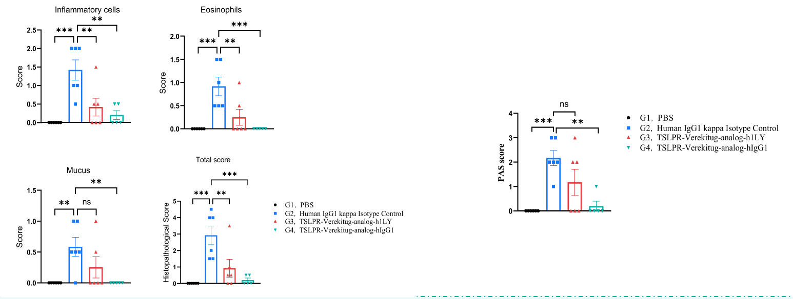
Hematoxylin and eosin (H&E) staining and Periodic acid–Schiff (PAS) staining of an asthma model in B-hTSLP/hTSLPR mice plus. Lung tissues were collected atthe studyendpoint and analyzed by H&E and PAS staining. Values are expressed as mean ± SEM. *P < 0.05, **P < 0.01, ***P < 0.001.

Q1: What are TSLP and TSLPR?
TSLP is an epithelial-derived cytokine, and TSLPR (CRLF2) is its receptor on immune cells. Together, they regulate inflammation and allergy responses.
Q2: Why use B-hTSLP/hTSLPR mice plus?
They uniquely co-express human TSLP and TSLPR, making them ideal for antibody validation and inflammatory disease modeling.
Q3: What diseases can be studied with this model?
Autoimmune and allergic diseases, including asthma, IBD, and atopic dermatitis.
Q4: Are these mice suitable for antibody validation?
Yes, they are widely used for in vivo validation of therapeutic antibodies targeting the TSLP–TSLPR pathway.
Q5: How do they compare with competitor models?
Unlike single humanized models, B-hTSLP/hTSLPR humanized mice offer dual-target humanization, providing higher translational relevance.
