


Gene Information: CD98HC, encoded by the SLC3A2 gene, is a critical type II transmembrane glycoprotein. It functions primarily as a heavy chain that covalently associates via a disulfide bond with various light chains, such as LAT1 or LAT2.
Protein Expression: In normal physiological conditions, CD98HC is expressed moderately across various tissues, including the kidneys, intestinal epithelium, and placenta, where it maintains regular metabolic and transport functions. However, its expression is markedly upregulated in proliferating cells, activated lymphocytes, and a wide array of human malignancies, including carcinomas and melanomas.
Signaling Pathway: CD98HC actively participates in intricate intracellular signaling networks by interacting directly with beta-1 and beta-3 integrins. This interaction modulates integrin-mediated signal transduction, promoting cell adhesion, migration, spreading, and survival.
Application: Given its prominent role in cancer cell metabolism and immune activation, CD98HC has emerged as a promising therapeutic target in oncology and immunology. CD98hc-targeting antibodies or ligands enable receptor-mediated transcytosis across the blood-brain barrier, serving as an efficient shuttle to transport therapeutic drugs, proteins, and nanoparticles directly into the central nervous system.


Species specific CD98HC gene expression in wild-type B-NDG mice and homozygous CD98HC humanized B-NDG mice by RT-PCR. Kidney and brain were collected from wild-type B-NDG mice (+/+) and homozygous B-NDG hCD98HC mice (H/H), then cDNA libraries were synthesized by reverse transcription, followed by PCR with mouse or human CD98HC primers.

Strain specific CD98HC expression analysis in wild-type B-NDG mice and homozygous CD98HC humanized B-NDG mice. Splenocytes were collected from wild-type B-NDG mice (+/+) and homozygous B-NDG hCD98HC mice (H/H). Protein expression was analyzed with anti-mouse CD98HC antibody (Biolegend, 128211) and anti-human CD98HC antibody (Biolegend, 315603) by flow cytometry. Mouse CD98HC was detectable in neutrophils, DCs, monocytes and macrophages of wild-type B-NDG mice, and human CD98HC was exclusively detectable in homozygous B-NDG hCD98HC mice.

Strain specific CD98HC expression analysis in wild-type B-NDG mice and homozygous CD98HC humanized B-NDG mice. Bone marrow cells were collected from wild-type B-NDG mice (+/+) and homozygous B-NDG hCD98HC mice (H/H). Protein expression was analyzed with anti-mouse CD98HC antibody (Biolegend, 128211) and anti-human CD98HC antibody (Biolegend, 315603) by flow cytometry. Mouse CD98HC was detectable in neutrophils, DCs, monocytes and macrophages of wild-type B-NDG mice, and human CD98HC was exclusively detectable in homozygous B-NDG hCD98HC mice.

Western blot analysis of human CD98HC protein expression in homozygous CD98HC humanized B-NDG mice. Various tissue lysates were collected from wild-type B-NDG and homozygous B-NDG hCD98HC mice (H/H), and then analyzed by western blot with species-specific anti-human CD98 antibody (Abcam, ab307587). 40 μg total protein was loaded for western blotting analysis.

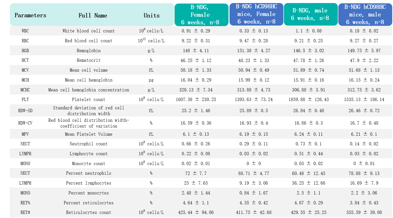
Complete blood count (CBC) of CD98HC humanized B-NDG mice.CD98HC humanization does not disrupt hematopoiesis or immune cell composition. Values are expressed as mean ± SD.
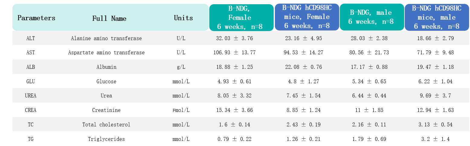
Biochemical test of CD98HC humanized B-NDG mice. CD98HC humanization does not adversely affect metabolic health or organ function. Values are expressed as mean ± SD.

Growth kinetics of B-Tg(Luc-EGFP) U-87 MG tumors determined by bioluminescence imaging (BLI). B-Tg(Luc-EGFP) U-87 MG cells (1x10⁵) were injected into the brain of B-NDG hCD98HC mice (female and male, 7-week-old, n=6). Signal intensity and body weight were measured twice a week. (A) Raw bioluminescence images. (B) Body weight. (C) Signal intensity. These results indicate that B-Tg(Luc-EGFP) U-87 MG cells (Cat. No 322265) can establish brain orthotopic tumors in B-NDG hCD98HC mice, and this strain of mice can be used for drug efficacy studies. Values are expressed as mean ± SEM.