on this page
Biocytogen provides comprehensive preclinical pharmacology services to evaluate drug efficacy, pharmacodynamic (PD) and pharmacokinetic (PK) profiles, biomarker responses, and non-GLP toxicity of new therapeutics. Our technical team brings extensive project experience and deep expertise in evaluating novel therapeutics across immuno-oncology, oncology, autoimmune and inflammatory diseases, and neurology applications.
Discover how our validated in vivo models—such as CDX, PDX, autoimmune, and metabolic disease models—can streamline and accelerate your drug discovery and development process.
See DetailsOur PD/PK services leverage cutting-edge technology platforms to offer customized and professional study designs, meeting the specific requirements of drug development.
See DetailsLeveraging advanced techniques such as flow cytometry, ELISA, HTRF, and Incucyte live-cell imaging, we offer comprehensive in vitro functional evaluation services.
See DetailsOur pathology platform leverages advanced techniques and AI-driven analysis to support non-GLP toxicology and safety studies, enhancing drug discovery and translational research efforts.
See DetailsOur pharmacology team has developed multiple disease models, including tumor models (syngeneic models, CDX and PDX models), metabolic disease models (NASH, obesity and diabetes, atherosclerosis, chronic kidney disease, fibrosis, etc.), inflammatory and autoimmune disease models (asthma, rheumatoid arthritis, atopic dermatitis, psoriasis, SLE, MS, IBD, etc.) and neurological disease models for preclinical drug efficacy evaluations.


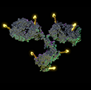
We have successfully supported our pharmaceutical and biotech customers by providing preclinical efficacy studies and toxicity studies for multiple biologics (including monoclonal antibodies, bispecific antibodies, and recombinant proteins), antibody-drug conjugates (ADCs), peptides, tumor vaccines, small molecules, CAR-T cell therapy, and oncolytic viruses.


Our experts are committed to your project's success.
We offer the following policies to earn the trust of your partnership and guarantee your satisfaction with our pharmacology services:

There will be no charges for any overages of mice supplied by Biocytogen, including both animal and service costs. Charges will only be applied to the actual number of enrolled animals in the study.
Eligible clients who have not previously run a study in a specific therapeutic area/disease model category have the opportunity to run a complimentary study with us free of charge or obligation. Eligibility is subject to approval; restrictions may apply.
We stand behind the quality of our services. Should you find any aspect of our project delivery less than satisfactory, we offer two options to ensure your complete satisfaction
Free Project Repeat: We will repeat the study at no additional cost to you.
Full Refund: We will provide a full refund of the project cost, no questions asked.
Policy Restrictions:
¹Eligibility: This policy is exclusively applicable to pharmacology services utilizing Biocytogen mice and excludes studies involving mice sourced from other vendors. Studies may only be repeated once under this policy. If the study protocol is altered to incorporate the additional mice into the study after the mice are shipped, the client will be charged accordingly.
²Data Restrictions Apply: In the event of a project repeat or refund, any data generated from the initial project cannot be utilized by the client under any circumstances. Refunds do not include the cost of the test article preparation.