Pulmonary fibrosis is a lung disease often triggered by chemicals (such as asbestos, coal dust or silica). Inhalation of these chemicals cause abnormal accumulation of the extracellular matrix and fibroblast proliferation, resulting in scarring and thickening of the lung tissue, which ultimately impairs normal breathing function. Pulmonary fibrosis can also occur as a result of other types of injury/inflammation.
In animals, exposure of mice to bleomycin is a common method to induce pulmonary fibrosis, as it causes inflammation resulting from cellular damage. The fibrosis model is accomplished via intratracheal administration of the drug, followed by monitoring for body weight, survival and assessment of fibrosis via chemical and histologic methods.
-
Pulmonary Fibrosis Model Build
-
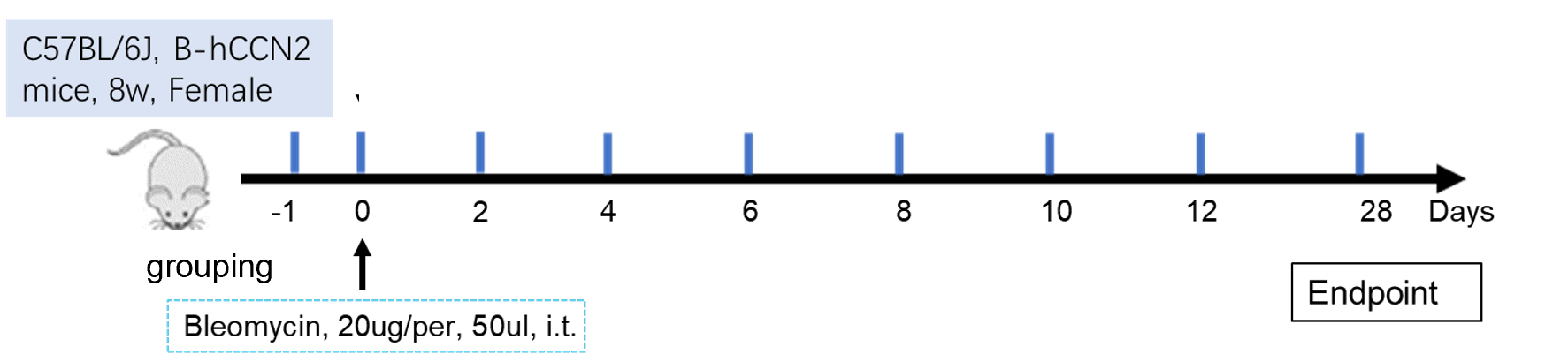
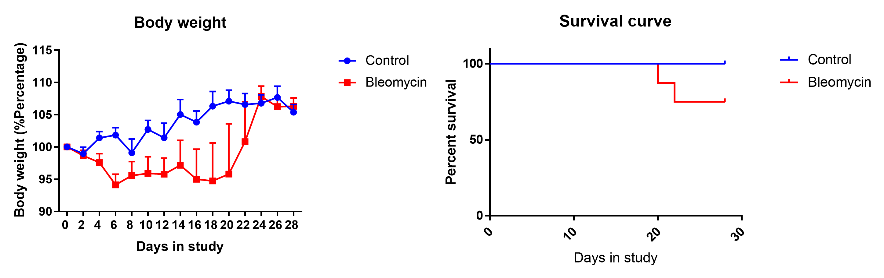
Bleomycin-induced Pulmonary Fibrosis. C57BL/6J mice were administered bleomycin (intratracheal) on day 0. Body weight and survival was monitored daily until the endpoint at day 28.
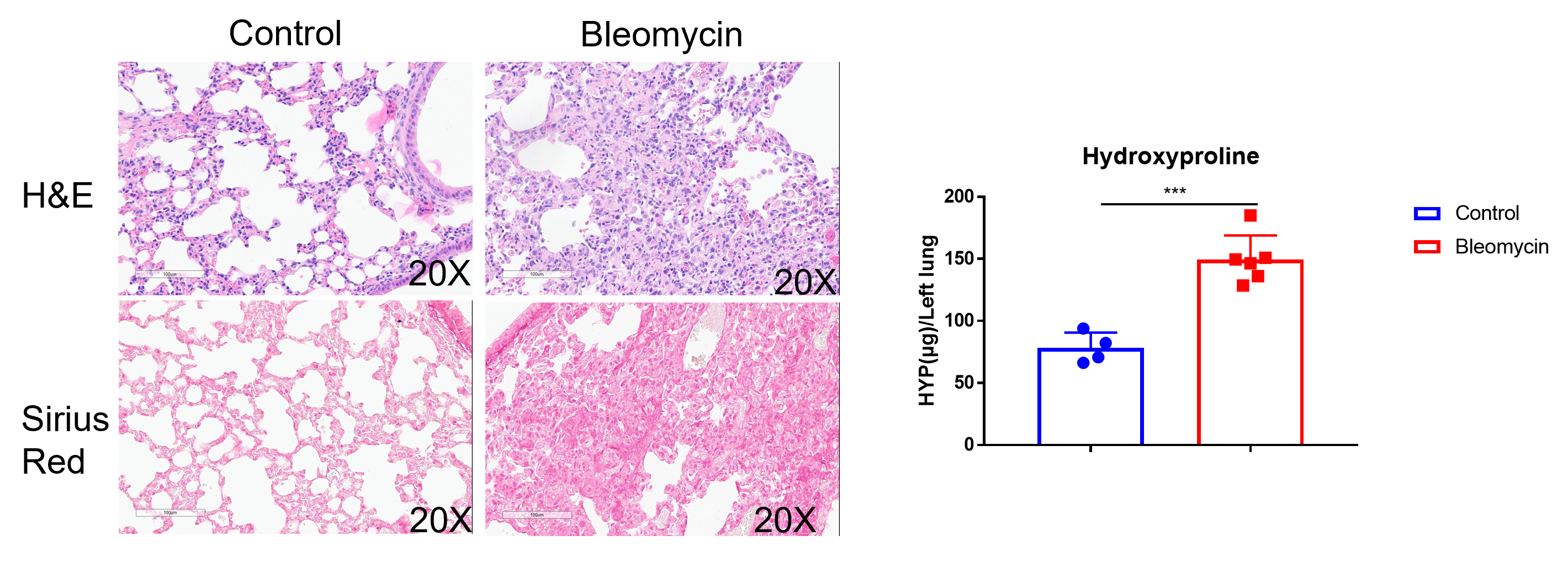
Histologic and chemical assessment of fibrosis. Lung tissue was collected at the end of the experiment; hydroxyproline in the lung was measured. Histologic analyses with Sirius Red indicate significant deposition of collagen, indicative of pulmonary fibrosis (left). Bleomycin treatment also induced an increase in hydroxyproline content, indicative of fibrosis (right).
-
Pamrevlumab (anti-CTGF) Efficacy in Pulmonary Fibrosis Model (B-hCCN2 mice)
-

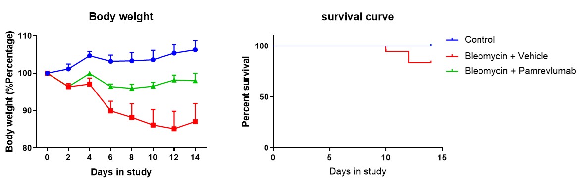
Bleomycin-induced Pulmonary Fibrosis with Pamrevlumab administration. B-hCCN2 mice were administered bleomycin (intratracheal) on day 0. Pamrevlumab (anti-CTGF) was administered (i.p., 10 mg/kg) daily for 7 days. Body weight and survival was monitored daily until the endpoint at day 14. Pamrevlumab treatment was able to improve bleomycin-induced weight loss in B-hCCN2 mice.
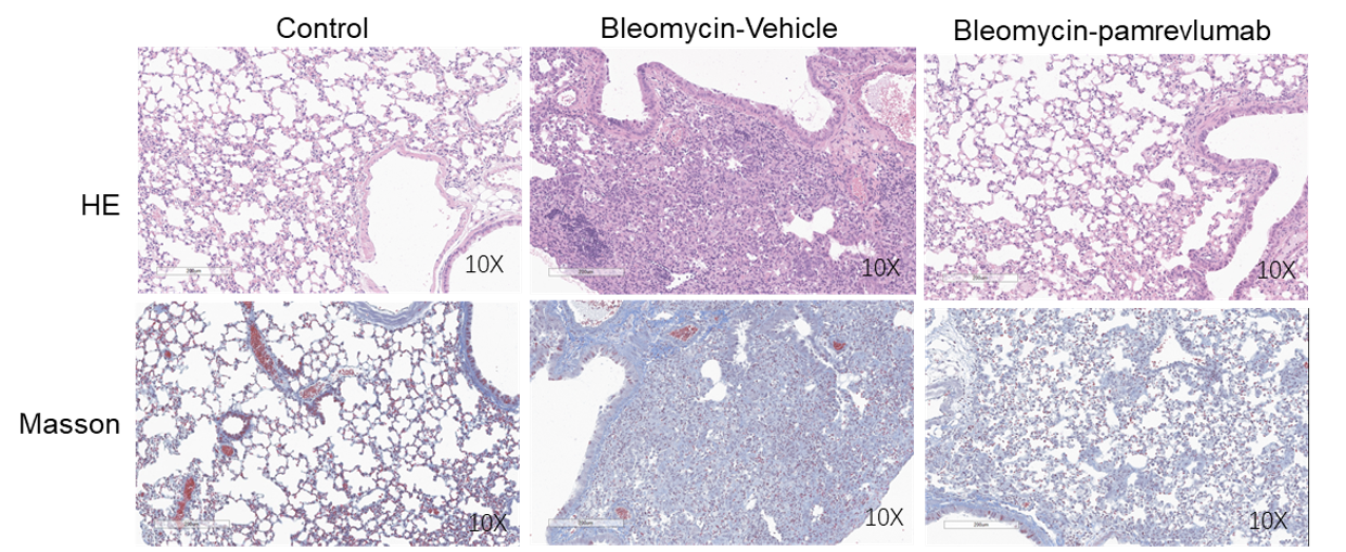
Histologic changes improve with pamrevlumab treatment. H&E and Masson trichrome staining of lung tissues indicate significant pathologic changes induced by bleomycin that associated with fibrosis in the lung. Tissue architecture is improved with antibody treatment.

Measurements of hydroxyproline improve with pamrevlumab treatment. Bleomycin treatment increases hydroxyproline content compared with controls. Compared with vehicle-treated bleomycin controls, pamrevlumab treatment resulted in a reduction in mean induced lung HYP content. Values are expressed as mean ± SEM. *** p<0.001, **p<0.01.
Our Pharmacology Service Guarantee