
Basic Information
-
Human CD34+ HSC engraftment for human immune system reconstitution

-
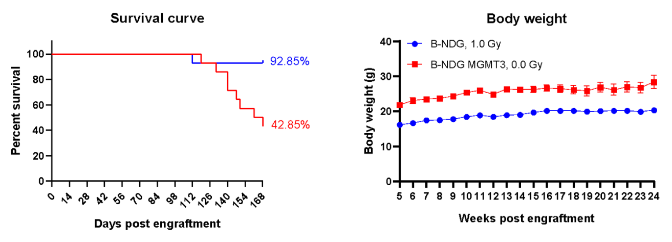
Human CD34+ HSCs (3E4) were intravenous (temporal vein) engrafted into wild-type B-NDG mice and homozygous B-NDG MGMT3 mice (both sex, 24-72 hr after birth, n=15). B-NDG mice were treated with 1.0 Gy-irradiation. B-NDG MGMT3 mice were not irradiated. A. Survival rates of the mice were analyzed with Kaplan Meier survival curves. B. Body weight. Results showed that the survival rate of B-NDG MGMT3 mice was similar to that of B-NDG mice until 18 weeks after human CD34+ HSCs engraftment and then decreased to 42.85% at 24 weeks post engraftment. But the body weight of B-NDG MGMT3 mice was significantly higher than that of B-NDG mice and increased steadily during the whole reconstitution. Values are expressed as mean ± SEM. HSCs: hematopoietic stem cells.
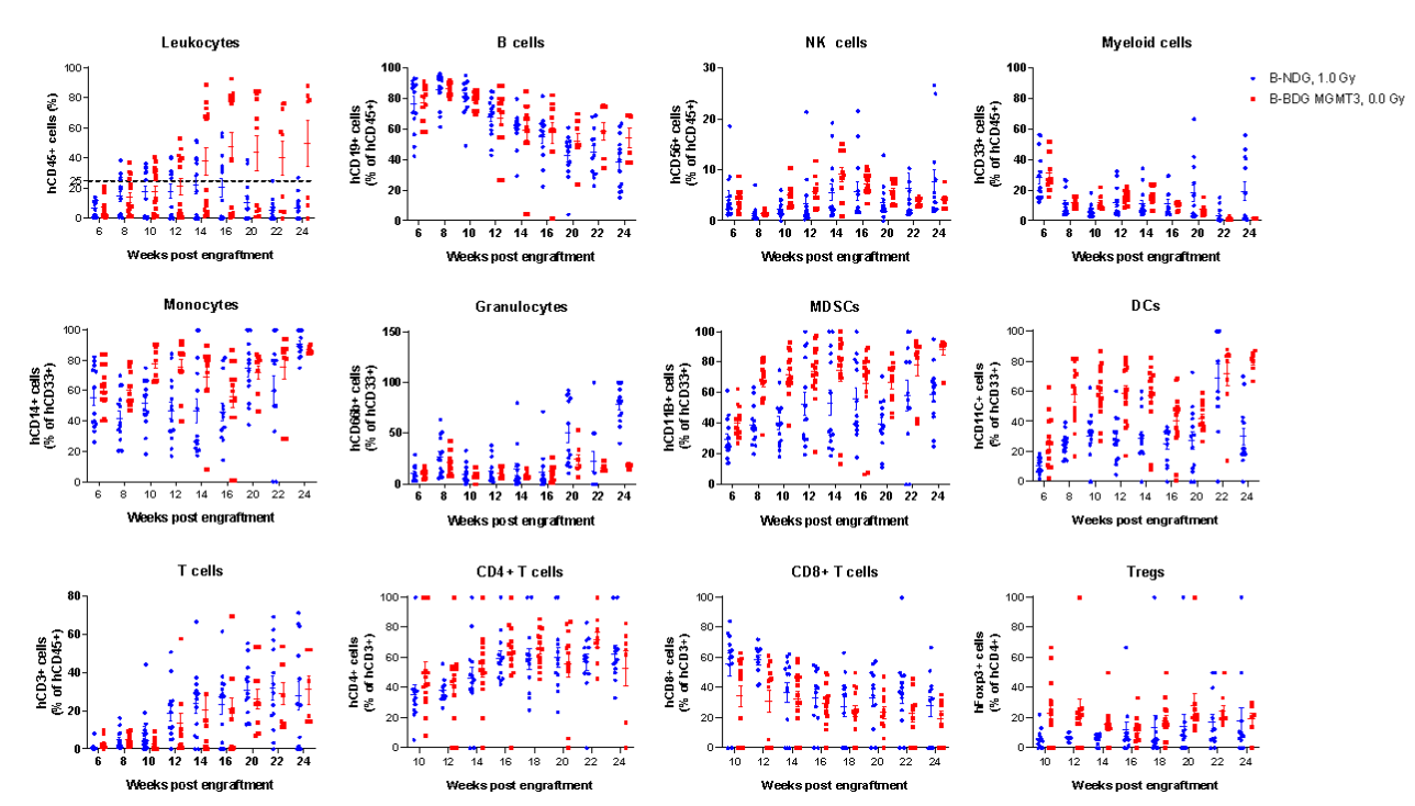
Human CD34+ HSCs (3E4) were intravenous (temporal vein) engrafted into wild-type B-NDG mice and homozygous B-NDG MGMT3 mice (both sex, 24-72 hr after birth, n=15). B-NDG mice were treated with 1.0 Gy-irradiation. B-NDG MGMT3 mice were not irradiated. Peripheral blood lymphocytes from the two mice after engraftment with human CD34+ HSCs were analyzed with flow cytometry. Results showed that the proportion of CD45+ cells in B-NDG MGMT3 mice reached 25% starting from 12 weeks after engraftment and continued to rise, significantly higher than that in B-NDG mice. The proportions of monocytes, MDSCs, DCs and Tregs in B-NDG MGMT3 mice were higher than that in B-NDG mice. Values are expressed as mean ± SEM.
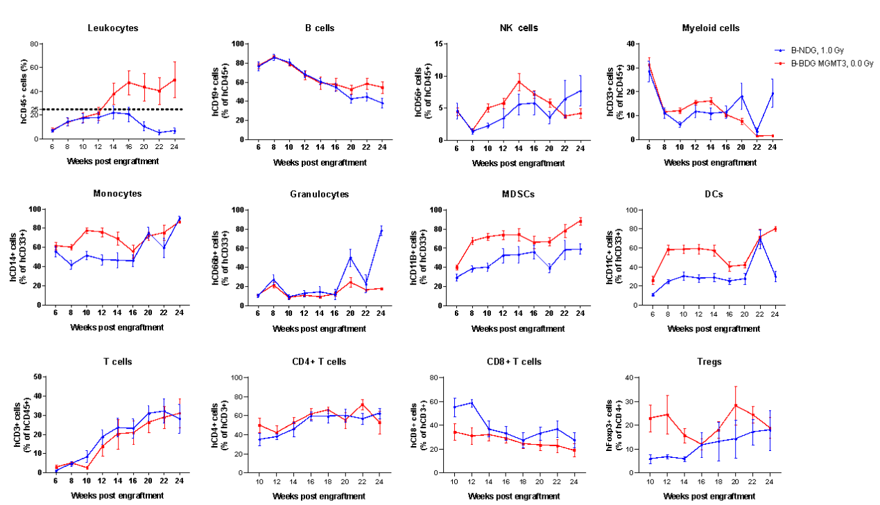
Human CD34+ HSCs (3E4) were intravenous (temporal vein) engrafted into wild-type B-NDG mice and homozygous B-NDG MGMT3 mice (both sex, 24-72 hr after birth, n=15). B-NDG mice were treated with 1.0 Gy-irradiation. B-NDG MGMT3 mice were not irradiated. Peripheral blood lymphocytes from the two mice after engraftment with human CD34+ HSCs were analyzed with flow cytometry. Results showed that the proportion of CD45+ cells in B-NDG MGMT3 mice reached 25% starting from 12 weeks after engraftment and continued to rise, significantly higher than that in B-NDG mice. The proportions of monocytes, MDSCs, DCs and Tregs in B-NDG MGMT3 mice were higher than that in B-NDG mice. Values are expressed as mean ± SEM.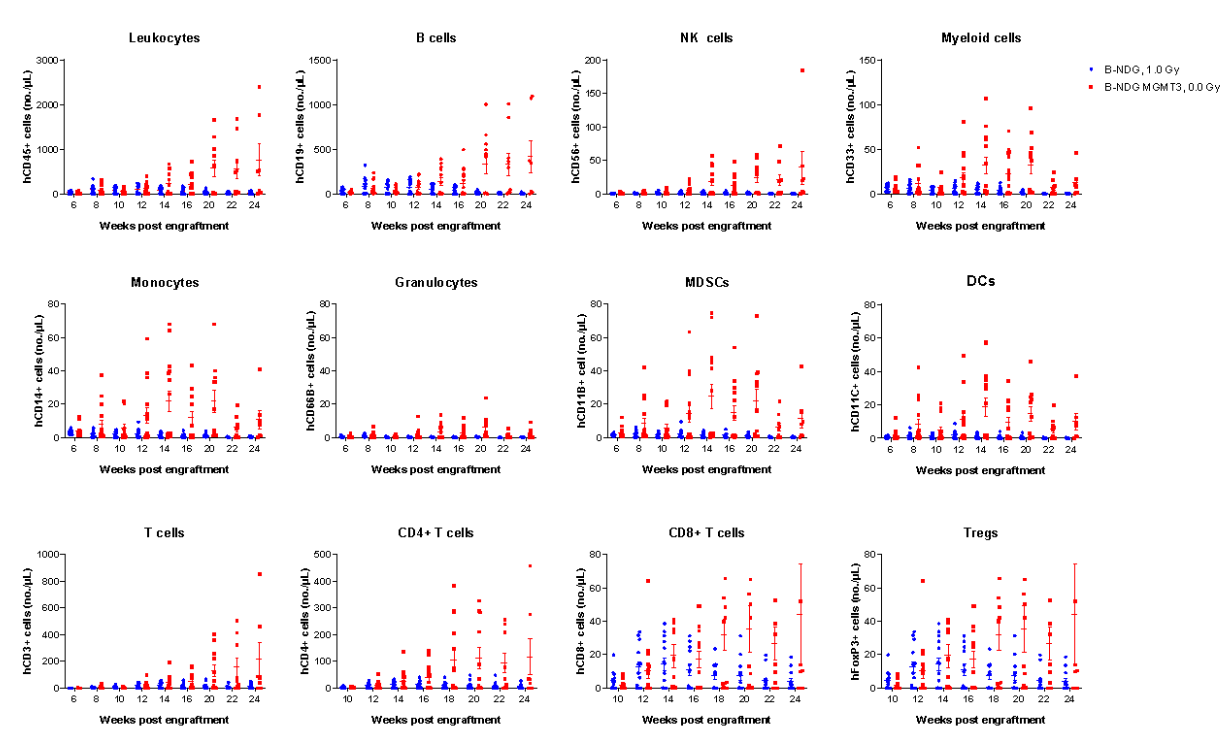
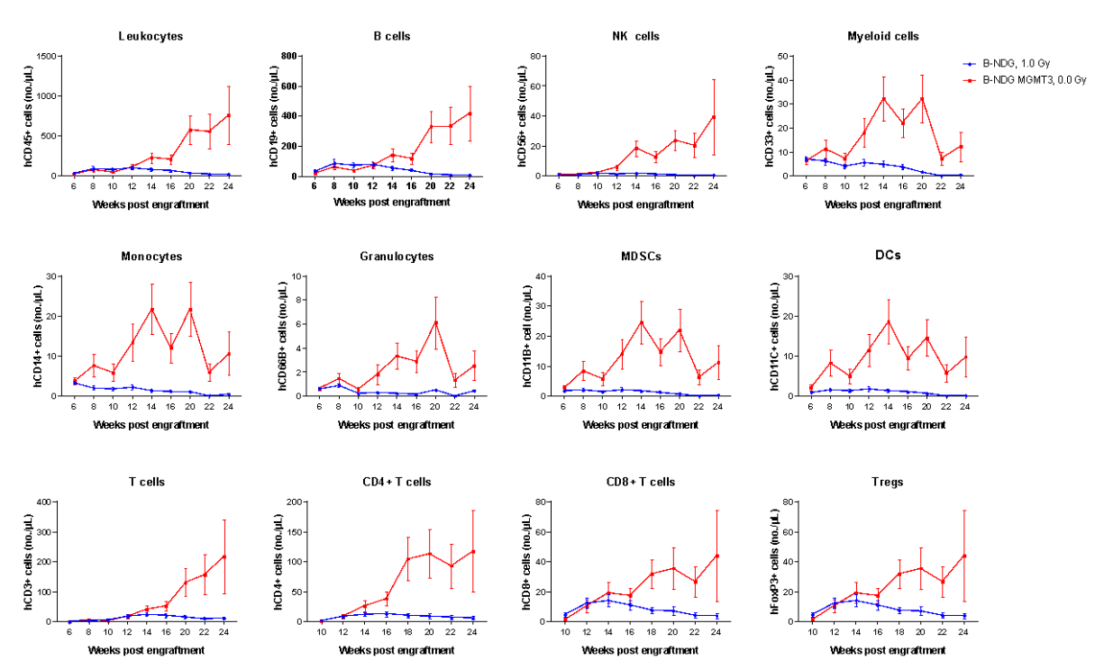
Human CD34+ HSCs (3E4) were intravenous (temporal vein) engrafted into wild-type B-NDG mice and homozygous B-NDG MGMT3 mice (both sex, 24-72 hr after birth, n=15). B-NDG mice were treated with 1.0 Gy-irradiation. B-NDG MGMT3 mice were not irradiated. Peripheral blood lymphocytes from the two mice after engraftment with human CD34+ HSCs were analyzed with flow cytometry. Results showed that the cell numbers of all the cells analyzed from 12 weeks after engraftment in B-NDG MGMT3 mice were higher than that in B-NDG mice. Values are expressed as mean ± SEM. Values are expressed as mean ± SEM.
Human CD34+ HSCs (3E4) were intravenous (temporal vein) engrafted into wild-type B-NDG mice and homozygous B-NDG MGMT3 mice (both sex, 24-72 hr after birth, n=15). B-NDG mice were treated with 1.0 Gy-irradiation. B-NDG MGMT3 mice were not irradiated. Peripheral blood lymphocytes from the two mice after engraftment with human CD34+ HSCs were analyzed with flow cytometry. Results showed that the cell numbers of all the cells analyzed from 12 weeks after engraftment in B-NDG MGMT3 mice were higher than that in B-NDG mice. Values are expressed as mean ± SEM. Values are expressed as mean ± SEM.
