
Basic Information
-
Targeting strategy

-
Gene targeting strategy for B-hIL17A mice.
The exons 1-3 of mouse Il17a gene that encode the extracellular domain were replaced by human IL17A exons 1-3 in B-hIL17A mice.
-
mRNA expression analysis
-
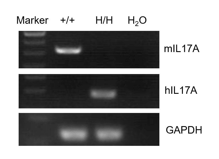
Strain specific analysis of IL17A gene expression in wild type (WT) mice and B-hIL17A mice by RT-PCR.
Mouse Il17a mRNA was detectable only in splenocytes of WT mice (+/+). Human IL17A mRNA was detectable only in homozygous B-hIL17A mice (H/H) but not in WT mice (+/+).
-
Protein Expression Analysis
-
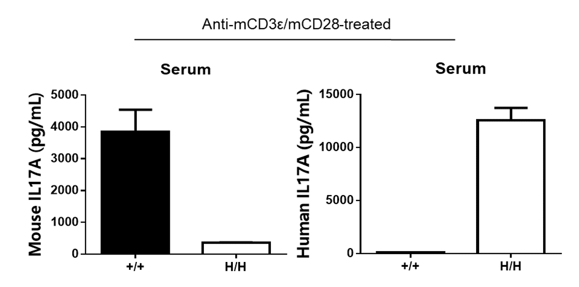
Strain specific IL17A expression analysis in wild type (WT) mice and B-hIL17A mice by ELISA.
Serum were collected from WT mice (+/+) and homozygous B-hIL17A mice (H/H) stimulated with anti-CD3ε (7.5 μg/mice, 2 hours, i.p.) and anti-CD28 (4 μg/mice, 2 hours, i.p.) in vivo, and analyzed by ELISA with species-specific IL17A ELISA kit. Mouse IL17A was detectable in WT mice. Human IL17A was detectable in homozygous B-hIL17A mice (H/H) but not WT mice (+/+) .
-
Analysis of spleen leukocytes cell subpopulations in B-hIL17A mice
-
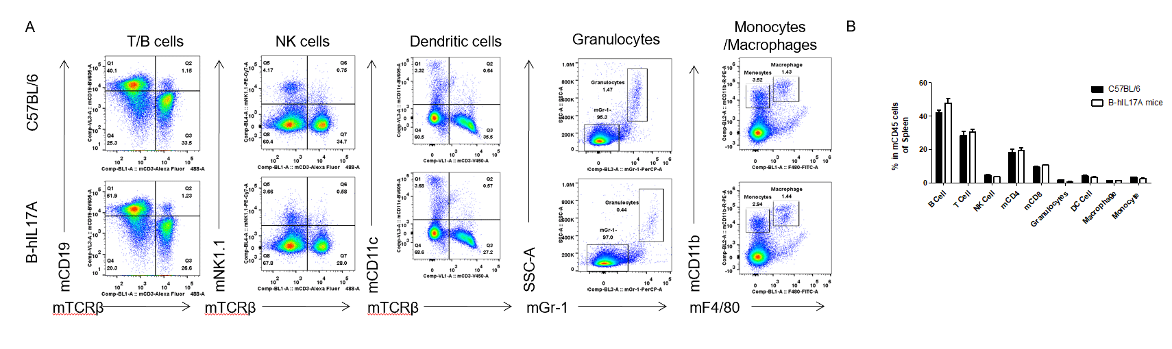
Analysis of spleen leukocyte subpopulations by FACS.
Splenocytes were isolated from female C57BL/6 and B-hIL17A mice (n=3, 6 week-old). Flow cytometry analysis of the splenocytes was performed to assess leukocyte subpopulations. A. Representative FACS plots. Single live cells were gated for CD45 population and used for further analysis as indicated here. B. Results of FACS analysis. Percent of T cells, B cells, NK cells, dendritic cells, granulocytes, monocytes and macrophages in homozygous B-hIL17A mice were similar to those in the C57BL/6 mice, demonstrating that introduction of hIL17A in place of its mouse counterpart does not change the overall development, differentiation or distribution of these cell types in spleen. Values are expressed as mean ± SEM.
-
Analysis of spleen T cell subpopulations in B-hIL17A mice
-
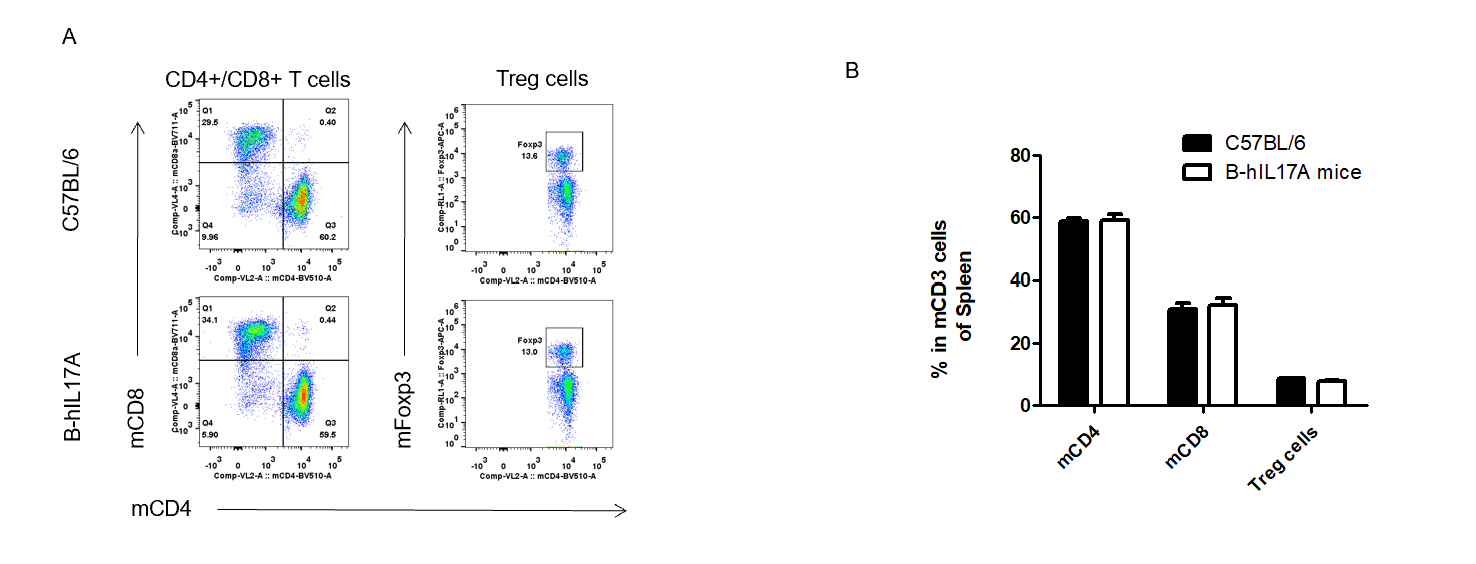
Analysis of spleen T cell subpopulations by FACS.
Splenocytes were isolated from female C57BL/6 and B-hIL17A mice (n=3, 6 week-old). Flow cytometry analysis of the splenocytes was performed to assess leukocyte subpopulations. A. Representative FACS plots. Single live CD45+ cells were gated for CD3 T cell population and used for further analysis as indicated here. B. Results of FACS analysis. Percent of CD8+ T cells, CD4+ T cells and Treg cells in homozygous B-hIL17A mice were similar to those in the C57BL/6 mice, demonstrating that introduction of hIL17A in place of its mouse counterpart does not change the overall development, differentiation or distribution of these T cell sub types in spleen. Values are expressed as mean ± SEM.
-
Analysis of blood leukocytes cell subpopulations in B-hIL17A mice
-
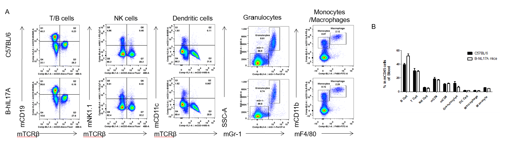
Analysis of blood leukocyte subpopulations by FACS.
Blood cells were isolated from female C57BL/6 and B-hIL17A mice (n=3, 6 week-old). Flow cytometry analysis of the blood leukocytes was performed to assess leukocyte subpopulations. A. Representative FACS plots. Single live cells were gated for CD45 population and used for further analysis as indicated here. B. Results of FACS analysis. Percent of T cells, B cells, NK cells, dendritic cells, granulocytes, monocytes and macrophages in homozygous B-hIL17A mice were similar to those in the C57BL/6 mice, demonstrating that introduction of hIL17A in place of its mouse counterpart does not change the overall development, differentiation or distribution of these cell types in blood. Values are expressed as mean ± SEM.
-
Analysis of blood T cell subpopulations in B-hIL17A mice
-
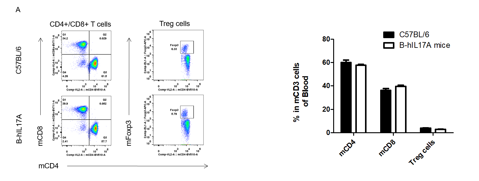
Analysis of blood T cell subpopulations by FACS.
Blood cells were isolated from female C57BL/6 and B-hIL17A mice (n=3, 6 week-old). Flow cytometry analysis of the leukocytes was performed to assess leukocyte subpopulations. A. Representative FACS plots. Single live CD45+ cells were gated for CD3 T cell population and used for further analysis as indicated here. B. Results of FACS analysis. Percent of CD8+ T cells, CD4+ T cells and Treg cells in homozygous B-hIL17A mice were similar to those in the C57BL/6 mice, demonstrating that introduction of hIL17A in place of its mouse counterpart does not change the overall development, differentiation or distribution of these T cell sub types in blood. Values are expressed as mean ± SEM.
-
Blood routine test in B-hIL17A mice
-
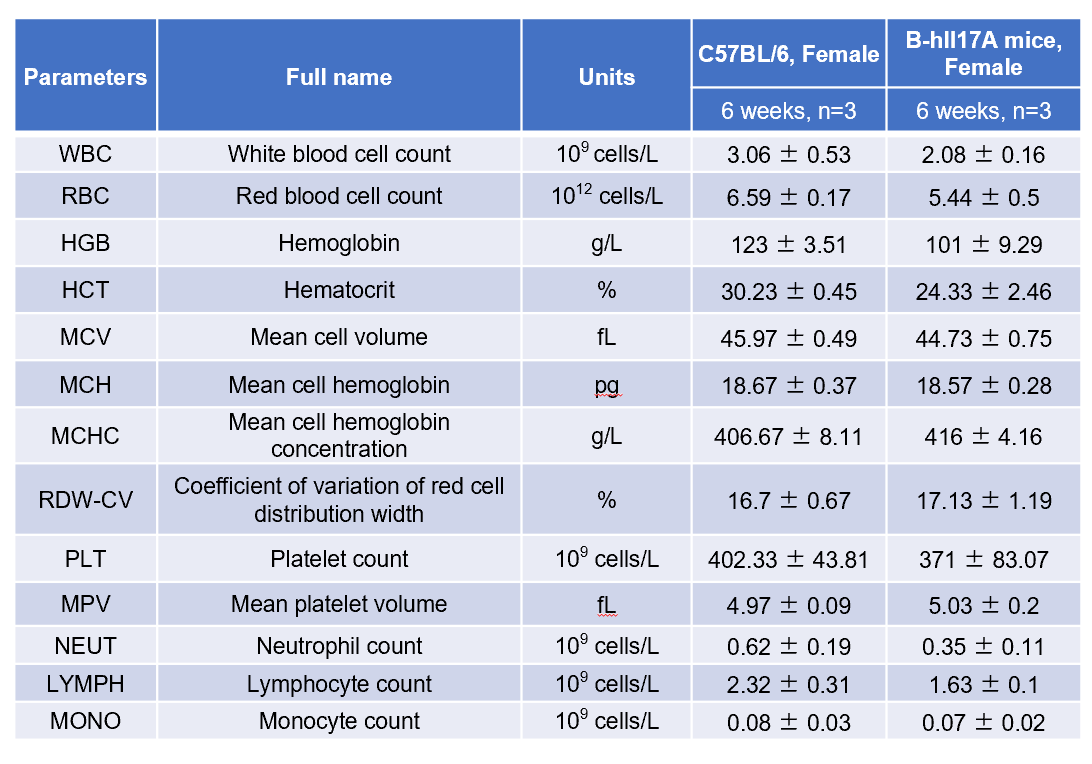
Complete blood count (CBC).
Blood from female C57BL/6 and B-hIL17A mice (n=3, 6 week-old) was collected and analyzed for CBC. There was no differences among any measurement between C57BL/6 and B-hIL17A mice, indicating that introduction of hIL17A in place of its mouse counterpart does not change blood cell composition and morphology. Values are expressed as mean ± SEM.
-
Blood chemistry of B-hIL17A mice
-
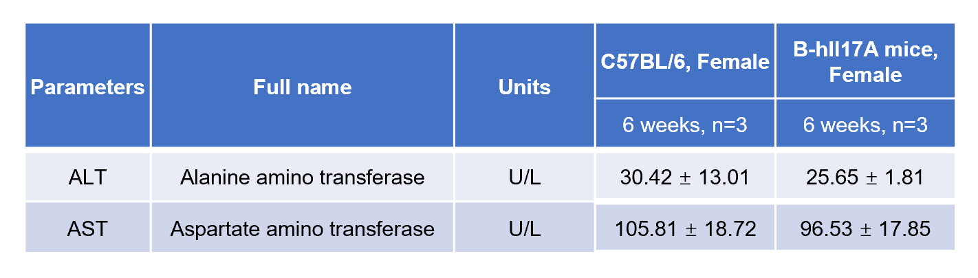
Blood chemistry tests of B-hIL17A mice.
Serum from female C57BL/6 and B-hIL17A mice (n=3, 6 week-old) was collected and analyzed for levels of ALT and AST. There was no differences on either measurement between C57BL/6 and B-hIL17A mice, indicating that introduction of hIL17A in place of its mouse counterpart does not change ALT and AST levels or health of liver. Values are expressed as mean ± SEM.
-
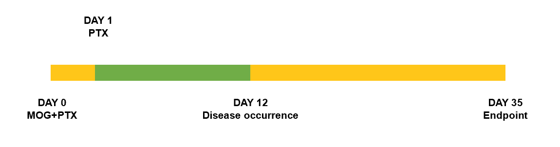
Schematic of EAE model
-
Experimental Autoimmune Encephalomyelitis (EAE) is an induced demyelinating disease model that closely resembles the progression and symptoms of the human neurological disease Multiple Sclerosis (MS).
-
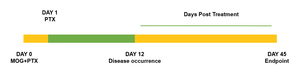
Clinical score of EAE model
-
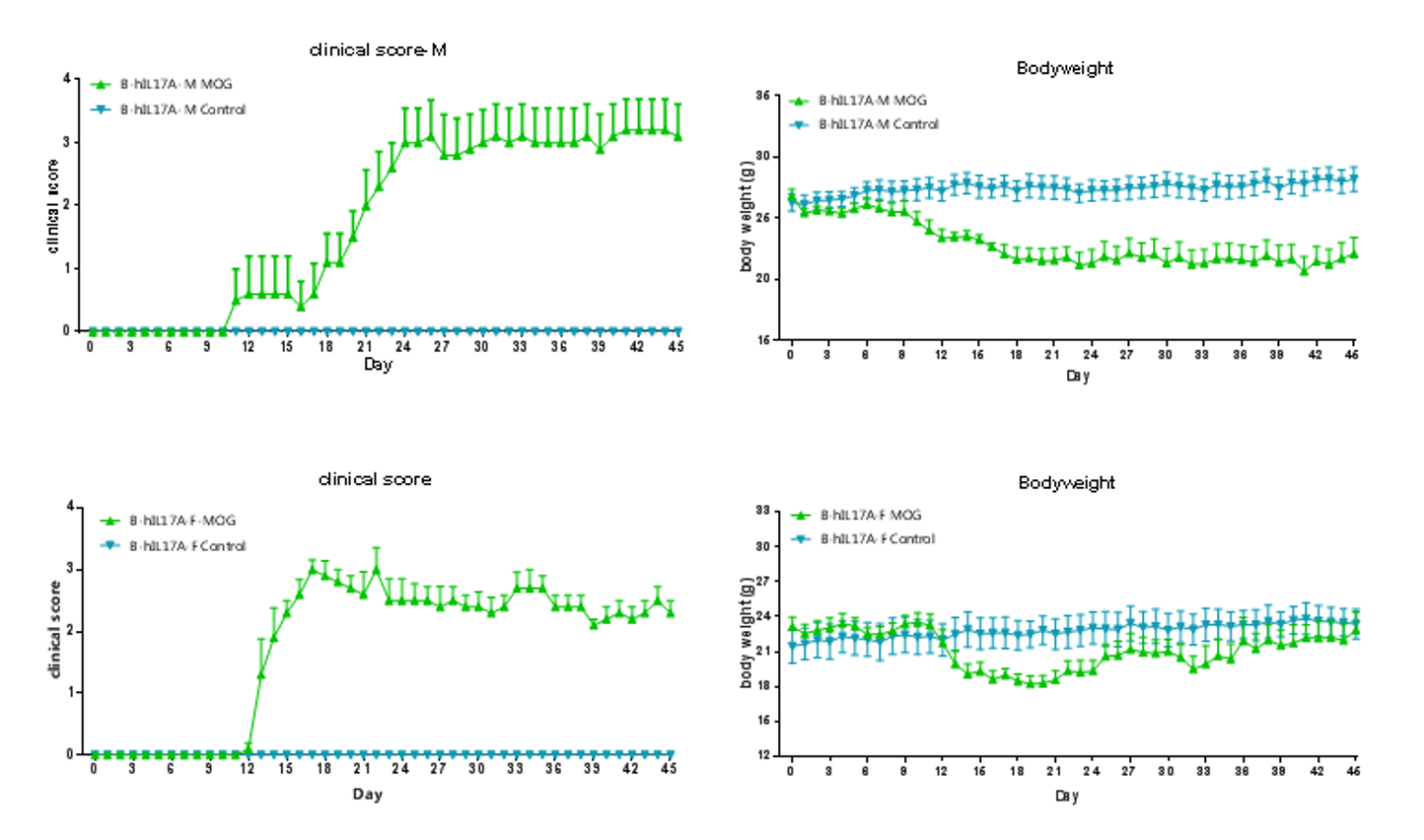
EAE induction by MOG35–55/CFA Emulsion PTX in 10-week-old B-hIL17A mice. Data are expressed as mean ± SEM from a typical experiment (n=5). MOG: myelin-oligodendrocyte glycoprotein; PTX: pertussis toxin. F: female; M: male.
-
H&E staining and IHC staining in mouse EAE model
-
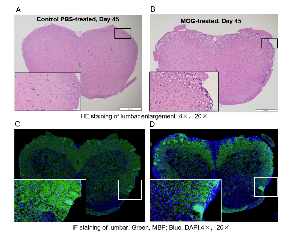
Local inflammation of the CNS in B-hIL17A mice (female,n=5) during EAE. On day 45 after MOG/CFA and PTX immunization, spinal cords were removed. The tissue sections were stained with H&E(A,B) and IHC (C,D)(Green, MBP; Blue, DAPI). The sections at the lumbar level are shown. The results showed that the infiltration of inflammatory cells in the MOG group was significantly increased, and the myelin protein was greatly reduced.
-
IL17A+ cells in lymph node of EAE model
-
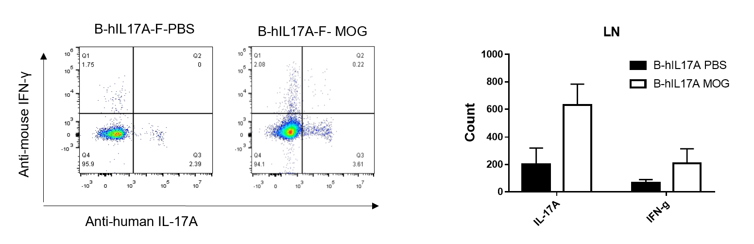
IL-17 was primarily produced by CD4 Th17 cells during the development of EAE. To detect IL-17 production, cells from lymph node of B-hIL17A mice(female,n=5) immunized with MOG/CFA were stimulated for 6 hours by PMA and ionomycin in the presence of brefeldin A. IL-17-producing cells were analyzed by FACS, along with IFNg. The percentage of IL-17+CD3+CD4+ T cells in CD3+CD4+ T cells was increased in response to MOG immunization in B-hIL17A mice (left panel). So was percentage of IFNg+ T cells.
-
IL17A+ cells in CNS of EAE model
-
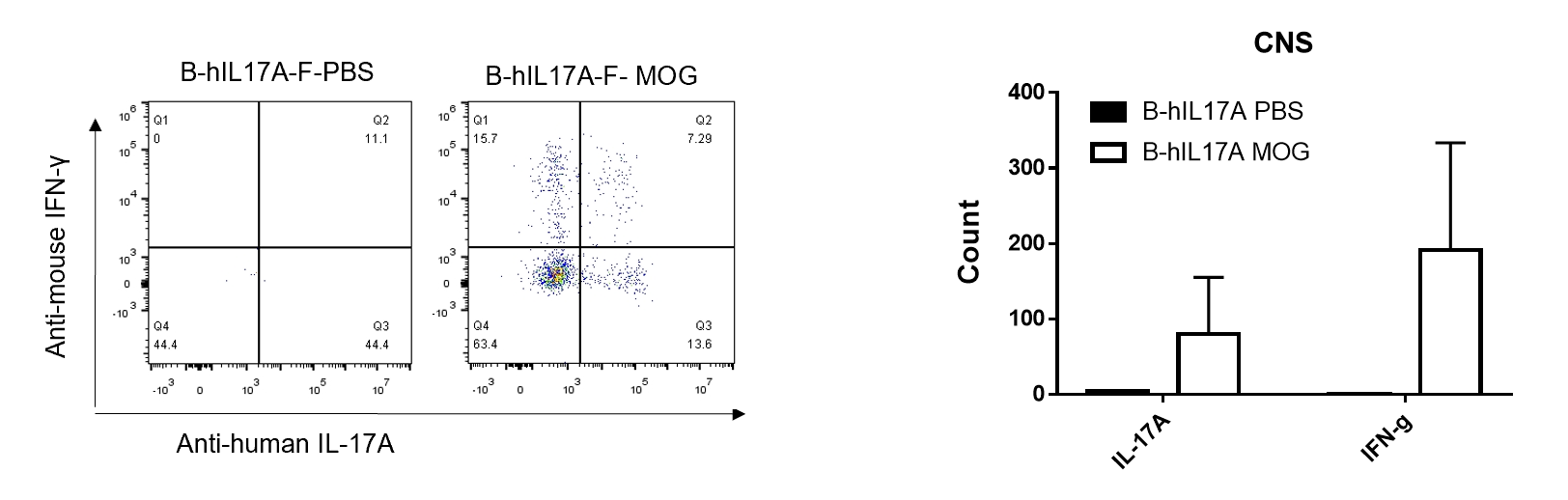
IL-17 was primarily produced by CD4 Th17 cells during the development of EAE. To detect IL-17 production, cells from CNS(Brain cell) of B-hIL17A mice(female,n=5) immunized with MOG/CFA were stimulated for 6 hours by PMA and ionomycin in the presence of brefeldin A. IL-17-producing cells were analyzed by FACS, along with IFNg. The percentage of IL-17+CD3+CD4+ T cells in CD3+CD4+ T cells was increased in response to MOG immunization in B-hIL17A mice (left panel). So was percentage of IFNg+ T cells.
-
In vivo efficacy of anti-human IL17A antibodies in EAE model of B-hIL17A mice
-
Model schematic
-
In vivo efficacy of anti-human IL17A antibodies in EAE model of B-hIL17A mice
-
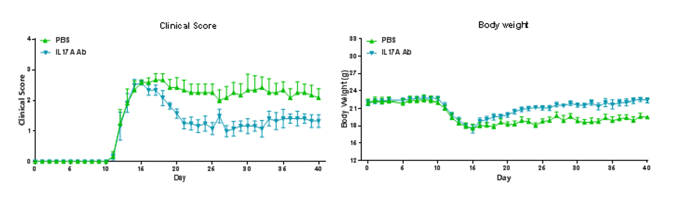
EAE induction by MOG35–55/CFA Emulsion PTX in B-hIL17A mice. The clinical score and body weight was collected after the treatment of anti-hIL17A antibody. After treatment of anti-hIL17A antibody, the clinical score level was much lower than the control in homozygous B-hIL17A mice. Data are expressed as mean ± SEM from a typical experiment (n = 6). MOG: myelin-oligodendrocyte glycoprotein; PTX: pertussis toxin.
-
Experimental schedule for induction of psoriasis-like skin lesions in B-hIL17A mice
-
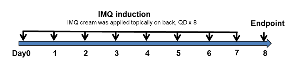
Experimental schedule for induction of psoriasis-like skin lesions in B-hIL17A mice.
Mice at 10 week-old of age received a daily topical of commercially available IMQ cream on the shaved back for 8 consecutive days to induce psoriasis-like skin lesions. Severity of skin inflammation was daily scored and back skin was collected at the endpoint. IMQ: imiquimod.
-
In vivo efficacy of anti-human IL17A antibodies in psoriasis model induced in B-hIL17A mice
-
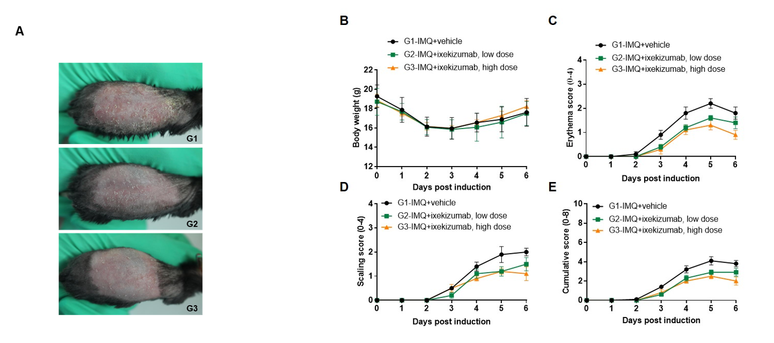
IMQ-induced skin inflammation in B-hIL17A mice phenotypically resembles psoriasis.
Mice (female, 10 week-old, n=5) were scored daily for up to 6 days for body weight and clinical signs of skin inflammation following treatment with imiquimod (IMQ) cream. Mice in each group were treated with different dose of ixekizumab produced in house. Doses are shown in legend. (A) Phenotypical presentation of mouse back skin after 6 days of treatment. (B) Body weight changes during treatment. (C-D) Erythema and scaling score of the back was scored daily on a scale from 0 to 4. Additionally, the cumulative score (erythema plus scaling) is depicted. Values are expressed as mean ± SEM.
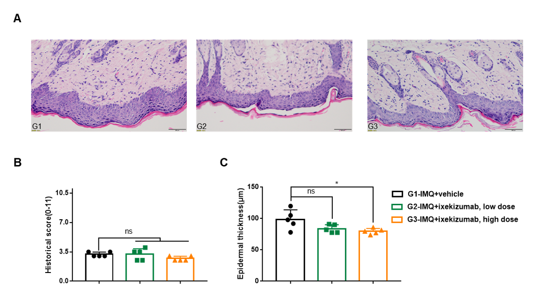
Dose dependent effects of antibody on keratinocyte proliferation and inflammatory cell infiltration in IMQ induced psoriasis-like skin lesions in B-hIL17A mice.
Back skin was collected at the endpoint and stained with Hematoxylin and eosin (H&E). (A) H&E staining of the back skin. (B) Histological changes were scored on a scale from 0 to 11. (C) Epidermal thickness of the mice. Results indicated that therapeutic effects of ixekizumab (in house) on psoriasis-like skin lesions in B-hIL17A mice were dose dependent, confirming that B-hIL17A mice provide a powerful model for in vivo evaluation of anti-human IL17A antibodies. Values are expressed as mean ± SEM.
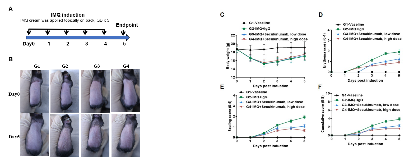
IMQ-induced skin inflammation in B-hIL17A mice phenotypically resembles psoriasis.
Mice (female, 8 week-old, n=6) were scored daily for up to 6 days for body weight and clinical signs of skin inflammation following treatment with imiquimod (IMQ) cream. Mice in each group were treated with different doses of Secukinumab (commercial drug). (A) Experimental schedule for induction of psoriasis-like skin lesions in B-hIL17A mice. (B) Phenotypical presentation of mouse back skin at day0 and day 5. (C) Body weight changes during treatment. (D-E) Erythema and scaling score of the back was scored daily. Additionally, the cumulative score (erythema plus scaling) is depicted. Values are expressed as mean ± SEM.
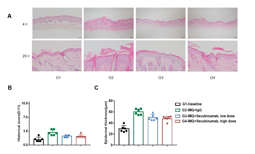
Dose dependent effects of antibodies on keratinocyte proliferation and inflammatory cell infiltration in IMQ induced psoriasis-like skin lesions in B-hIL17A mice.
Back skin was collected at the endpoint and stained with Hematoxylin and eosin (H&E). (A) H&E staining of the back skin. (B) Histological changes were scored. (C) Epidermal thickness of the mice. Results indicated that therapeutic effects of Secukinumab (commercial drug) on psoriasis-like skin lesions in B-hIL17A mice were dose dependent, confirming that B-hIL17A mice provide a powerful model for in vivo evaluation of anti-human IL17A antibodies. Values are expressed as mean ± SEM.
