Basic Information
-
Targeting strategy
-
Gene targeting strategy for B-hNKG2D mice plus. The 5’UTR and exons 2~8 of mouse Nkg2d gene encoding the full-length were replaced by 5’UTR and exons 2~8 of human NKG2D in B-hNKG2D mice plus.
-
Protein expression analysis in CD4+T cells
-
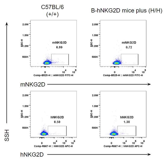
Strain specific analysis of NKG2D expression in wild-type (WT) mice (+/+) and homozygous B-hNKG2D mice plus(H/H) by flow cytometry. Splenocytes were collected from wild-type C57BL/6 mice (+/+) and homozygous B-hNKG2D mice plus (H/H), and analyzed by flow cytometry with species-specific anti-NKG2D antibody. mNKG2D and hNKG2D were not detectable in CD4+T cells of wild-type and homozygous B-hNKG2D mice plus.
-
Protein expression analysis in CD8+T cells
-
Strain specific analysis of NKG2D expression in wild-type (WT) mice (+/+) and homozygous B-hNKG2D mice plus(H/H) by flow cytometry. Splenocytes were collected from wild-type C57BL/6 mice (+/+) and homozygous B-hNKG2D mice plus(H/H), and analyzed by flow cytometry with species-specific anti-NKG2D antibody. mNKG2D was not detectable in CD8+T cells of wild-type C57BL/6 mice, while hNKG2D was detectable in homozygous B-hNKG2D mice plus.
-
Protein expression analysis in NK cells
-
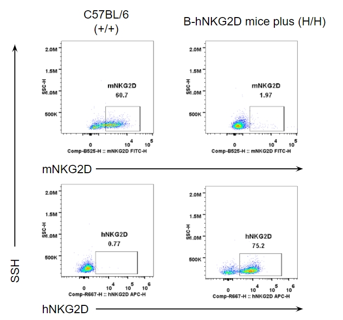
Strain specific analysis of NKG2D expression in wild-type (WT) mice (+/+) and homozygous B-hNKG2D mice plus(H/H) by flow cytometry. Splenocytes were collected from wild-type C57BL/6 mice (+/+) and homozygous B-hNKG2D mice plus(H/H), and analyzed by flow cytometry with species-specific anti-NKG2D antibody. mNKG2D was detectable in NK cells of wild-type C57BL/6 mice and homozygous B-hNKG2D mice plus, while hNKG2D was only detectable in homozygous B-hNKG2D mice plus.
-
Protein expression analysis
-
Protein expression analysis in NK cells
Strain specific NKG2D expression analysis in wild-type C57BL/6JNifdc mice and homozygous NKG2D humanized mice by flow cytometry. Splenocytes were collected from wild-type C57BL/6JNifdc mice (female, n=3, 6-week-old), homozygous B-hNKG2D mice (female, n=3, 6-week-old) and homozygous B-hNKG2D mice plus (female, n=5, 6-week-old), and analyzed by flow cytometry with anti-mouse NKG2D (BioLegend, 115711) and anti-human NKG2D (eBioscience, 46-5878-42) antibody. NKG2D expression was analyzed in NK cells gated by NK1.1+. Mouse NKG2D was only detectable in wild-type C57BL/6JNifc mice. Human NKG2D was exclusively detectable in NKG2D humanized mice, and NKG2D expression levels in five B-hNKG2D mice plus was consistent, and was higher than that in B-hNKG2D mice.
Protein expression analysis in CD8+T cells
Strain specific NKG2D expression analysis in wild-type C57BL/6JNifdc mice and homozygous NKG2D humanized mice by flow cytometry. Splenocytes were collected from wild-type C57BL/6JNifdc mice (female, n=3, 6-week-old), homozygous B-hNKG2D mice (female, n=3, 6-week-old) and homozygous B-hNKG2D mice plus (female, n=5, 6-week-old), and analyzed by flow cytometry with anti-mouse NKG2D (BioLegend, 115711) and anti-human NKG2D (eBioscience, 46-5878-42) antibody. NKG2D expression was analyzed in CD8+ T cells gated from T cells. Mouse NKG2D was not detectable in CD8+ T cells from wild-type C57BL/6JNifc mice. Human NKG2D was exclusively detectable B-hNKG2D mice plus but not in B-hNKG2D mice.
Protein expression analysis in CD4+T cells
Strain specific NKG2D expression analysis in wild-type C57BL/6JNifdc mice and homozygous NKG2D humanized mice by flow cytometry. Splenocytes were collected from wild-type C57BL/6JNifdc mice (female, n=3, 6-week-old), homozygous B-hNKG2D mice (female, n=3, 6-week-old) and homozygous B-hNKG2D mice plus (female, n=5, 6-week-old), and analyzed by flow cytometry with anti-mouse NKG2D (BioLegend, 115711) and anti-human NKG2D (eBioscience, 46-5878-42) antibody. NKG2D expression was analyzed in CD4+ T cells gated from T cells. Both mouse NKG2D and human NKG2D were not detectable in CD4+ T cells.
Protein expression analysis in NK1.1, NKP46 and KLRG1
Protein expression analysis of NK1.1, NKP46 and KLRG1 in wild-type C57BL/6JNifdc mice and homozygous NKG2D humanized mice by flow cytometry. Splenocytes were collected from wild-type C57BL/6JNifdc mice (female, n=3, 6-week-old), homozygous B-hNKG2D mice (female, n=3, 6-week-old) and homozygous B-hNKG2D mice plus (female, n=5, 6-week-old), and analyzed by flow cytometry with anti-mouse NK1.1 (BioLegend, 108745), anti-mouse NKP46 (BioLegend, 137612) and anti-KLRG1 (BioLegend, 138416) antibody. mNKP46 and mKLRG1 expression were analyzed from NK1.1+ cell populations. NK1.1 and KLRG1 expression was consistent between NKG2D humanized mice and wild-type mice. But mNKP46 was not detectable in three of the five B-hNKG2D mice plus.
Protein expression analysis in NK and CD8+ T cells
Human NKG2D is ubiquitously expressed on NK cells(A) and CD8+ T cells(B) in B-hNKG2D mice plus females bearing MC38-hTAA tumors. Blood, spleen, bone marrow(BM), lung, liver and tumor were collected from homozygous B-hNKG2D mice plus (H/H), and analyzed by flow cytometry with species-specific anti-human NKG2D antibody. Note: This experiment was performed by the client using B-hNKG2D mice plus. All the other materials were provided by the client.
-
Analysis of leukocytes cell subpopulation in spleen
-
Frequency of leukocyte subpopulations in spleen by flow cytometry. Splenocytes were isolated from wild-type C57BL/6N mice (male, n=3, 7-week-old) and homozygous B-hNKG2D mice plus (male, n=3, 7-week-old). A. Flow cytometry analysis of the splenocytes was performed to assess the frequency of leukocyte subpopulations. B. Frequency of T cell subpopulations. Percentages of T cells, B cells, NK cells, dendritic cells, monocytes, macrophages, neutrophils, CD4+ T cells, CD8+ T cells and Tregs in B-hNKG2D mice plus were similar to those in C57BL/6N mice. Values are expressed as mean ± SEM. Significance was determined by two-way ANOVA test. *P < 0.05, **P < 0.01, ***p < 0.001.
-
Treatment with NK cell engagers demonstrated significant anti-tumor activity against MC38-hTAA tumors B-hNKG2D mice plus
-
Treatment with NK cell engagers demonstrated significant anti-tumor activity against MC38-hTAA tumors B-hNKG2D mice plus. Female B-hNKG2D mice plus(n=8 in each treatment group) were inoculated with 5×10⁵ MC38-hTAA tumor cells subcutaneously in the right flank. On day 0, 3 and 6 mice received 5 mg/kg of NKG2D/TAA intravenously (i.v). Mean tumor volume ± SEM presented. TGI: Tumor growth inhibition; TAA: tumor associated antigen; RSV: respiratory syncytial virus; SEM: standard error mean. Note: This experiment was performed by the client using B-hNKG2D mice plus. All the other materials were provided by the client.