
Basic Information
-
Targeting Strategy

-
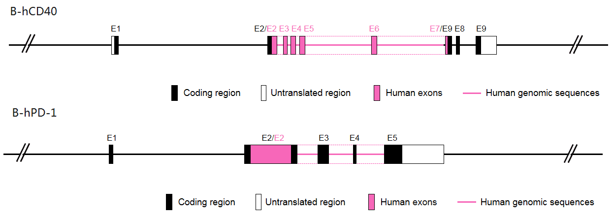
Gene targeting strategy for B-hPD-1/hCD40 mice. The exon 2 of mouse Pd-1 gene that encodes the extracellular domain was replaced by human PD-1 exon 2 in B-hPD-1/hCD40 mice. The exons 2-7 of mouse Cd40 gene that encode the extracellular domain were replaced by human CD40 exons 2-7 in B-hPD-1/hCD40 mice. The B-hPD-1/hCD40 two knock-in model, was developed by breeding the B-hPD-1 mice and the B-hCD40 mice, has a functional mouse immune system.
-
Protein expression analysis
-
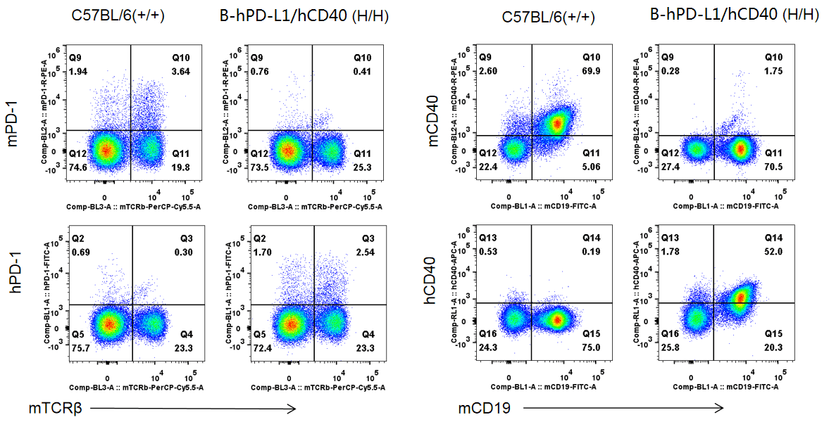
Strain specific CD40 and PD-1 expression analysis in homozygous B-hPD-1/hCD40 mice by flow cytometry. Splenocytes were collected from WT and homozygous B-hPD-1/hCD40 (H/H) mice analyzed by flow cytometry with species-specific anti-PD-1 antibody and anti-CD40 antibody. Mouse CD40 and PD-1 were detected in WT. Human CD40 and PD-1 were exclusively detected in H/H B-hPD-1/hCD40 but not WT mice.
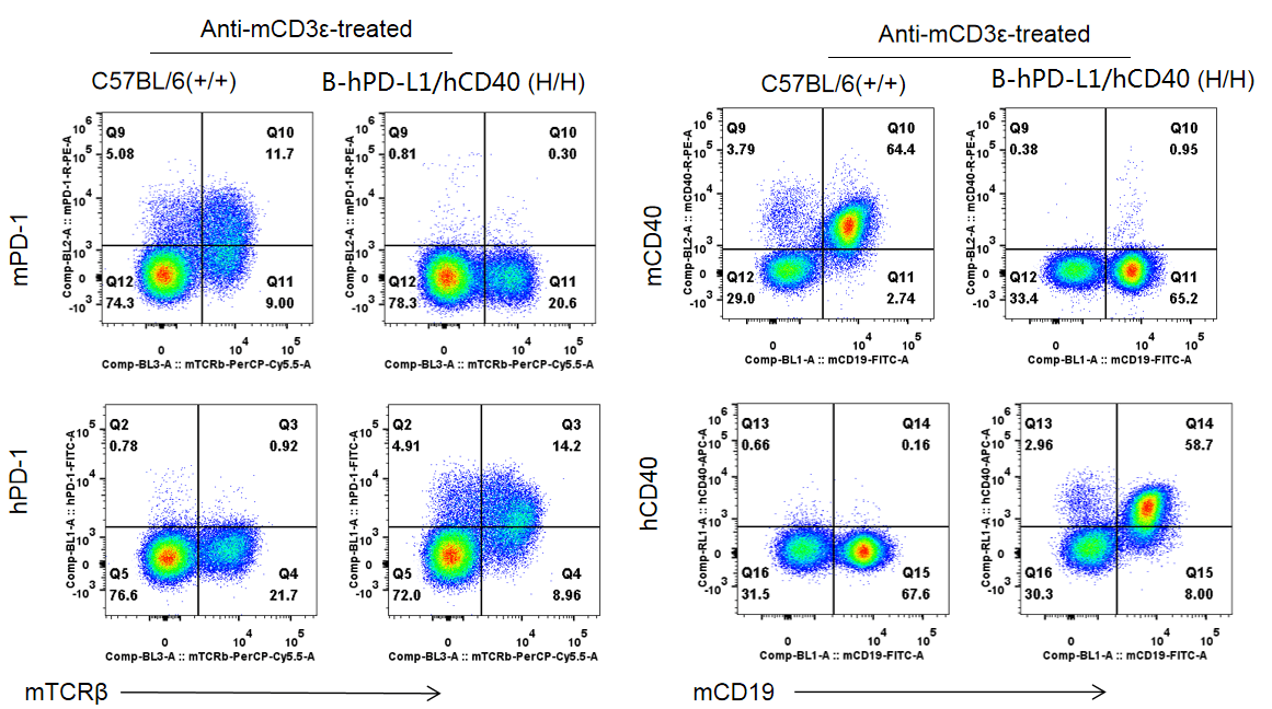
Strain specific CD40 and PD-1 expression analysis in homozygous B-hPD-1/hCD40 mice by flow cytometry. Splenocytes were collected from WT and homozygous B-hPD-1/hCD40 (H/H) mice stimulated with anti-CD3ε in vivo (7.5 μg/mice), and analyzed by flow cytometry with species-specific anti-PD-1 antibody. Mouse CD40 and PD-1 were detected in WT. Human CD40 and PD-1 were exclusively detected in H/H B-hPD-1/hCD40 but not WT mice.
-
Combination therapy of PD-1 mAb (keytruda) and CD40 mAb
-
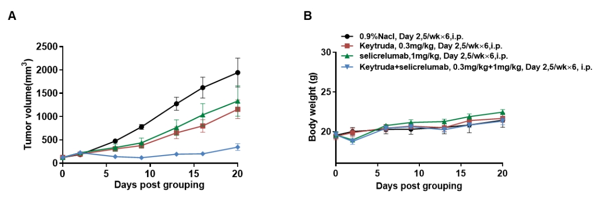
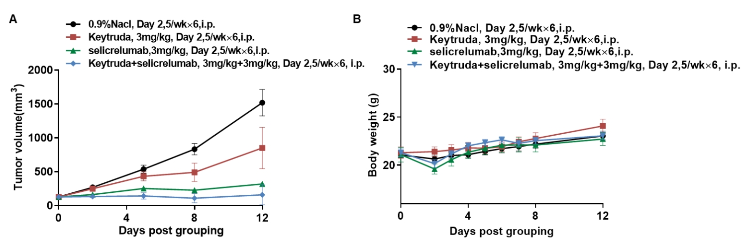
Antitumor activity of anti-hCD40 antibody Selicrelumab (in house) combined with anti-hPD-1 antibody keytruda in B-hPD-1/hCD40 mice. (A) Anti-hCD40 antibody Selicrelumab (in house) combined with anti-hPD-1 antibodies keytruda inhibited MC38-hPD-L1 tumor growth in B-hPD-1/hCD40 mice. Murine colon cancer MC38-hPD-L1 cells (5×10⁵) were subcutaneously implanted into homozygous B-hPD-1/hCD40 mice (female, 6 week-old, n=8). Mice were grouped when tumor volume reached approximately 100-150 mm3, at which time they were treated with antibody Selicrelumab (in house) combined with anti-hPD-1 antibody keytruda with doses and schedules indicated in panel (B) Body weight changes during treatment. As shown in panel A, combination of anti-hCD40 and anti-hPD-1 antibody shows more inhibitory effects than individual groups, demonstrating that the B-hPD-1/hCD40 mice provide a powerful preclinical model for in vivo evaluating combination therapy efficacy of hCD40 antibodies and hPD-1 antibodies . Values are expressed as mean ± SEM.

Antitumor activity of anti-hCD40 antibody Selicrelumab (in house) combined with anti-hPD-1 antibody keytruda in B-hPD-1/hCD40 mice. (A) Anti-hCD40 antibody Selicrelumab (in house) combined with anti-hPD-1 antibodies keytruda inhibited MC38 tumor growth in B-hPD-1/hCD40 mice. Murine colon cancer MC38 cells (5×10⁵) were subcutaneously implanted into homozygous B-hPD-1/hCD40 mice (female, 4 week-old, n=8). Mice were grouped when tumor volume reached approximately 100-150 mm3, at which time they were treated with antibody Selicrelumab (in house) combined with anti-hPD-1 antibody keytruda with doses and schedules indicated in panel (B) Body weight changes during treatment. As shown in panel A, combination of anti-hCD40 and anti-hPD-1 antibody shows more inhibitory effects than individual groups, demonstrating that the B-hPD-1/hCD40 mice provide a powerful preclinical model for in vivo evaluating combination therapy efficacy of hCD40 antibodies and hPD-1 antibodies . Values are expressed as mean ± SEM.
Antitumor activity of anti-hCD40 antibody Selicrelumab (in house) combined with anti-hPD-1 antibody keytruda in B-hPD-1/hCD40 mice. (A) Anti-hCD40 antibody Selicrelumab (in house) combined with anti-hPD-1 antibodies keytruda inhibited B16F10-hPD-L1 tumor growth in B-hPD-1/hCD40 mice. Murine colon cancer B16F10-hPD-L1 cells (5×10⁵) were subcutaneously implanted into homozygous B-hPD-1/hCD40 mice (female, 8 week-old, n=8). Mice were grouped when tumor volume reached approximately 100-150 mm3, at which time they were treated with antibody Selicrelumab (in house) combined with anti-hPD-1 antibody keytruda with doses and schedules indicated in panel (B) Body weight changes during treatment. As shown in panel A, combination of anti-hCD40 and anti-hPD-1 antibody shows more inhibitory effects than individual groups, demonstrating that the B-hPD-1/hCD40 mice provide a powerful preclinical model for in vivo evaluating combination therapy efficacy of hCD40 antibodies and hPD-1 antibodies . Values are expressed as mean ± SEM.
