
Basic Information
-
Targeting strategy

-
Gene targeting strategy for B-hPD-1 plus/hPD-L1 mice. Human PD-1 gene encoding the extracellular region and mouse PD-1 gene encoding the transmembrane and cytoplasmic region were inserted after the initiation codon ATG of mouse PD-1 gene in B-hPD-1 mice plus. The exon 3 of mouse Pdl1 gene that encodes the IgV domain was replaced by human PD-L1 exon 3 in B-hPD-1 plus/hPD-L1 mice. B-hPD-1 plus/hPD-L1 mice was developed by cross-mating the B-hPD-1 mice plus and the B-hPD-L1 mice together.
-
In vivo efficacy of anti-human PD-1 antibody
-
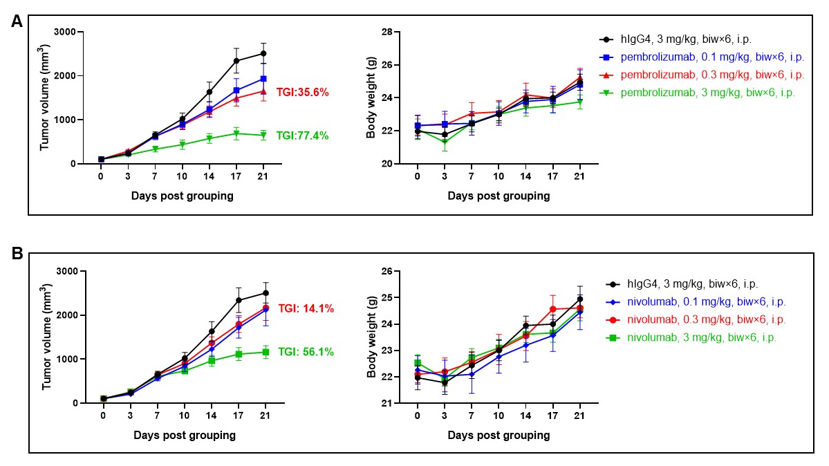
Antitumor activity of anti-human PD-1 antibodies in B-hPD-1 plus/PD-L1 mice. Anti-human PD-1 antibody pembrolizumab (in house) and nivolumab (in house) inhibited the growth of B-hPD-L1 MC38 plus in B-hPD-1 plus/PD-L1 mice. Murine colon cancer B-hPD-L1 MC38 plus cells were subcutaneously implanted into B-hPD-1 plus/PD-L1 mice (female, 8-week-old, n=6). Mice were grouped when tumor volume reached approximately 80-120 mm3, at which time they were treated with anti-human PD-1 antibodies with doses indicated in panel. (A) Tumor volume and body weight of mice treated with pembrolizumab. (B) Tumor volume and body weight of mice treated with nivolumab. Results showed that pembrolizumab and nivolumab were both efficacious in controlling tumor growth in B-hPD-1 plus/PD-L1 mice. The tumor inhibitory effects were dose-dependent. The results demonstrate that the B-hPD-1 plus/PD-L1 mice provide a powerful preclinical model for in vivo evaluation of anti-human PD-1 antibodies. Values are expressed as mean ± SEM.
-
Percentages of leukocytes in CD45+ TILs
-
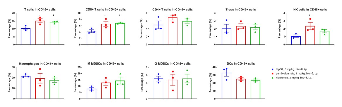
Flow cytometry analysis of tumor infiltrating lymphocytes (TILs). Tumor cells were harvested at the endpoint of experiment (n=3). Flow cytometry analysis of the lymphocytes were performed to assess cell proportion changes in anti-human PD-1 antibodies (in house) treated groups compared to the group treated with isotype antibody. Values are expressed as mean ± SEM.
-
Percentages of leukocyte subpopulations in TILs
-
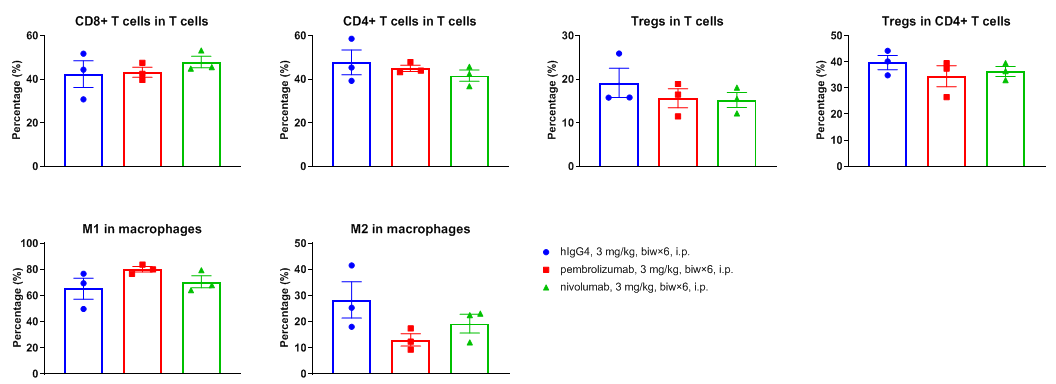
Flow cytometry analysis of tumor infiltrating lymphocytes (TILs). Tumor cells were harvested at the endpoint of experiment (n=3). Flow cytometry analysis of the lymphocytes were performed to assess cell proportion changes in anti-human PD-1 antibodies (in house) treated groups compared to the group treated with isotype antibody. Values are expressed as mean ± SEM.
-
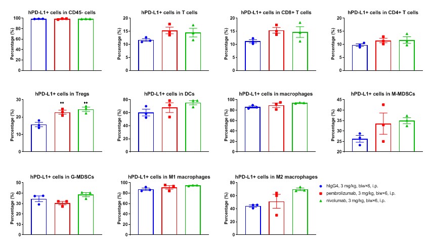
Percentages of hPD-1+ cells in TILs
-
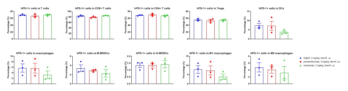
Flow cytometry analysis of tumor infiltrating lymphocytes (TILs). Tumor cells were harvested at the endpoint of experiment (n=3). Flow cytometry analysis of the lymphocytes were performed to assess cell proportion changes in anti-human PD-1 antibodies (in house) treated groups compared to the group treated with isotype antibody. Values are expressed as mean ± SEM.
-
Percentages of hPD-L1+ cells in tumor tissue
-
Flow cytometry analysis of tumor infiltrating lymphocytes (TILs). Tumor cells were harvested at the endpoint of experiment (n=3). Flow cytometry analysis of the lymphocytes were performed to assess cell proportion changes in anti-human PD-1 antibodies (in house) treated groups compared to the group treated with isotype antibody. Values are expressed as mean ± SEM.
-
Percentages of activated cells
-
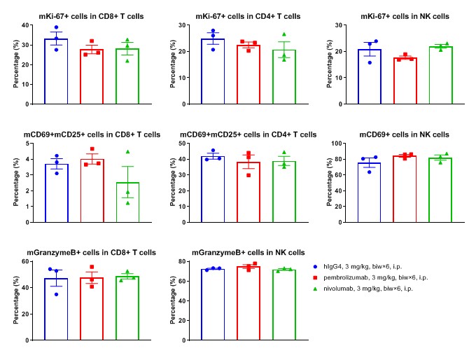
Flow cytometry analysis of tumor infiltrating lymphocytes (TILs). Tumor cells were harvested at the endpoint of experiment (n=3). Flow cytometry analysis of the lymphocytes were performed to assess cell proportion changes in anti-human PD-1 antibodies (in house) treated groups compared to the group treated with isotype antibody. Values are expressed as mean ± SEM.
-
Expression levels of hPD-1 on TILs
-
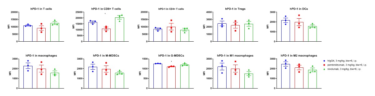
Flow cytometry analysis of tumor infiltrating lymphocytes (TILs). Tumor cells were harvested at the endpoint of experiment (n=3). Flow cytometry analysis of the lymphocytes were performed to assess cell proportion changes in anti-human PD-1 antibodies (in house) treated groups compared to the group treated with isotype antibody. Values are expressed as mean ± SEM.
-
Expression level of hPD-L1 on TILs
-
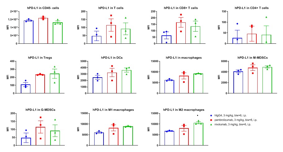
Flow cytometry analysis of tumor infiltrating lymphocytes (TILs). Tumor cells were harvested at the endpoint of experiment (n=3). Flow cytometry analysis of the lymphocytes were performed to assess cell proportion changes in anti-human PD-1 antibodies (in house) treated groups compared to the group treated with isotype antibody. Values are expressed as mean ± SEM.
