
Basic Information
-
Targeting Strategy

-
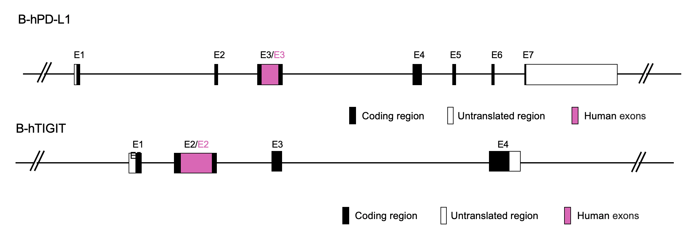
Gene targeting strategy of B-hPD-L1/hTIGIT mice. The exon 3 of mouse Pd-l1 gene that encodes the extracellular domain was replaced by human PD-L1 exon 3 in B-hPD-L1/hTIGIT mice. The exon 2 of mouse Tigit gene that encodes the extracellular domain was replaced by human TIGIT exon 2 in B-hPD-L1/hTIGIT mice.
-
Details
-
Phenotype
Protein Expression Analysis
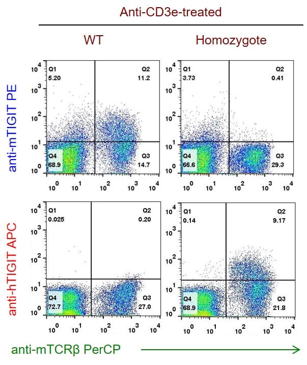
Splenocytes from B-hPD-L1/hTIGIT mice were analyzed by flow cytometry. mTIGIT+ cells were detectable in C57BL/6 mice, while hTIGIT+ cells were detectable in the homozygous B-hPD-L1/hTIGIT mice.
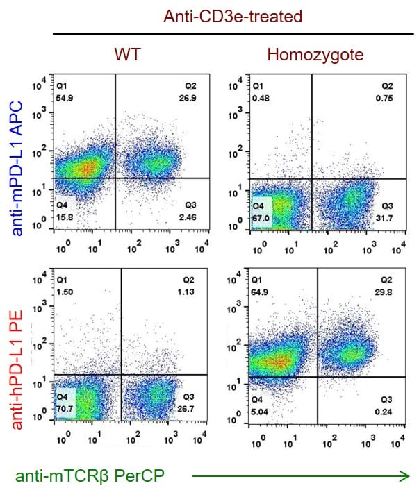
Splenocytes from B-hPD-L1/hTIGIT mice were analyzed by flow cytometry. mPD-L1+ cells were detectable in C57BL/6 mice, while hPD-L1+ cells were detectable in the homozygous B-hPD-L1/hTIGIT mice.
Application
Combination therapy of hTIGIT Ab and PD-L1 Ab
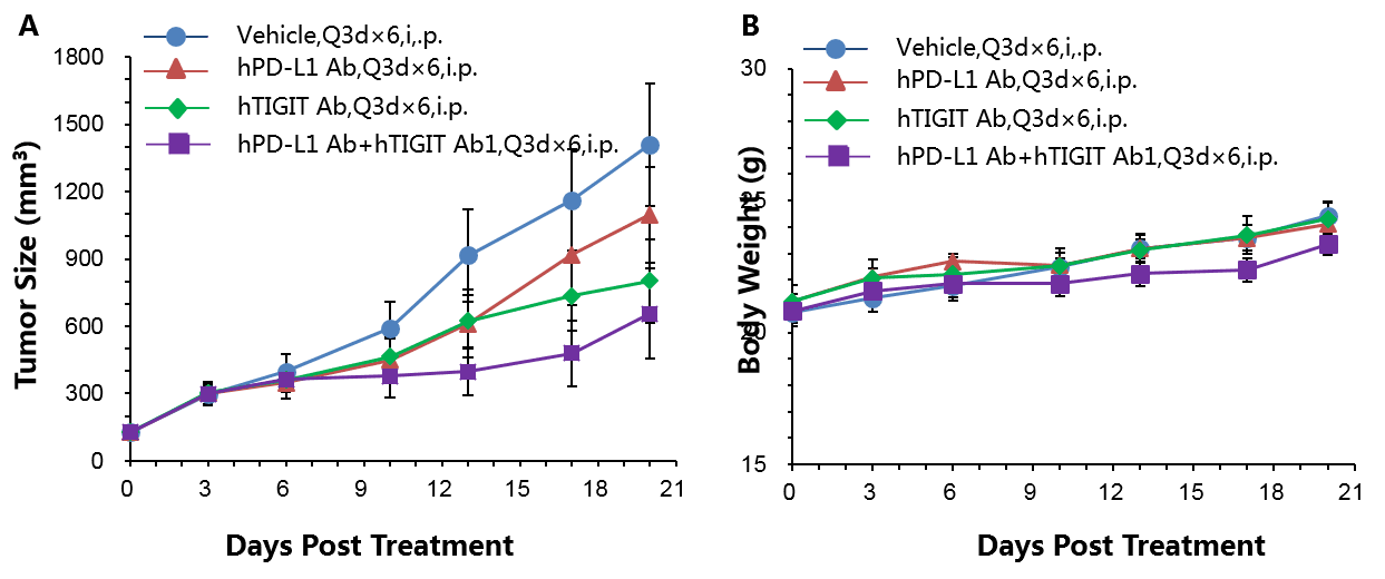
Antitumor activity of anti-hTIGIT antibody combined with anti-hPD-L1 antibody in B-hPD-L1/hTIGIT mice. (A) Anti-hTIGIT antibody combined with anti-hPD-L1 antibody inhibited MC38-hPD-L1 tumor growth in B-hPD-L1/hTIGIT mice. Murine colon cancer MC38-hPD-L1 cells (5×10⁵) were subcutaneously implanted into homozygous B-hPD-L1/hTIGIT mice (female, 5-8 week-old, n=7). Mice were grouped when tumor volume reached approximately 150±50 mm3, at which time they were treated with anti-hTIGIT antibody combined with anti-hPD-L1 antibody and schedules indicated in panel (B) Body weight changes during treatment. As shown in panel A, combination of anti-hTIGIT and anti-hPD-L1 antibody shows more inhibitory effects than individual groups, demonstrating that the B-hPD-L1/hTIGIT mice provide a powerful preclinical model for in vivo evaluating combination therapy efficacy of hTIGIT antibodies and hPD-L1 antibodies . Values are expressed as mean ± SEM.
-
References
-
- J Clin Invest. 2015 May;125(5):2046-58. doi: 10.1172/JCI80445. Epub 2015 Apr 13.
- Immunol Rev. 2017 Mar;276(1):112-120. doi: 10.1111/imr.12518.
- Cancer Cell. 2014 Dec 8;26(6):923-937. doi: 10.1016/j.ccell.2014.10.018. Epub 2014 Nov 26.
- J Exp Med. 2015 Nov 16;212(12):2165-82. doi: 10.1084/jem.20150792. Epub 2015 Nov 9.
