Q1: What are B-hTNFA/hTNFR2/hTNFR1 mice?
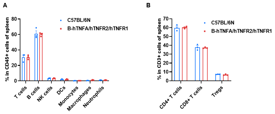
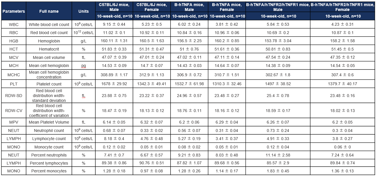
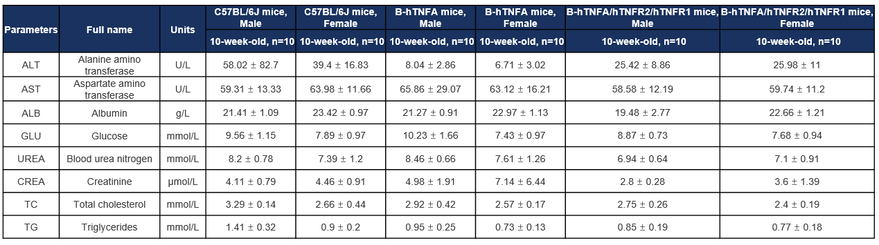
B-hTNFA/hTNFR2/hTNFR1 mice are triple gene-humanized mice expressing human TNFα, humanized TNFR2, and humanized TNFR1 in a C57BL/6 background for TNF pathway drug development.
Q2: Why are TNFα, TNFR1, and TNFR2 important therapeutic targets?
TNFα signaling through TNFR1 and TNFR2 regulates inflammation, immune activation, autoimmunity, tissue injury, and tumor immunity, making this pathway important for inflammatory disease and oncology therapeutics.
Q3: How were TNFα, TNFR1, and TNFR2 validated in this model?
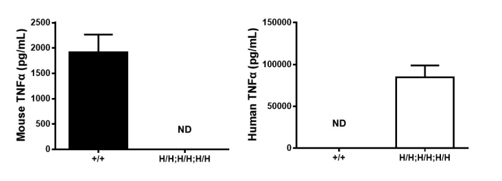
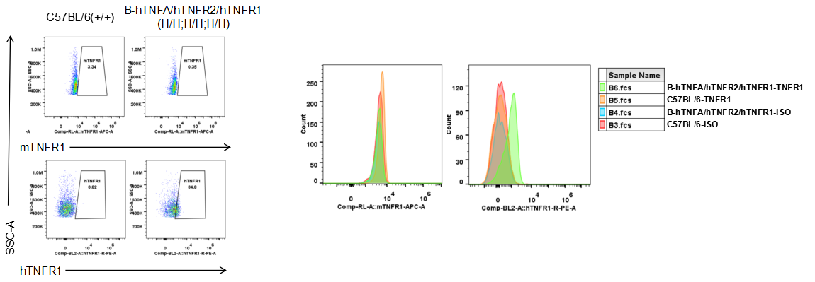
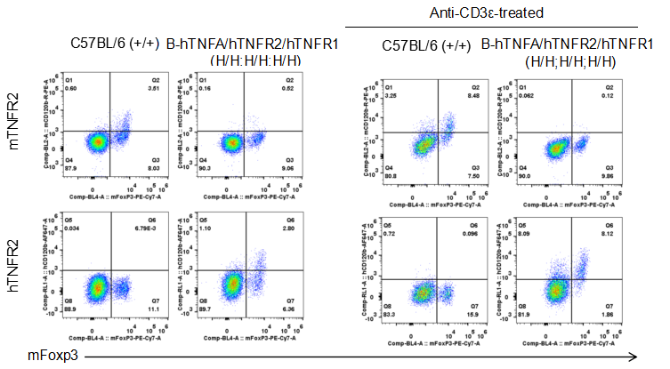
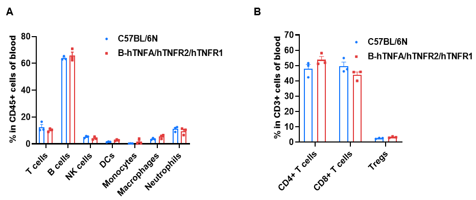
Human TNFα was validated by ELISA after LPS stimulation, while human TNFR1 and human TNFR2 were validated by flow cytometry using species-specific antibodies.
Q4: Can B-hTNFA/hTNFR2/hTNFR1 mice be used for in vivo antibody efficacy studies?
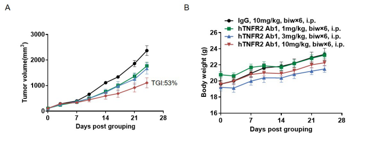
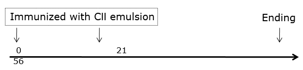
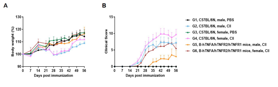
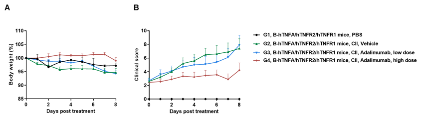
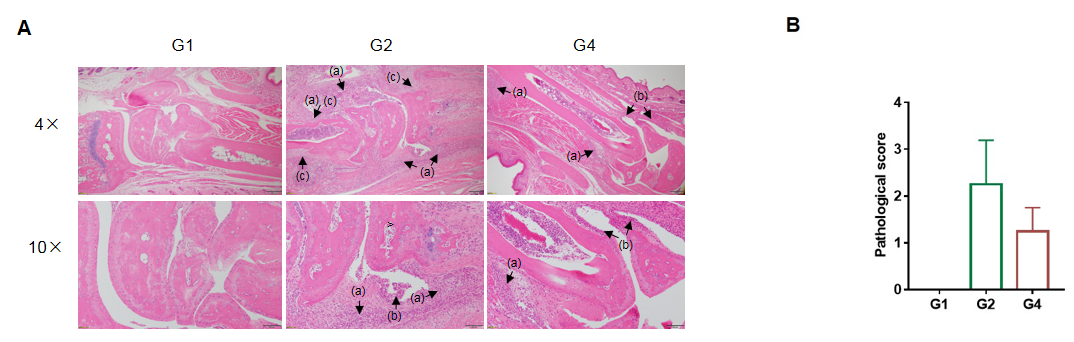
Yes. The model supports adalimumab evaluation in LPS-induced response and CIA arthritis models, and anti-human TNFR2 antibody evaluation in MC38 tumor-bearing mice.
Q5: What are the main applications of B-hTNFA/hTNFR2/hTNFR1 mice?
Applications include anti-human TNFα antibody studies, anti-human TNFR2 antibody research, TNFR1/TNFR2 pathway pharmacology, LPS inflammation models, MC38 tumor studies, and collagen-induced arthritis drug development.