
Basic Information
Description
The mouse Pdl1 gene was replaced by human PD-L1 coding sequence in B-hPD-L1 plus/hHER2 MC38 cells. Human PD-L1 is highly expressed on the surface of B-hPD-L1 plus/hHER2 MC38 cells.
The mouse Erbb2 gene was replaced by the coding sequence composed of human ERBB2 extracellular domain, mouse ERBB2 transmembrane domain and mouse ERBB2 intracellular domain in B-hPD-L1 plus/hHER2 MC38 cells. Human ERBB2 is highly expressed on the surface of B-hPD-L1 plus/hHER2 MC38 cells.
-
Targeting strategy

-
Gene targeting strategy for B-hPD-L1 plus/hHER2 MC38 cells.
The exogenous promoter and human PD-L1 coding sequence was inserted to replace part of murine exon 3. The insertion disrupts the endogenous murine Pdl1 gene, resulting in a non-functional transcript.
The exogenous CAG promoter and the coding sequence composed of human ERBB2 extracellular domain, mouse ERBB2 transmembrane domain and mouse ERBB2 intracellular domain were inserted to replace part of murine exon 2 and all of exons 3-7. The insertion disrupts the endogenous murine Erbb2 gene, resulting in a non-functional transcript.
-
Protein Expression Analysis
-
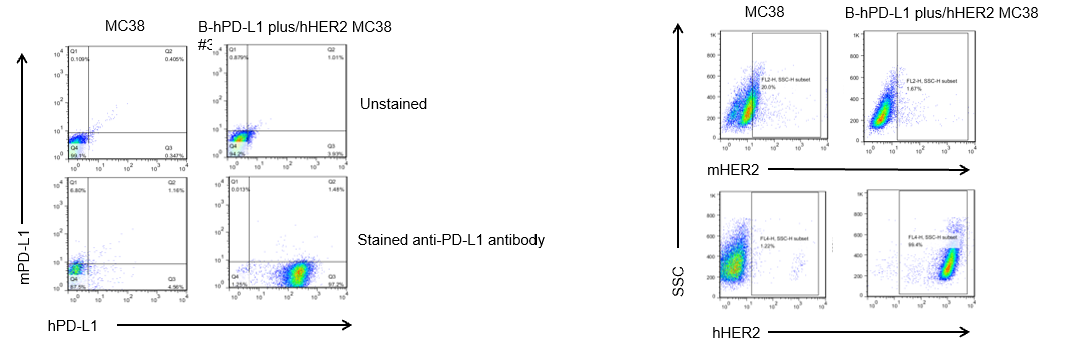
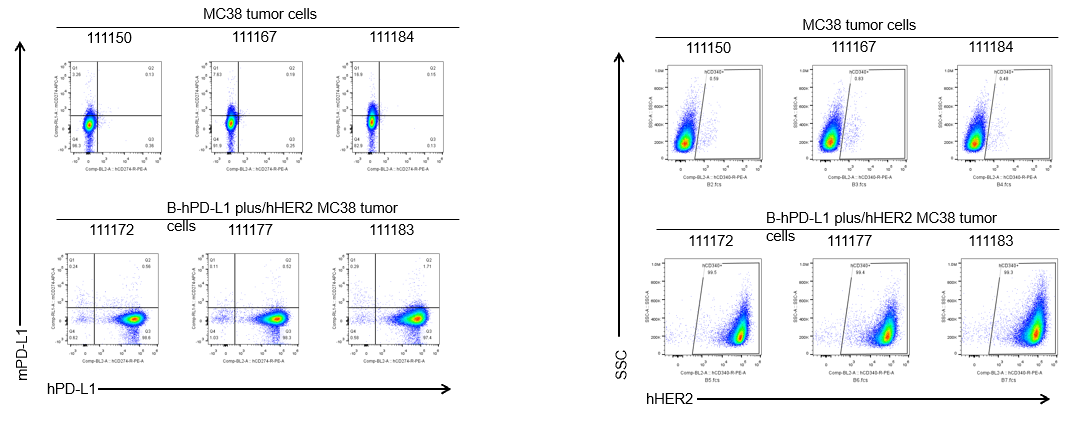
PD-L1 and HER2 expression analysis in B-hPD-L1 plus/hHER2 MC38 cells by flow cytometry.
Single cell suspensions from wild-type MC38 and B-hPD-L1 plus/hHER2 MC38 cultures were stained with species-specific anti-PD-L1 and anti-HER2 antibody. Human PD-L1 and HER2 were detected on the surface of B-hPD-L1 plus/hHER2 MC38 cells but not wild-type MC38 cells. The 3-B08 clone of B-hPD-L1 plus/hHER2 MC38 cells was used for in vivo tumor growth assays.
-
Tumor growth curve & Body weight changes
-
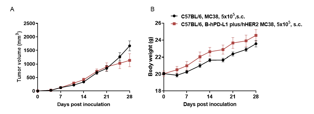
Subcutaneous homograft tumor growth of B-hPD-L1 plus/hHER2 MC38 cells.
B-hPD-L1 plus/hHER2 MC38 cells (5×10⁵) and wild-type MC38 cells (5×10⁵) were subcutaneously implanted into C57BL/6 mice (female, 9-week-old, n=6). Tumor volume and body weight were measured twice a week. (A) Average tumor volume ± SEM. (B) Body weight (Mean ± SEM). Volume was expressed in mm3 using the formula: V=0.5 X long diameter X short diameter2. As shown in panel A, B-hPD-L1 plus/hHER2 MC38 cells were able to form tumors in vivo and can be used for efficacy studies.
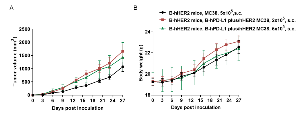
Subcutaneous homograft tumor growth of B-hPD-L1 plus/hHER2 MC38 cells.
B-hPD-L1 plus/hHER2 MC38 cells (2×10⁵, 5×10⁵) and wild-type MC38 cells (5×10⁵) were subcutaneously implanted into homozygous B-hHER2 mice (female, 5-week-old, n=6). Tumor volume and body weight were measured twice a week. (A) Average tumor volume ± SEM. (B) Body weight (Mean ± SEM). Volume was expressed in mm3 using the formula: V=0.5 X long diameter X short diameter2. As shown in panel A, B-hPD-L1 plus/hHER2 MC38 cells were able to form tumors in vivo and can be used for efficacy studies.
-
Protein expression analysis of tumor cells
-
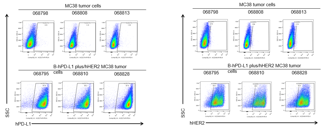
B-hPD-L1 plus/hHER2 MC38 cells were subcutaneously transplanted into C57BL/6 mice (n=6). At the end of the experiment, tumor cells were harvested and assessed for human PD-L1 and HER2 expression by flow cytometry. As shown, human PD-L1 and HER2 were highly expressed on the surface of tumor cells. Therefore, B-hPD-L1 plus/hHER2 MC38 cells can be used for in vivo efficacy studies of PD-L1 and HER2 therapeutics.
B-hPD-L1 plus/hHER2 MC38 cells were subcutaneously transplanted into homozygous B-hHER2 mice (n=6). At the end of the experiment, tumor cells were harvested and assessed for human PD-L1 and HER2 expression by flow cytometry. As shown, human PD-L1 and HER2 were highly expressed on the surface of tumor cells. Therefore, B-hPD-L1 plus/hHER2 MC38 cells can be used for in vivo efficacy studies of PD-L1 and HER2 therapeutics.
