
Basic Information
Description
This B-luc Daudi cell line expresses firefly luciferase as a marker of Daudi cells. Luminescence can be observed B-luc Daudi cells.
-
Targeting Strategy

-
Gene targeting strategy for B-luc Daudi. The luciferase cDNA sequence with CAG promoter was inserted in the AAVS1 locus intron1 of wild-type Daudi .
-
Phenotypic Analysis
-
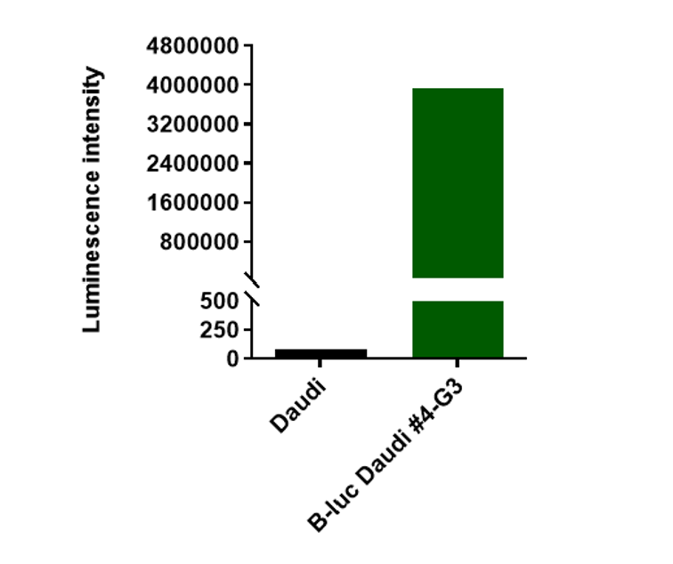
Luminescence signal intensity of B-luc Daudi cells. Luminescence intensity was measured using the Bright-GloTM luciferase Assay (Promega, Cat E2610). B-luc Daudi cells have a strong luminescence signal. The 4-G3 clone of B-luc Daudi cells can be used in vivo experiments.
-
Tumor Growth Curve & Body Weight Changes
-
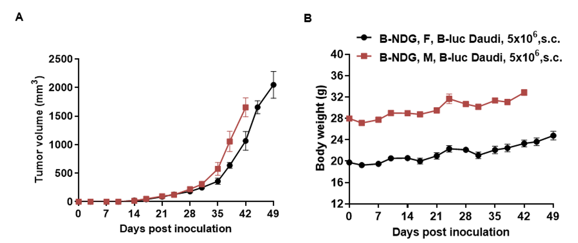
Subcutaneous xenograft tumor growth of B-luc Daudi cells. B-luc Daudi cells (5×10⁶) were subcutaneously implanted into the B-NDG mice (female and male, 7-week-old, n=8).Tumor size and mice body weight were measured twice a week. (A) Tumor average volume ± SEM, (B) Mice body weight (Mean± SEM). Volume was expressed in mm3 using the formula: V=0.5a X b2, where a and b were the long and short diameters of the tumor, respectively. As shown in panel A, B-luc Daudi were able to establish tumor in vivo and can be used for efficacy study.
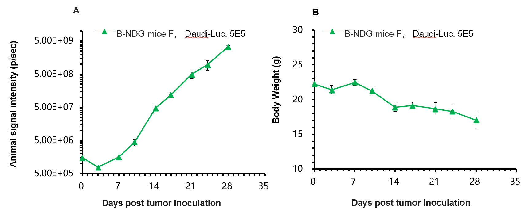
A. B-luc Daudi cells were inoculated into the tail vein of B-NDG mice, and imaging was performed on days 0, 3, 7, 10, 14, 17, 21, 24, and 28 for detection and tumor imaging signal intensity analysis
B. Animal weight monitoring after B-NDG mice were inoculated with B-luc Daudi cells.
Results: B-luc Daudi cells were able to establish tumor model steadily and track tumor volume effectively .
