Basic Information
-
mRNA expression analysis
-
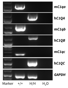
Strain-specific analysis of C1QA, C1QB, and C1QC gene expression in wild-type C57BL/6 mice and homozygous B-hC1Q mice by RT-PCR. Thymus were collected from wild-type C57BL/6 mice (+/+) and homozygous B-hC1Q mice (H/H). Mouse C1qa, C1qb, and C1qc mRNA were detectable in the thymus of wild-type C57BL/6 mice. Human C1QA, C1QB, and C1QC mRNA were detectable only in homozygous B-hC1Q mice but not in wild-type mice.
-
Protein expression analysis
-
Strain-specific C1Q expression analysis in wild-type C57BL/6 mice and homozygous B-hC1Q mice by ELISA. Serum was collected from wild-type C57BL/6 mice (+/+) (female, n=4, 8 weeks old; male, n=4, 8 weeks old) and homozygous B-hC1Q mice (H/H) (female, n=4, 8 weeks old; male, n=4, 8 weeks old). Mouse C1Q was only detectable in wild-type mice. C1Q was detectable in wild-type C57BL/6 mice and homozygous B-hC1Q mice. Therefore, it is speculated that this anti-human C1Q antibody is cross-reactive between human and mice. Values are expressed as mean ± SEM. Significance was determined by unpaired t-test. ns: non-significant, *p < 0.05, **p< 0.01, ***p < 0.001.
-
Functional activity of the classical pathway
-
Functional activity of the classical pathway of homozygous B-hC1Q mice. Serum was collected from wild-type C57BL/6JNifdc mice (+/+) (male, n=3, 7 weeks old; female, n=3, 7 weeks old) and homozygous B-hC1Q mice (H/H) (male, n=5, 7 weeks old; female, n=5, 7 weeks old) and evaluate the functional activity with the complement kit (Classical Complement Pathway Mouse Assay, Hycult Biotech, HIT420). B-hC1Q mice showed consistent complement activity compared to wild-type C57BL/6JNifdc mice. Values are expressed as mean ± SEM. Significance was determined by Ordinary one-way ANOVA, ns: non-significant, *p < 0.05, **p< 0.01, ***p < 0.001.
-
In vivo efficacy of anti-human CD38 antibodies in B-hC1Q mice
-
Antitumor activity of anti-human CD38 antibody in B-hC1Q mice. (A) Anti-human CD38 antibody inhibited B-hCD38-luc E.G7-OVA lymphoma growth in B-hC1Q mice. B-hCD38-luc E.G7-OVA cells (1×10⁴) were injected by tail vein into B-hC1Q mice (male, 8 week-old, n=10). Mice were grouped on the day of tail vein injection, and treated with anti-hCD38 antibody (i.p., 90 μg/mouse) or mIgG1 (i.p., 100 μg/mouse) in panel A. (B) Body weight changes during treatment. (C) Raw bioluminescence images. As shown in panel A, the anti-hCD38 antibody Daratumumab (from a collaborator) was efficacious in controlling tumor growth in B-hC1Q mice. Values are expressed as mean ± SEM. (Note: A collaboration data shared by a client.)
-
In vivo efficacy of anti-human CD38 antibodies in B-hC1Q mice individual tumor growth curves
-
Antitumor activity of anti-human CD38 antibody in B-hC1Q mice. Anti-human CD38 antibody inhibited B-hCD38-luc E.G7-OVA lymphoma growth in B-hC1Q mice. B-hCD38-luc E.G7-OVA cells (1×10⁴) were injected by tail vein into B-hC1Q mice (male, 8 week-old, n=10). Mice were grouped on the day of tail vein injection, and treated with anti-hCD38 antibody (from a collaborator). (A) Individual tumor growth curves in control group in B-hC1Q mice. (B) Individual tumor growth curves in Daratumumab (from a collaborator) group in B-hC1Q mice. Values are expressed as mean ± SEM. (Note: A collaboration data shared by a client.)
-
Tumor growth curve & Body weight changes
-
Growth kinetics of B-hCD38-luc E.G7-OVA tumors determined by bioluminescence imaging (BLI). B-hCD38-luc E.G7-OVA cells (1×10⁵, 2×10⁵, 5×10⁵) were injected into the tail vein of B-hC1Q mice (male, 8-week-old, n=6). Signal intensity and body weight were measured twice a week. (A) Signal intensity. (B) Body weight (Mean ± SEM). (C) Raw bioluminescence images. These results indicate that B-hCD38-luc E.G7-OVA cells can be used for in vivo efficacy evaluation. Values are expressed as mean ± SEM. (Note: A collaboration data shared by a client.)