

C57BL/6N-B2mtm2(B2M/HLA-A2.1/H2-D)Bcgen/Bcgen • 110110

Key Advantages
Validation
Application

In B-HLA-A2.1 mice, mouse B2m exons 1-3 were replaced by sequences encompassing human B2M CDS and the HLA-A*0201 gene. The inserted HLA-A2.1 sequence includes the leader sequence, α1 and α2 domains, and a fragment of murine H-2Db containing the α3, transmembrane, and cytoplasmic domains.

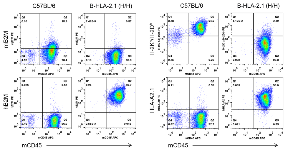
Strain-specific B2M and HLA expression analysis in homozygous B-HLA-A2.1 mice by flow cytometry. Splenocytes from wild-type C57BL/6N (+/+) and homozygous B-HLA-A2.1 mice (H/H) were analyzed with anti-mouse B2M (BioLegend, 154503), anti-human B2M (BioLegend, 316305), anti-H-2Kb/H-2Db (BioLegend, 114607), and anti-HLA-A2 (BioLegend, 343305). Mouse B2M and H-2Kb/H-2Db were detected in wild-type mice, while human B2M and HLA-A2.1 were detected in homozygous B-HLA-A2.1 mice.
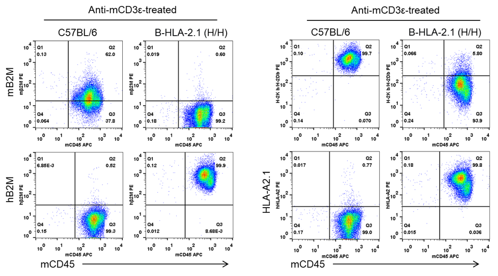
Splenocytes from wild-type C57BL/6N (+/+) and homozygous B-HLA-A2.1 mice (H/H) were analyzed after anti-CD3ε stimulation in vivo. B2M and HLA expression were analyzed by flow cytometry with species-specific antibodies. Human B2M and HLA-A2.1 expression was detected in homozygous B-HLA-A2.1 mice after anti-mCD3ε stimulation.
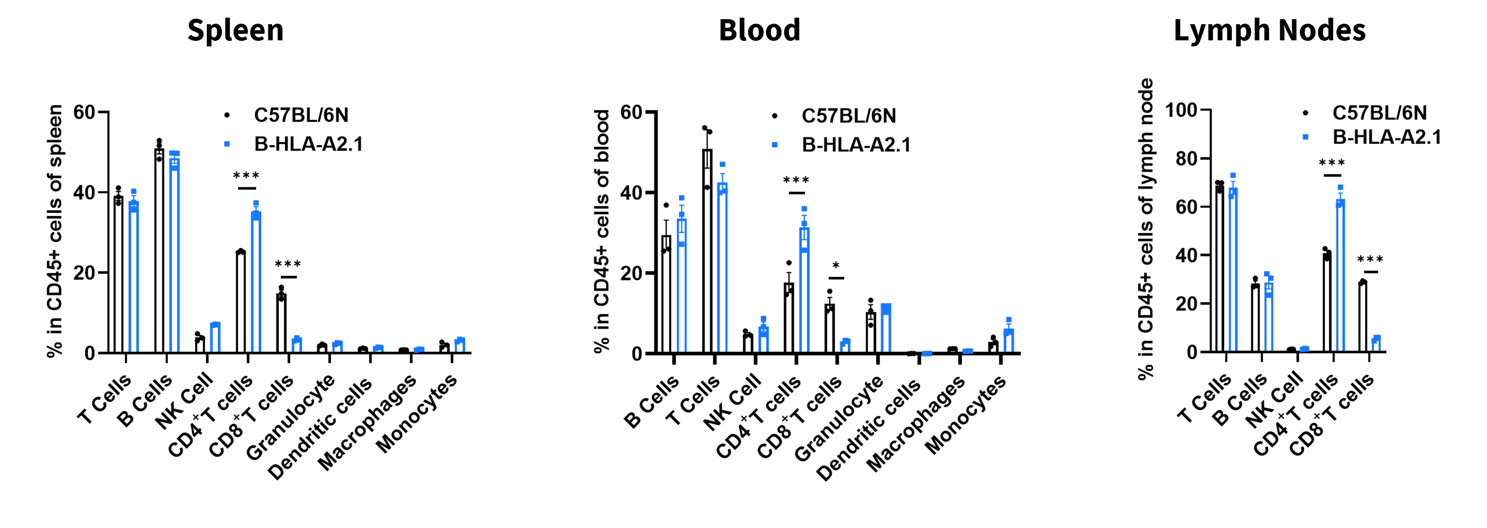
Leukocyte subpopulations were analyzed by flow cytometry in spleen, peripheral blood, and lymph nodes from female C57BL/6N and B-HLA-A2.1 mice. B cells, NK cells, DCs, neutrophils, monocytes, granulocytes, and macrophages were similar between strains. CD8+ T-cell frequency was significantly decreased and CD4+ T-cell frequency was significantly increased.

T-cell subpopulations were analyzed by flow cytometry in spleen, peripheral blood, and lymph nodes from female C57BL/6N and B-HLA-A2.1 mice. Treg proportions were comparable between strains, while CD8+ T-cell frequency was significantly decreased and CD4+ T-cell frequency was significantly increased.
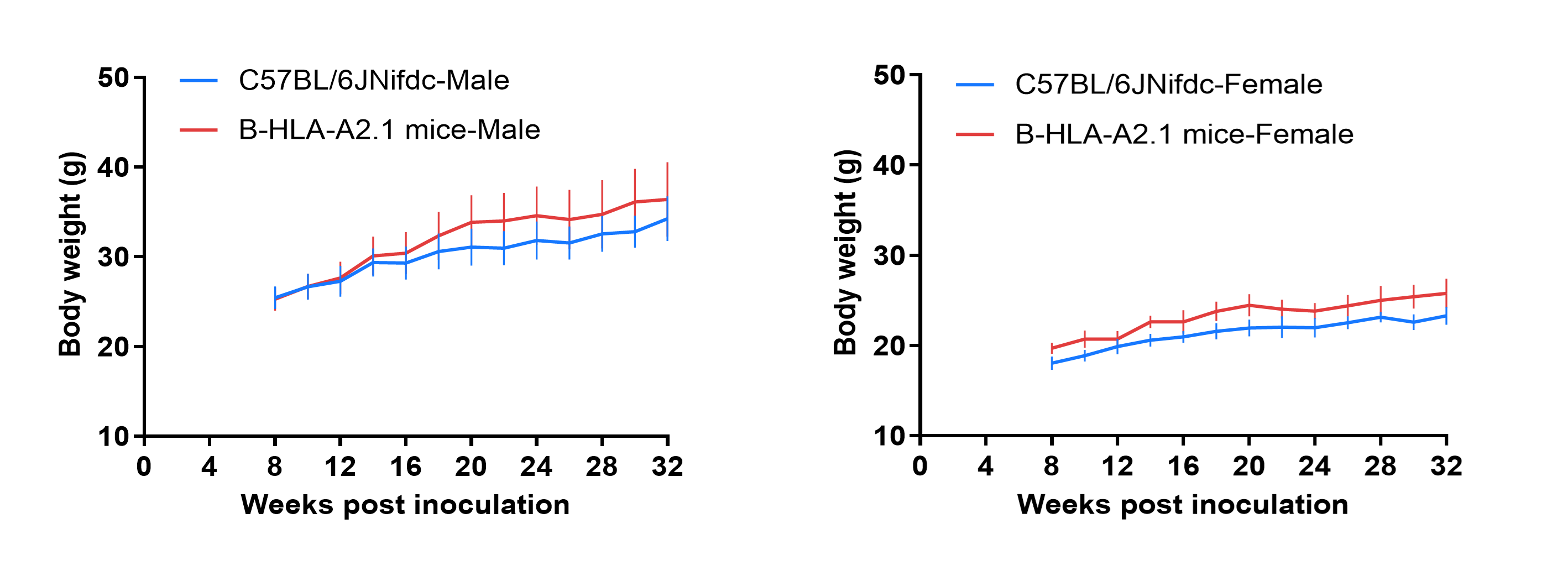
Growth curve of wild-type C57BL/6JNifdc and B-HLA-A2.1 mice. Eight-week-old mice were grouped by sex (10 males and 10 females), and body weight was measured every two weeks until 32 weeks. Growth of B-HLA-A2.1 mice was similar to C57BL/6JNifdc mice.
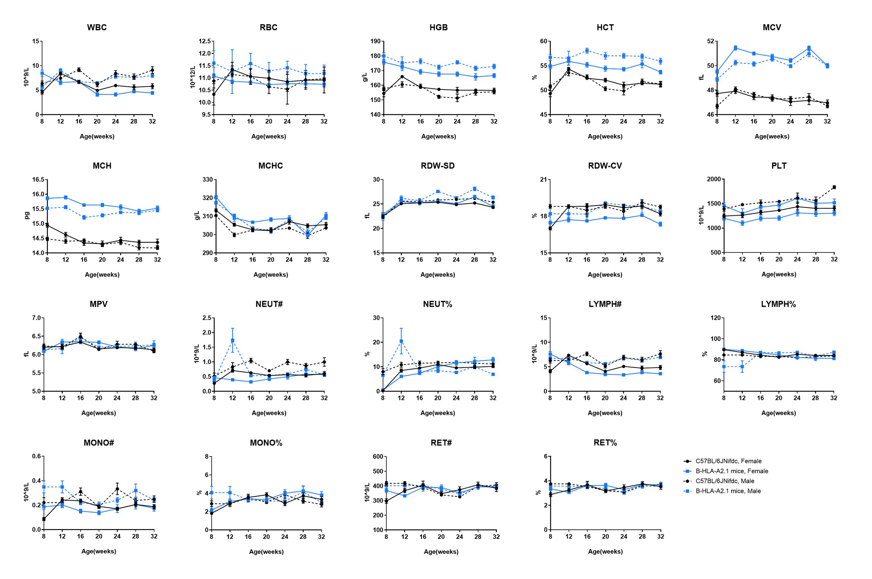
Complete blood count (CBC) of B-HLA-A2.1 mice. Hematology parameters were evaluated as part of baseline physiological characterization.
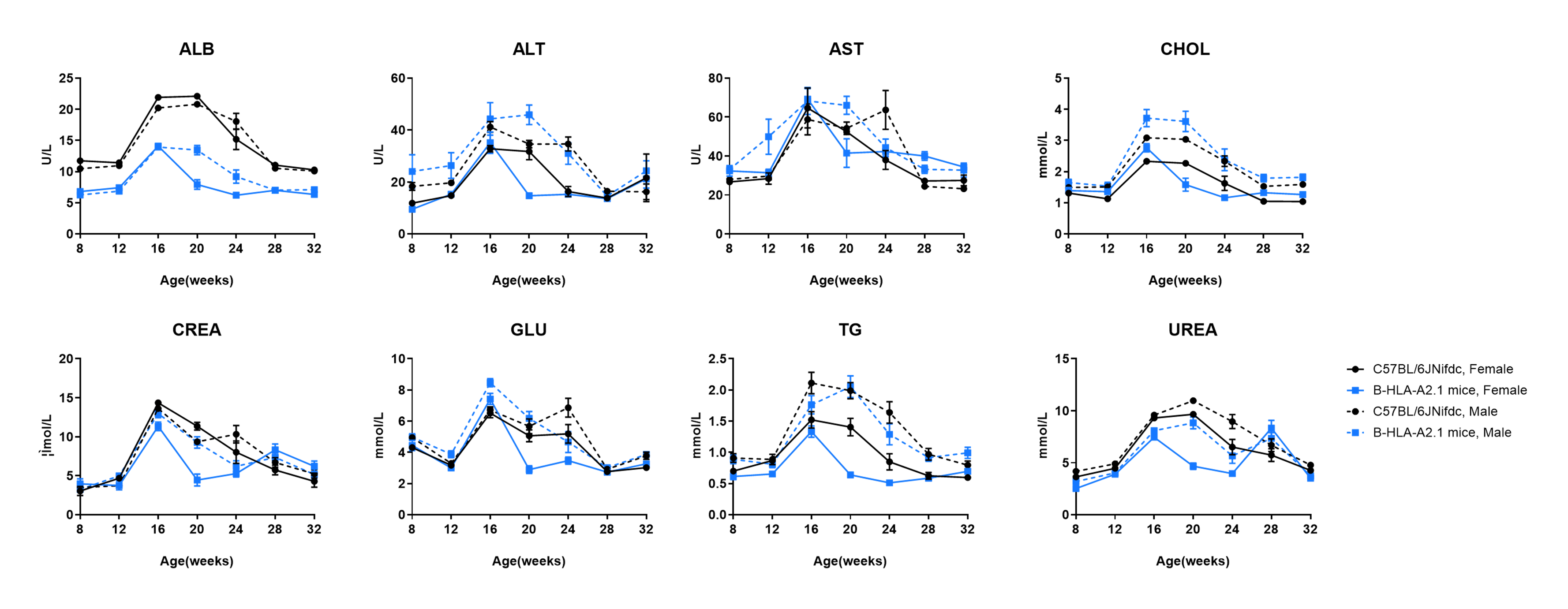
Blood biochemical parameters of B-HLA-A2.1 mice. No significant differences were observed compared with wild-type mice.
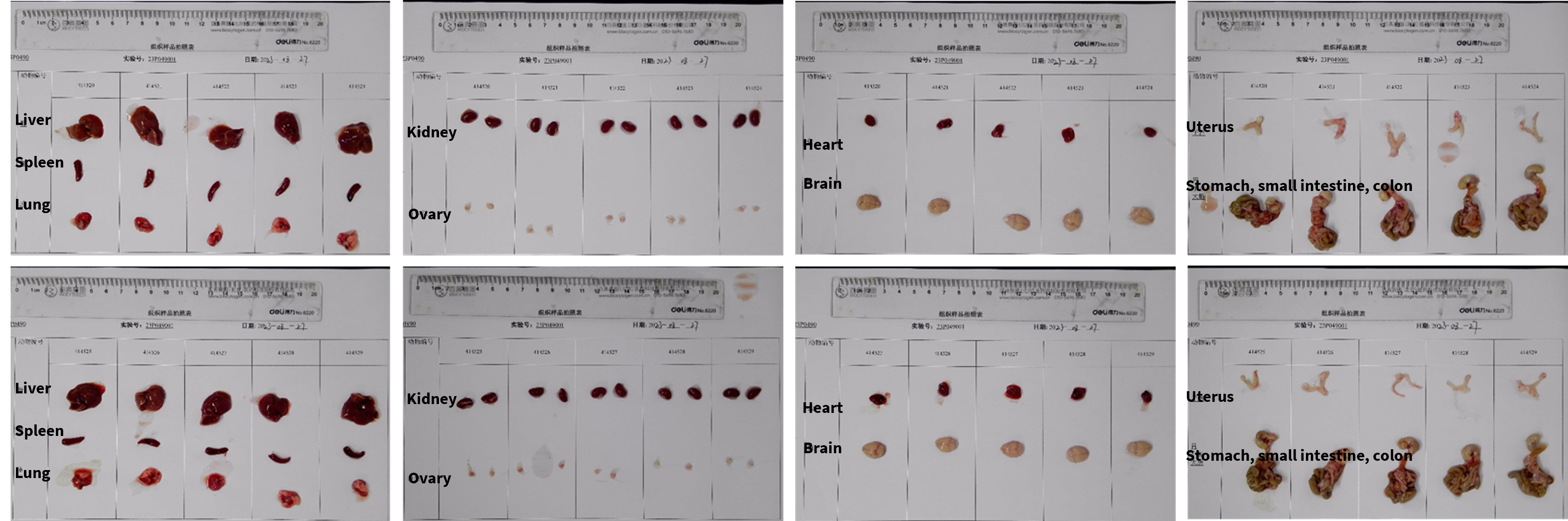
Gross organ anatomy of female B-HLA-A2.1 mice. Organs were collected from female B-HLA-A2.1 mice at 8 weeks of age (n=10). No abnormalities were observed.
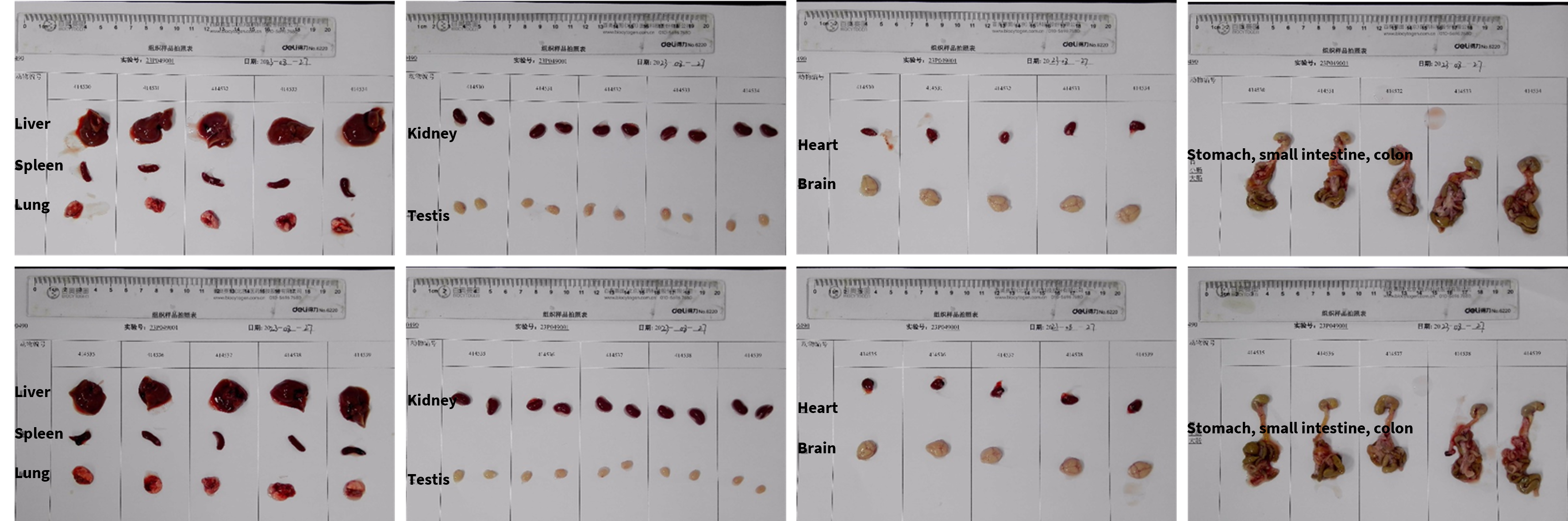
Gross organ anatomy of male B-HLA-A2.1 mice. Organs were collected from male B-HLA-A2.1 mice at 8 weeks of age (n=10). No abnormalities were observed.
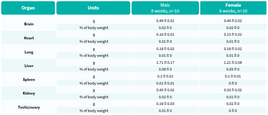
Average weights of major organs in B-HLA-A2.1 mice. Organ weight analysis was performed as part of baseline physiological characterization. No abnormalities were observed.
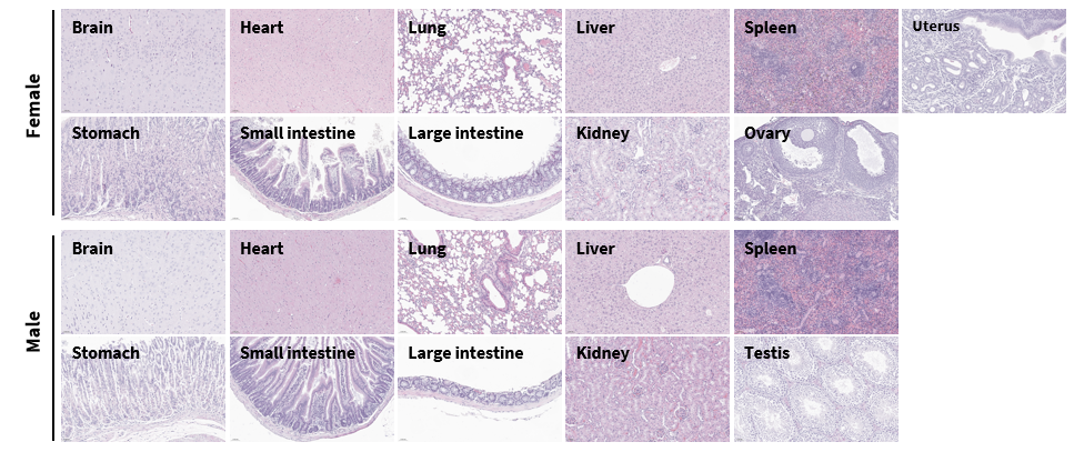
Histopathological analysis of organs in B-HLA-A2.1 mice. Major organs from B-HLA-A2.1 mice were collected at 32 weeks and analyzed by H&E staining (male, n=10; female, n=10). No obvious abnormalities were observed in the examined organs.
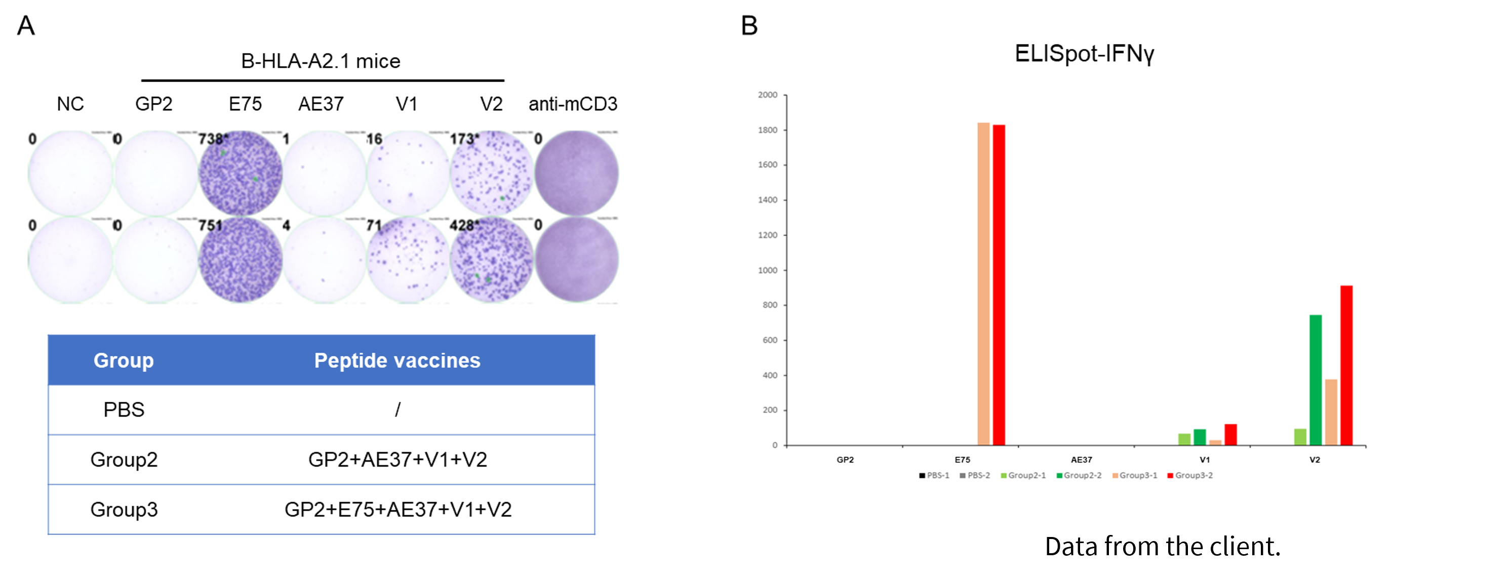
Detection of vaccine-induced immune responses in B-HLA-A2.1 mice by IFN-γ ELISpot assay. Female B-HLA-A2.1 mice aged 9-10 weeks were divided into PBS, Group 2, and Group 3, and inoculated with PBS or HER2-targeted peptide vaccines. Splenocytes were stimulated with individual peptide, negative control peptide, or anti-CD3 positive control, then measured for IFN-γ secretion. No significant body-weight difference was shown.
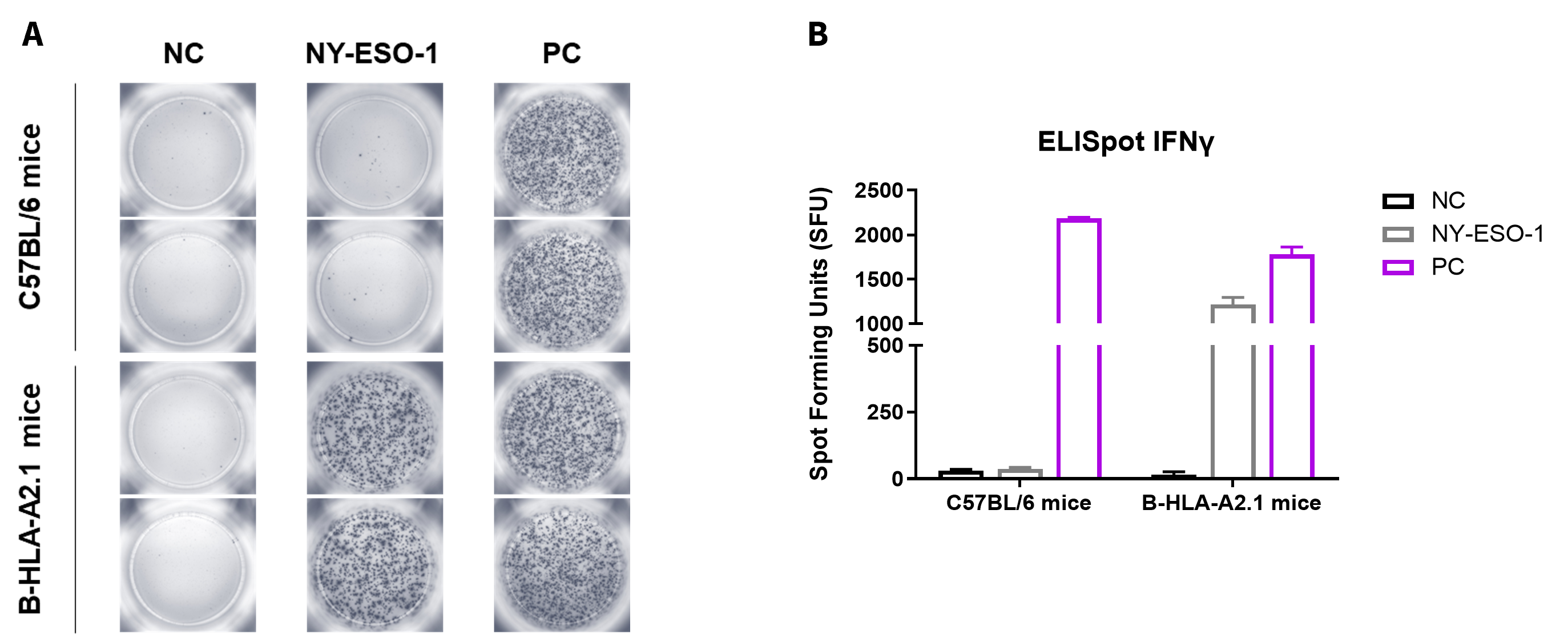
Detection of vaccine-induced immune responses in B-HLA-A2.1 mice by IFN-γ ELISpot assay. Male B-HLA-A2.1 mice aged 9-10 weeks were divided into PBS and NY-ESO-1 groups (n=3). One week after the last immunization, splenocytes were stimulated with individual peptide, negative control peptide, or PMA/Ionomycin positive control and measured for IFN-γ secretion.

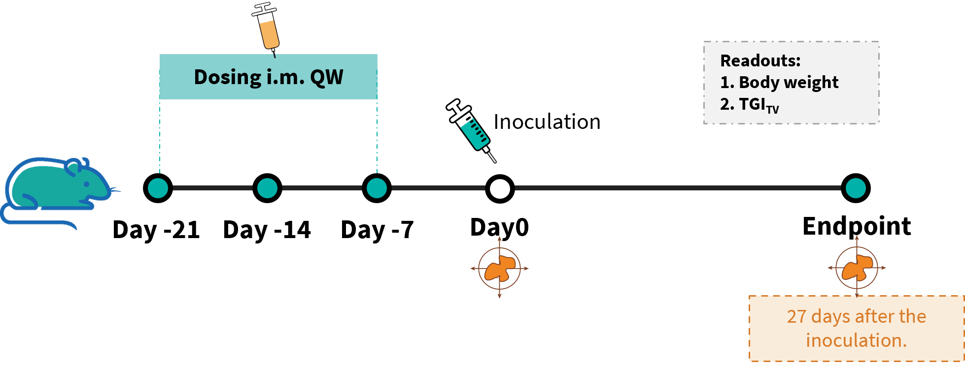
In vivo efficacy study of an anti-human WT1 peptide in a B-HLA-A2.1/hWT1 MC38 model. B-HLA-A2.1/hWT1 MC38 cells were implanted subcutaneously into homozygous B-HLA-A2.1 mice (male, 8-weeks-old, n=8).
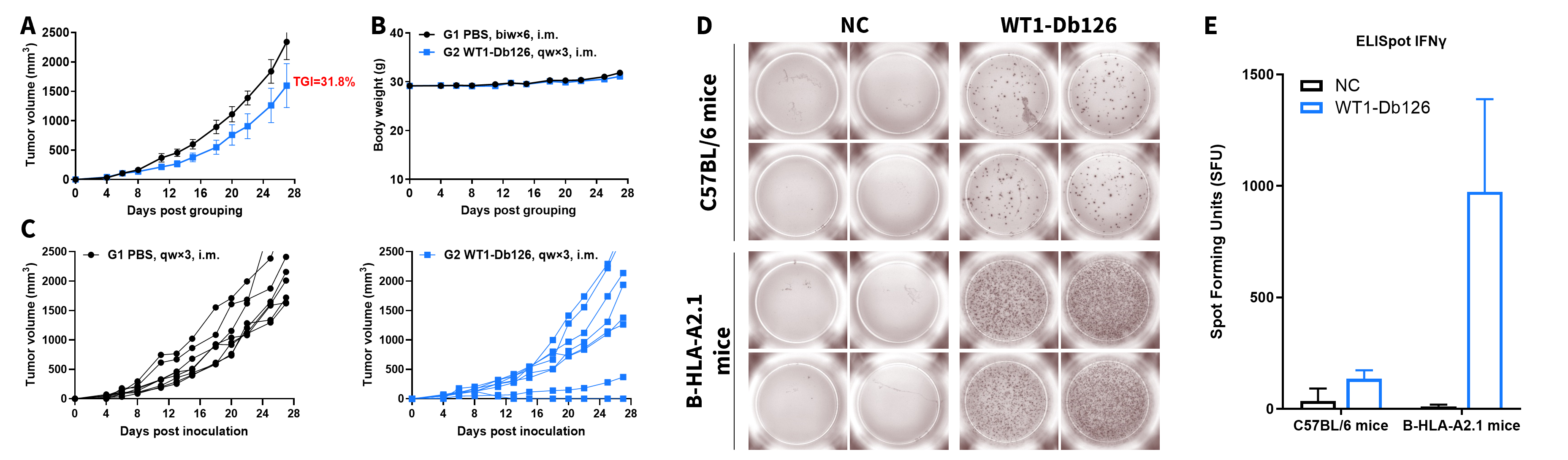
Antitumor activity of WT1-Db126 against syngeneic tumors. Readouts include tumor growth curves, body weight changes, individual tumor growth, ELISpot stimulation results, and ELISpot quantification of IFN-γ-secreting cells. B-HLA-A2.1 mice provide a preclinical model for vaccine evaluation. The overage of this tumor model is 40%.
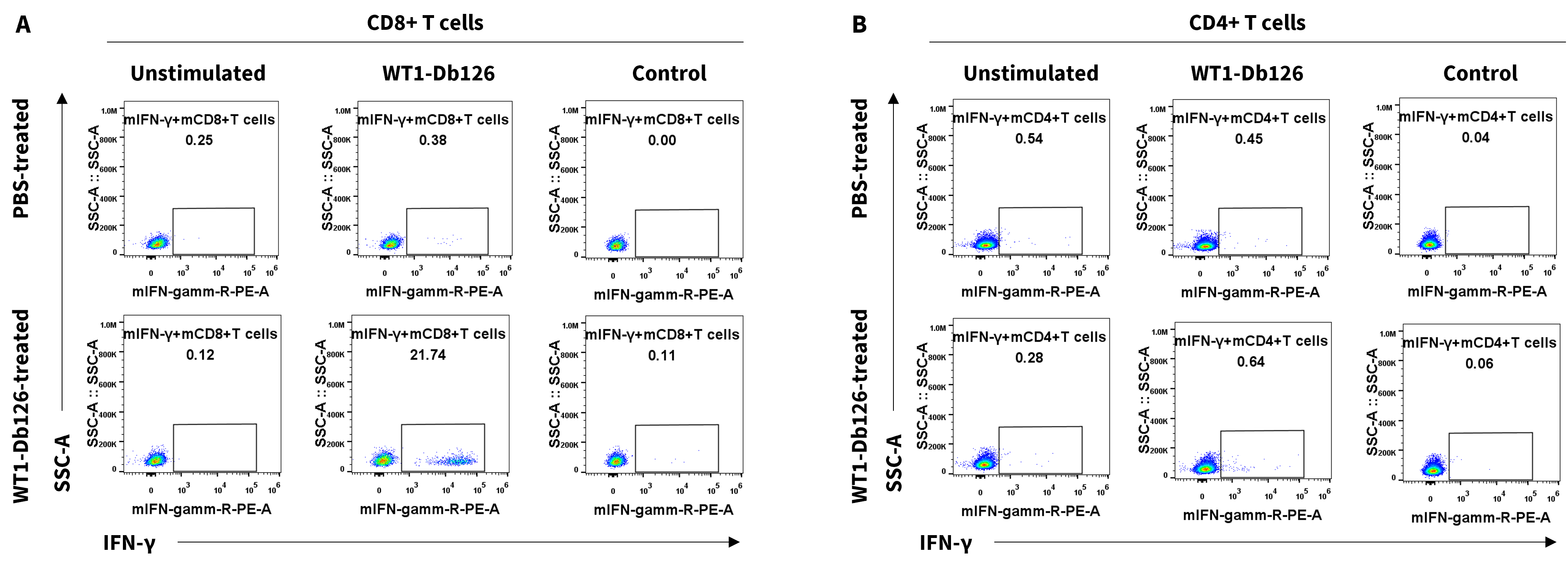
T-cell intracellular cytokine staining (ICS) assays in B-HLA-A2.1 mice. FACS plots demonstrated WT1-Db126-specific CD4+ and CD8+ T cells. IFN-γ was produced by CD8+ T cells but not CD4+ T cells.
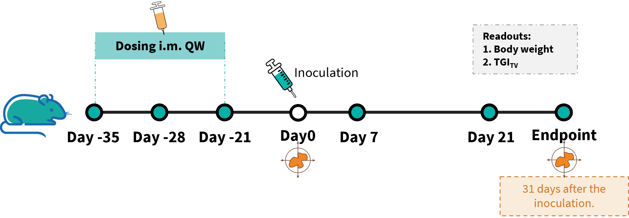
In vivo efficacy study of anti-human NY-ESO-1 peptides in B-HLA-A2.1/hNY-ESO-1 MC38 model . B-HLA-A2.1/hNY-ESO-1 MC38 cells were implanted subcutaneously into homozygous B-HLA-A2.1 mice.

Antitumor activity of NY-ESO-1 peptides against syngeneic tumors. Readouts include tumor growth curves, body weight changes, and individual tumor growth. The overage of this tumor model is 40%.
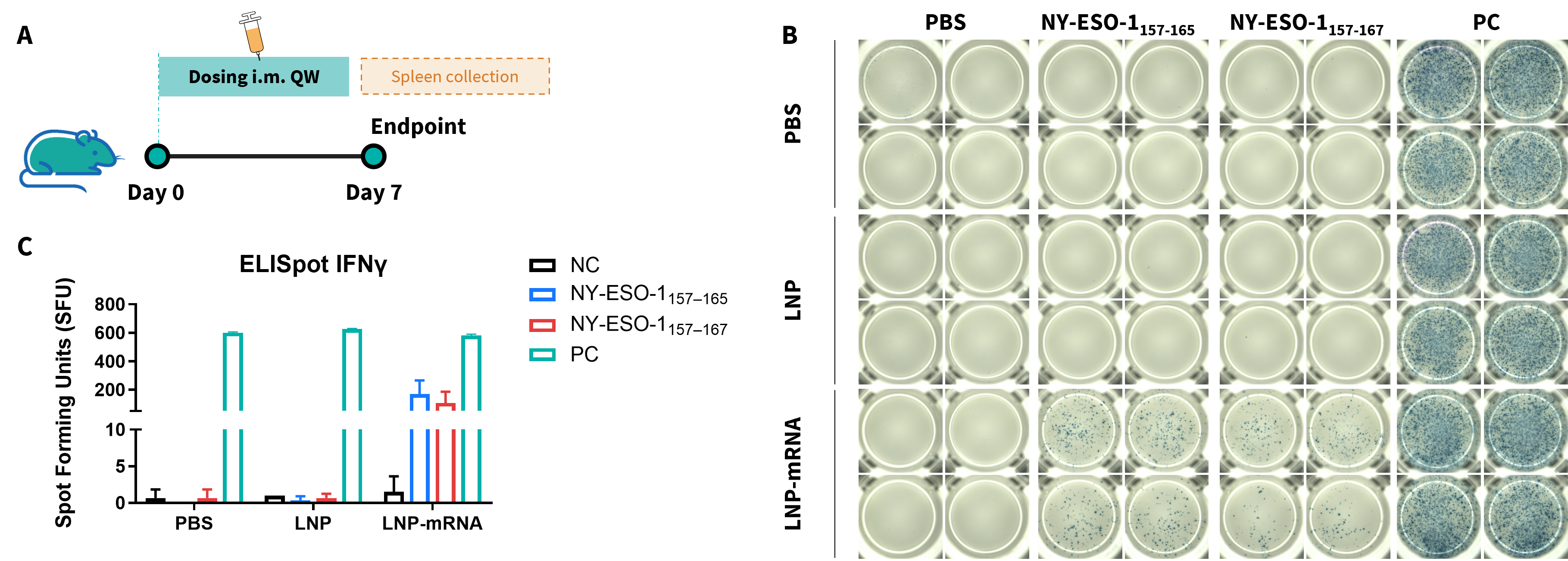
One dose of vaccine-induced immune responses in B-HLA-A2.1 mice by IFN-γ ELISpot assay. (A) Scheme of vaccination and testing. Male B-HLA-A2.1 mice (9-10-week-old, n=3) were immunized via intramuscular injection in both hind legs with PBS, empty LNPs, or LNP-mRNA. One week post-immunization, splenocytes were harvested and stimulated with specific peptides, no peptide (NC), or PMA/Ionomycin (PC) to measure IFN-γ secretion. (B) ELISpot Results: Representative assay wells. (C) ELISpot Quantification: Statistical summary of IFN-γ-secreting cells. NC: negative control. PC: positive control.
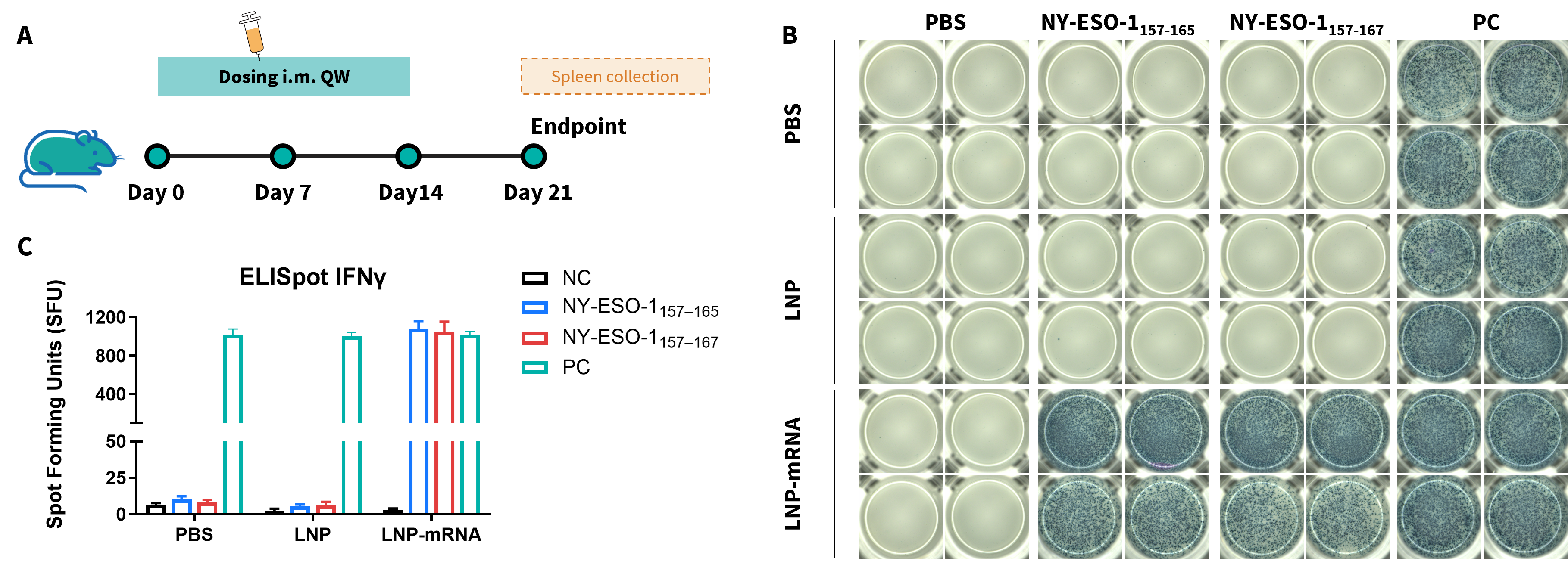
Three doses of vaccine-induced immune responses in B-HLA-A2.1 mice by IFN-γ ELISpot assay. (A) Scheme of vaccination and testing. Male B-HLA-A2.1 mice (9-10-week-old, n=3) were immunized via intramuscular injection in both hind legs with PBS, empty LNPs, or LNP-mRNA. One week post-immunization, splenocytes were harvested and stimulated with specific peptides, no peptide (NC), or PMA/Ionomycin (PC) to measure IFN-γ secretion. (B) ELISpot Results: Representative assay wells. (C) ELISpot Quantification: Statistical summary of IFN-γ-secreting cells. NC: negative control. PC: positive control.
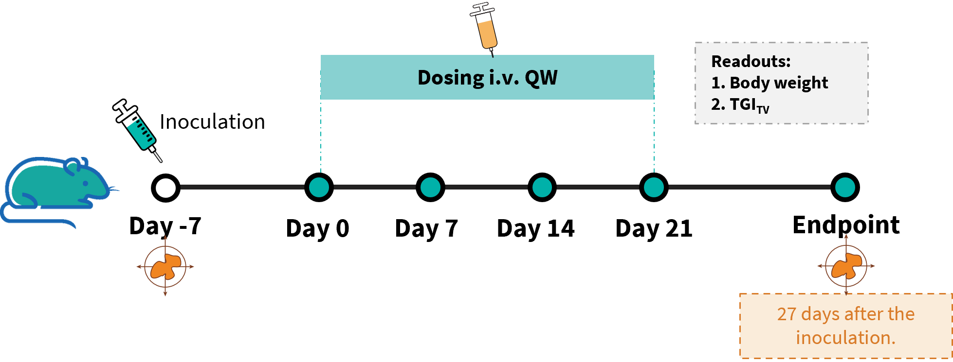
Establishment of a B-HLA-A2.1/hNY-ESO-1 MC38 model and in vivo efficacy study of an anti-human NY-ESO-1 LNP mRNA. B-HLA-A2.1/hNY-ESO-1 MC38 cells were implanted subcutaneously into homozygous B-HLA-A2.1 mice (male, 9-weeks-old, n=6).

Antitumor activity of NY-ESO-1 mRNA vaccine against syngeneic tumors. (A) Tumor growth curves. (B) Body weight changes during treatment. (C) Tumor cells growth of individual mouse.

LDH release assay for cytotoxicity of CTLs from B-HLA-A2.1 mice immunized with PBS, LNP or LNP-mRNA against the B-HLA-A2.1/hNY-ESO-1 MC38 cell line. Cytotoxic activities of isolated single splenocytes of immunized mice against NY-ESO-1 peptides pulsed B-HLA-A2.1/hNY-ESO-1 MC38 cell line were detected by LDH release assay at effector-to-target ratio of 50:1 or 100:1 with two different peptide concentrations, 10 μg/ml or 50 μg/ml.
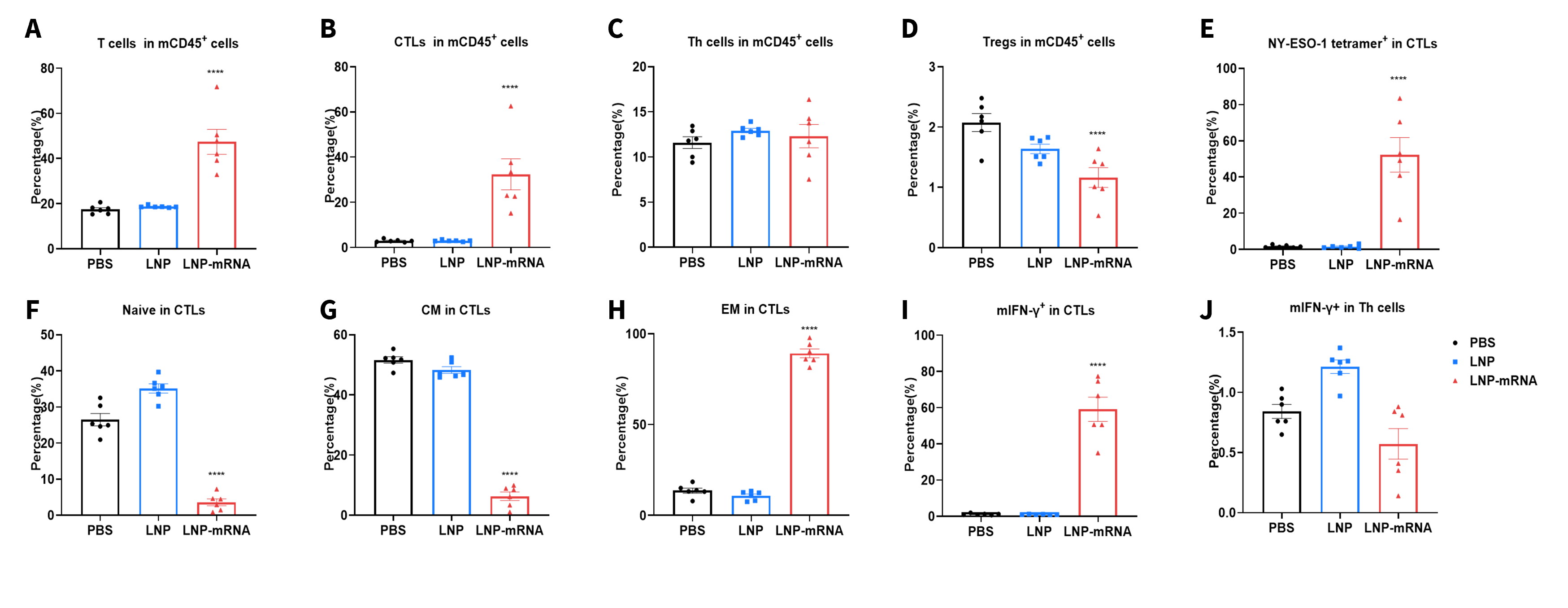
Vaccination LNP-mRNA generates specific effector CD8+ T cells in spleens. Spleens from B-HLA-A2.1/hNY-ESO-1 MC38 tumor-bearing mice that were immunized with the PBS, empty LNP or LNP-mRNA were analyzed on day 27. Analysis of CD8+T, CD4+ T and Treg cells in the spleens determined by the flow cytometric assay. For spleen T(A) and CD8+T cells(B), the percentage(in CD45+ cells) were significantly elevated. LNP-mRNA generated frequencies of tetramer+ CD8+ T cells at approximately 50% of total CD8+ T cells in spleens(E). The CD8+ T cells had an obviously lower frequency in the naïve(F) and central memory(G) and were mainly localized in the effector memory(H). The IFNγ were mainly secreted by CD8+ (I) but not CD4+T(J).
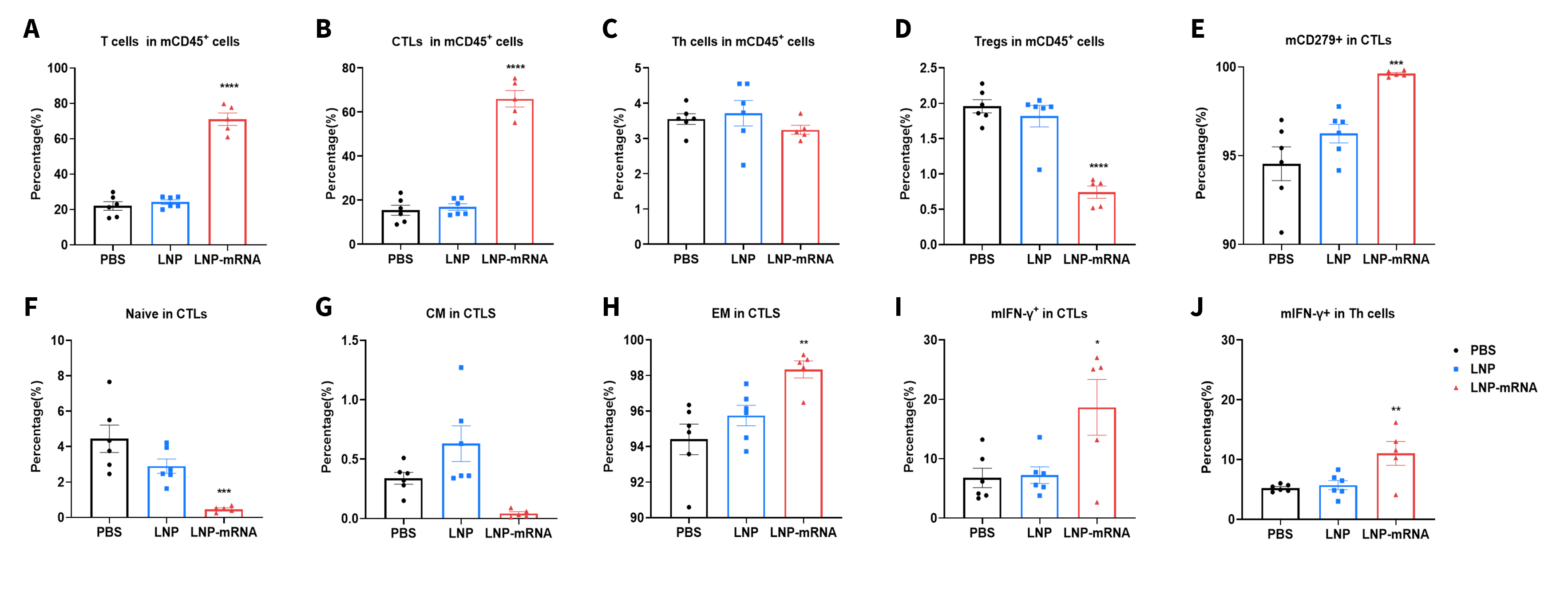
LNP-mRNA enhances beneficial repertoire of anti-tumor T cells. Tumors from B-HLA-A2.1/hNY-ESO-1 MC38 tumor-bearing mice that were immunized with the PBS, empty LNP or LNP-mRNA were analyzed on day 27. Analysis of CD8+T, CD4+ T and Treg cells in the tumors determined by the flow cytometric assay. For tumor-infiltrated T (A) and CD8+T cells (B), the percentage (in CD45+ cells) were significantly elevated. In contrast, the frequencies of Tregs was significantly diminished in LNP-mRNA group (D). Tumor-infiltrated CD8+ T cells had an obviously lower frequency in the naïve (F) and the effector memory(EM) CD8+T cells was increased significantly (H). The IFNγ in Both CD8+ T cells (I) and CD4+T cells (J) produced IFNγ.
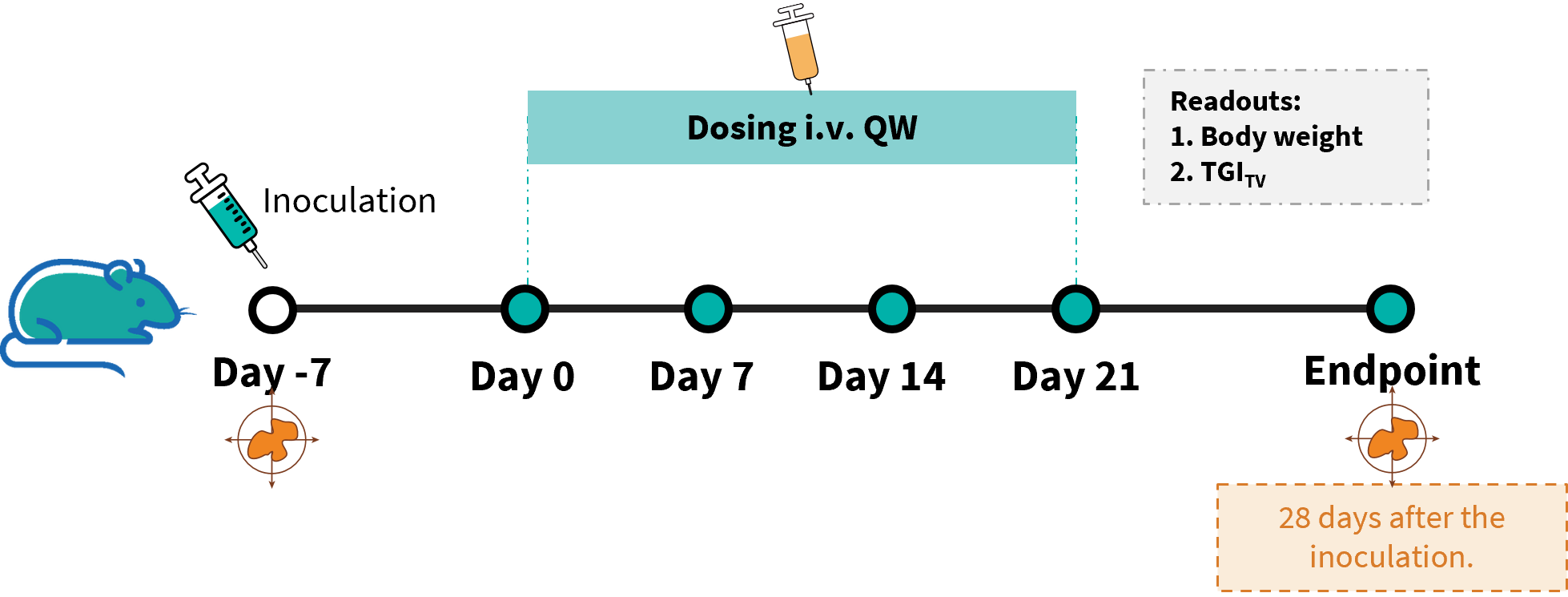
Establishment of a B-HLA-A2.1/hWT1 MC38 model and in vivo efficacy study of an anti-human WT1 LNP mRNA. B-HLA-A2.1/hWT1 MC38 cells were implanted subcutaneously into homozygous B-HLA-A2.1 mice (female, 9-weeks-old, n=8).

Antitumor activity of WT1 mRNA vaccine against syngeneic tumors. (A) Tumor growth curves. (B) Body weight changes during treatment. (C) Tumor cells growth of individual mouse. These results demonstrate that B-HLA-A2.1 mice provide a powerful preclinical model for in vivo evaluation of LNP-mRNA vaccines.
The overage of this tumor model is 40%.

LDH release assay for cytotoxicity of CTLs from B-HLA-A2.1 mice immunized with PBS, LNP or LNP-mRNA against the B-HLA-A2.1/hWT1 MC38 cell line. Cytotoxic activities of isolated single splenocytes of immunized mice against WT1 peptides pulsed B-HLA-A2.1/hWT1 MC38 cell line were detected by LDH release assay at effector-to-target ratio of 50:1 or 100:1 with 10 μg/ml peptides.
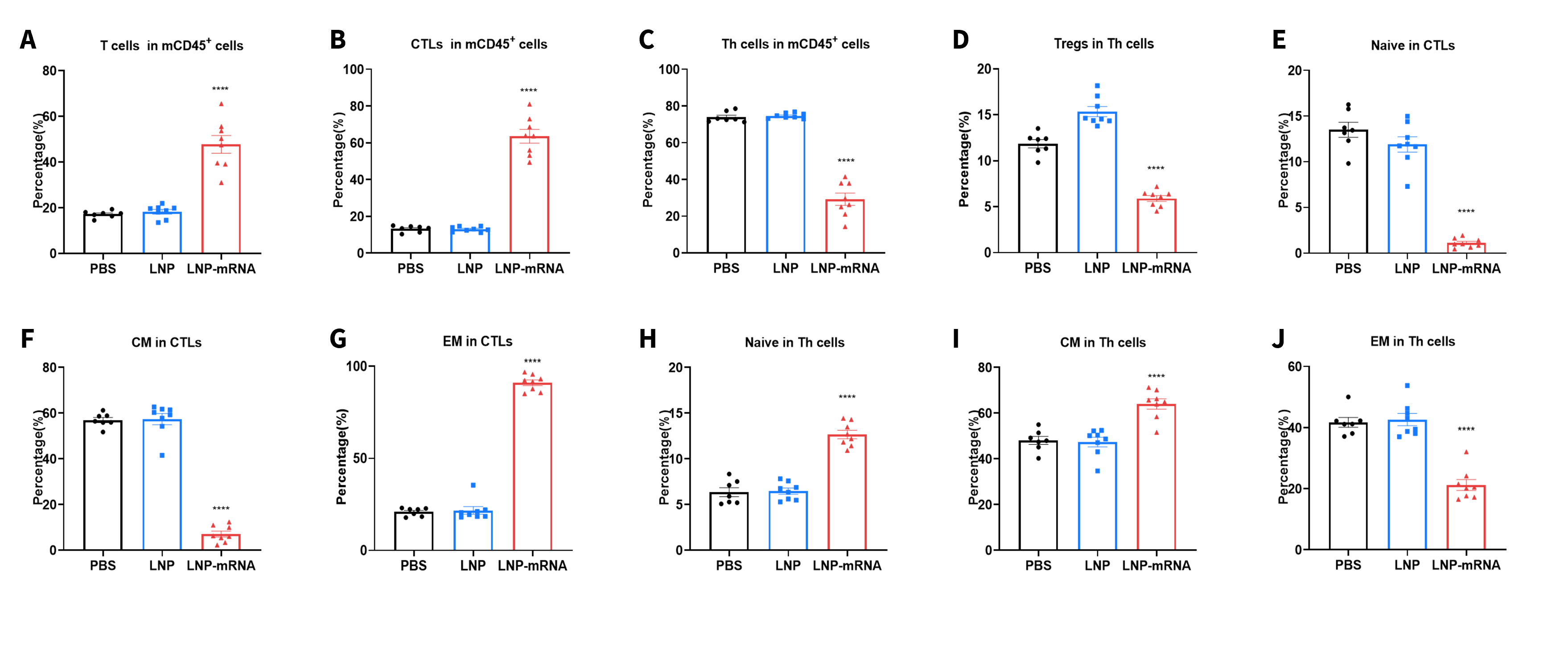
Vaccination LNP-mRNA generates specific effector CD8+ T cells in spleens. Spleens from B-HLA-A2.1/hWT1 MC38 tumor-bearing mice that were immunized with the PBS, empty LNP or LNP-mRNA were analyzed on day 28. Analysis of CD8+T, CD4+ T and Treg cells in the spleens determined by the flow cytometric assay. For spleen T cells(A), and CD8+T cells(B), the percentage(in CD45+ cells) were significantly elevated. The CD8+ T cells had an obviously lower frequency in the naïve(E) and central memory(F) and were mainly localized in the effector memory(G), while the trend of Th cells was opposite to that of CD8+T cells(H-J).
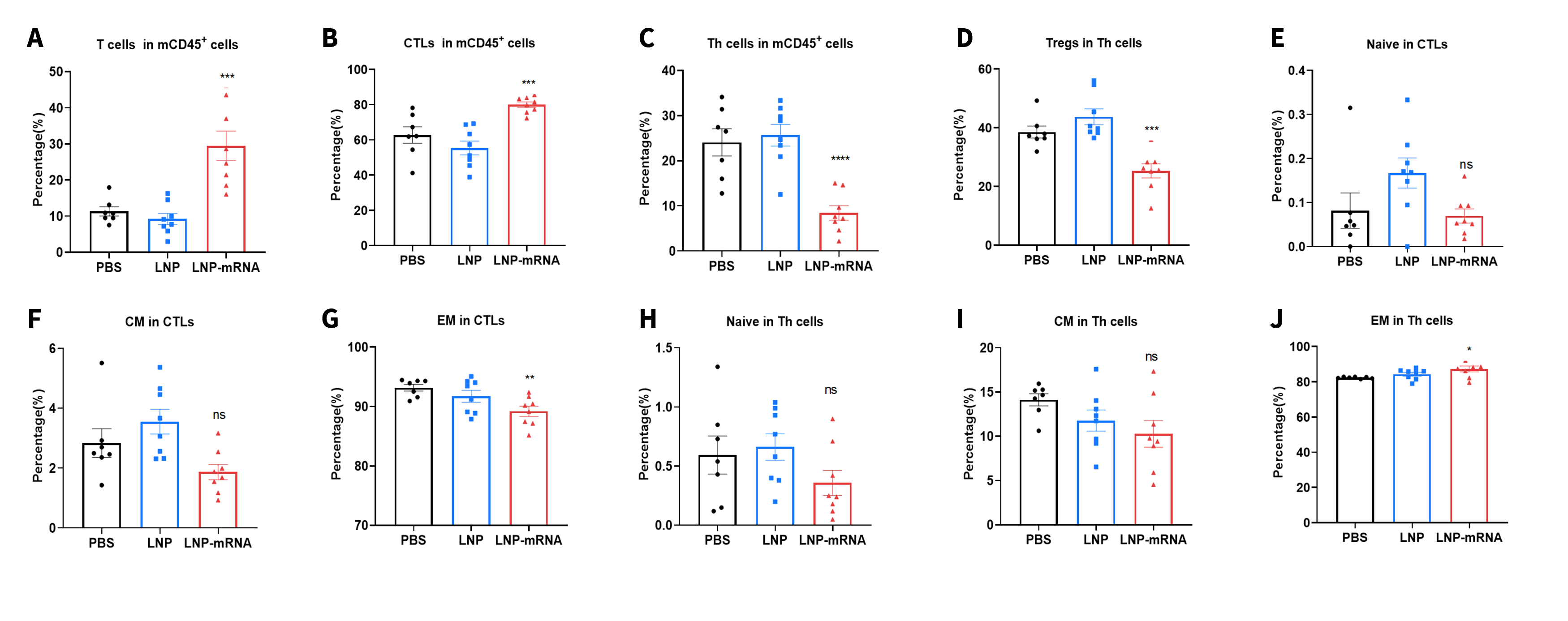
LNP-mRNA enhances beneficial repertoire of anti-tumor T cells. Tumors from B-HLA-A2.1/hWT1 MC38 tumor-bearing mice that were immunized with the PBS, empty LNP or LNP-mRNA were analyzed on day 28. Analysis of CD8+T, CD4+ T and Treg cells in the tumors determined by the flow cytometric assay. For tumor-infiltrated T(A) and CD8+T cells(B), the percentage(in CD45+ cells) were significantly elevated. In contrast, the frequencies of Tregs was significantly diminished in LNP-mRNA group(D). Tumor-infiltrated effector memory(EM) CD4+T cells was increased significantly(J).
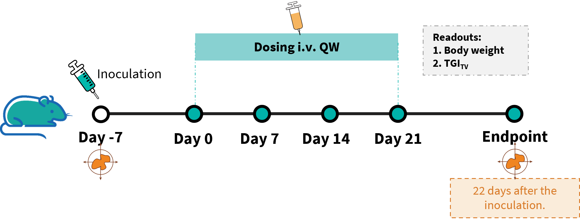
Establishment of a B-HLA-A2.1/hGP100 MC38 model and in vivo efficacy study of an anti-human GP100 LNP mRNA. B-HLA-A2.1/hGP100 MC38 cells were implanted subcutaneously into homozygous B-HLA-A2.1 mice (female, 8-weeks-old, n=8).

Antitumor activity of GP100 mRNA vaccine against syngeneic tumors. (A) Tumor growth curves. (B) Body weight changes during treatment. (C) Tumor cells growth of individual mouse. The overage of this tumor model is 40%.
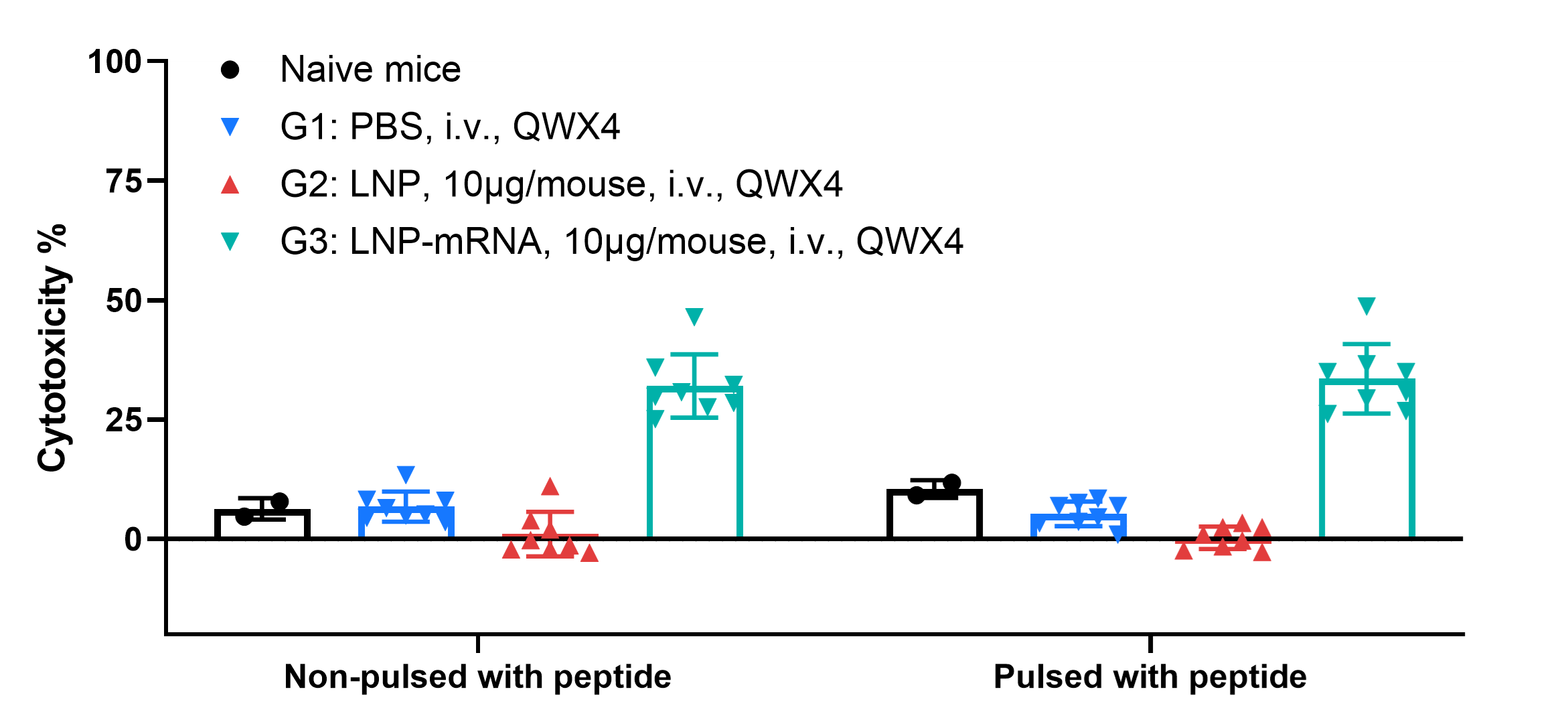
LDH release assay for cytotoxicity of CTLs from B-HLA-A2.1 mice immunized with PBS, LNP or LNP-mRNA against the B-HLA-A2.1/hGP100 MC38 cell line. Cytotoxic activities of isolated single splenocytes of immunized mice with or without GP100 peptides pulsed B-HLA-A2.1/hGP100 MC38 cell line were detected by LDH release assay at effector-to-target ratio of 100:1 with 10 μg/ml peptides.
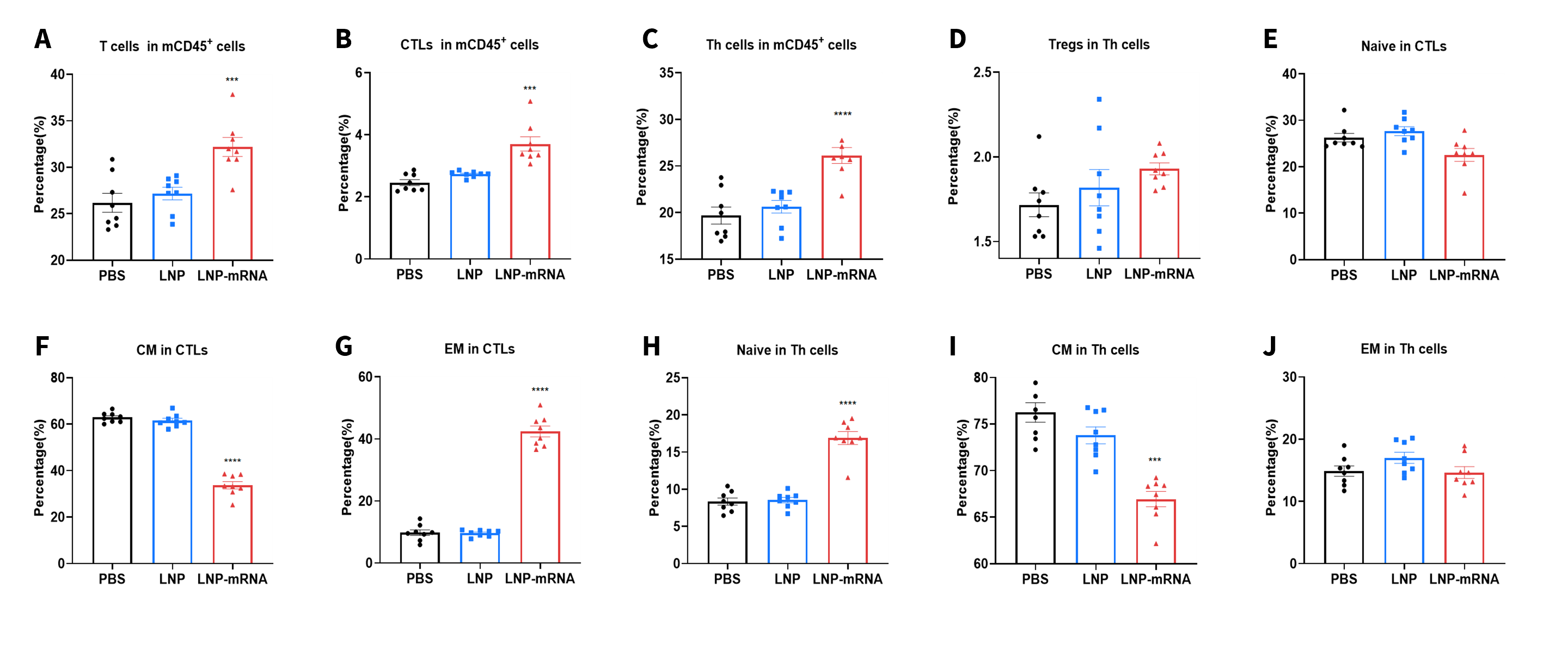
Vaccination LNP-mRNA generates specific effector CD8+ T cells in spleens. Spleens from B-HLA-A2.1/hGP100 MC38 tumor-bearing mice that were immunized with the PBS, empty LNP or LNP-mRNA were analyzed on day 22. Analysis of CD8+ T, CD4+ T and Treg cells in the spleens determined by the flow cytometric assay. For spleen T cells(A), and CD8+ T cells(B), the percentages (in CD45+ cells) were significantly elevated. The CD8+ T cells had an obviously lower frequency in the central memory(F) and were mainly localized in the effector memory(G), while the trend of naïve Th cells was opposite to that of CD8+T cells(H).
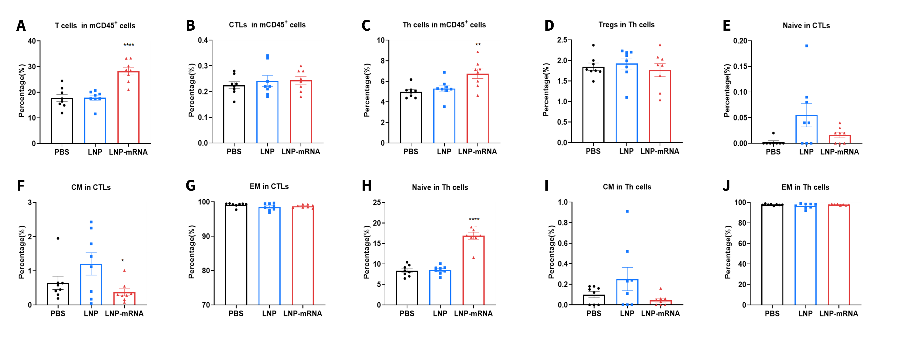
LNP-mRNA enhances beneficial repertoire of anti-tumor T cells. Tumors from B-HLA-A2.1/hGP100 MC38 tumor-bearing mice that were immunized with the PBS, empty LNP or LNP-mRNA were analyzed on day 22. Analysis of CD8+ T, CD4+ T and Treg cells in the tumors determined by the flow cytometric assay. For tumor-infiltrated T(A) and CD4+ T cells(C), the percentages(in CD45+ cells) were significantly elevated. In contrast, the frequencies of CD8+ T(B) and Tregs(D) were not significantly changed in LNP-mRNA group. Tumor-infiltrated naïve CD4+T cells was increased significantly(H).
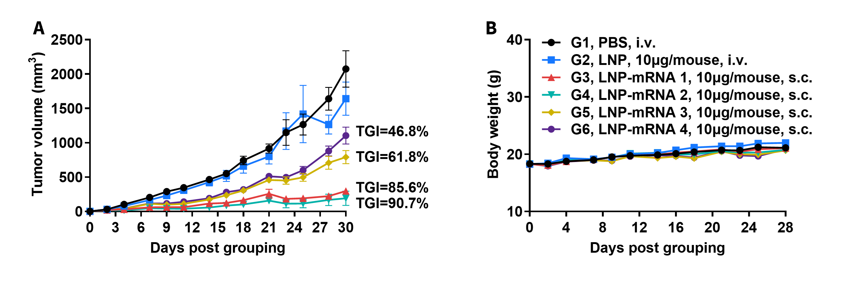
Antitumor activity of MAGEA3 mRNA vaccine against syngeneic tumors. (A) Tumor growth curves. (B) Body weight changes during treatment. These results demonstrate that B-HLA-A2.1 mice provide a powerful preclinical model for in vivo evaluation of LNP-mRNA vaccines.
The overage of this tumor model is 40%.
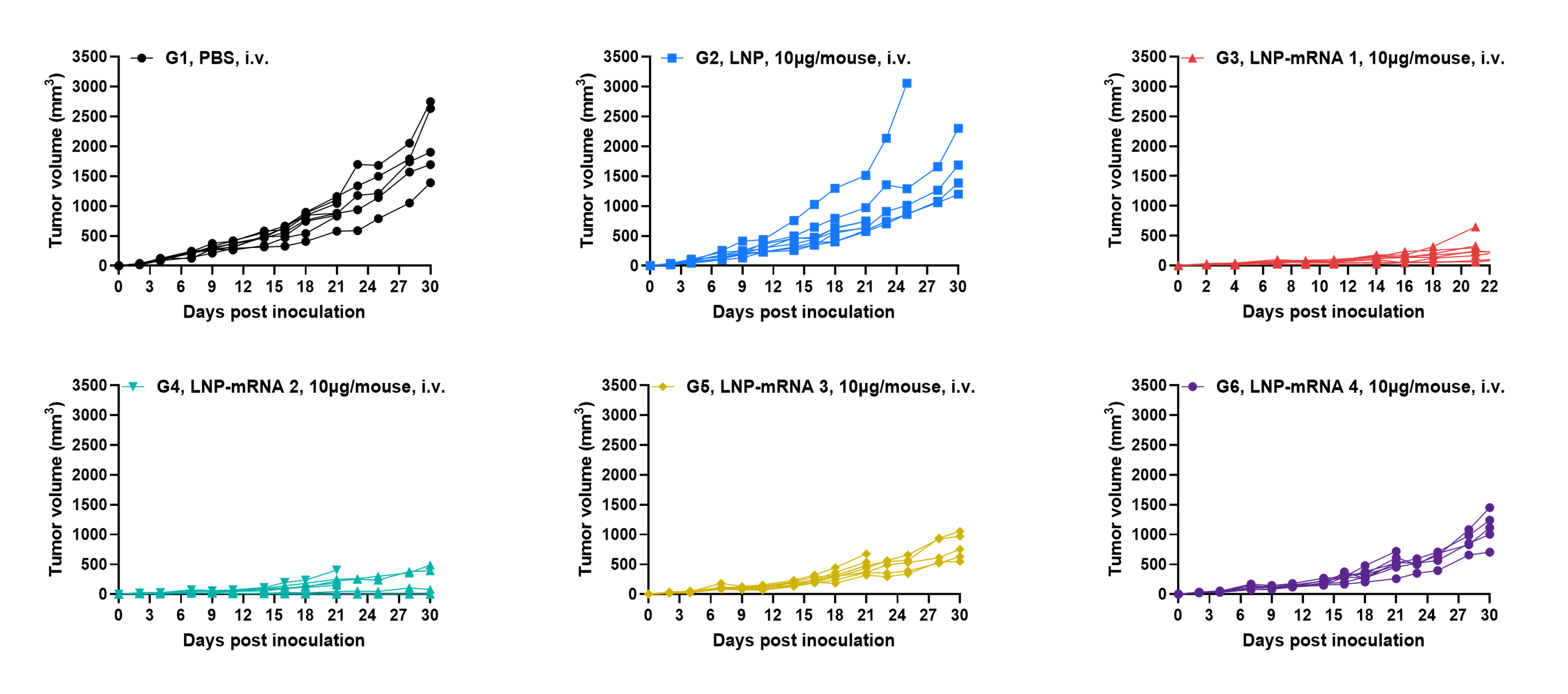
Antitumor activity of MAGEA3 mRNA vaccine against syngeneic tumors. Tumor cells growth of individual mouse. These results demonstrate that B-HLA-A2.1 mice provide a powerful preclinical model for in vivo evaluation of LNP-mRNA vaccines.
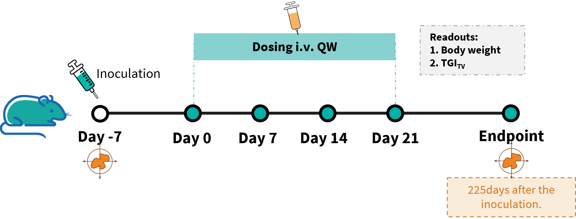
In vivo efficacy study of anti-human AFP LNP-mRNA in B-HLA-A2.1/hAFP MC38 model. B-HLA-A2.1/hAFP MC38 cells were implanted subcutaneously into homozygous B-HLA-A2.1 mice (female, 8-week-old, n=8).

Antitumor activity of AFP mRNA vaccine against syngeneic tumors. Readouts include tumor growth curves(A), body weight changes(B), and individual tumor growth(C). The overage of this tumor model is 40%.
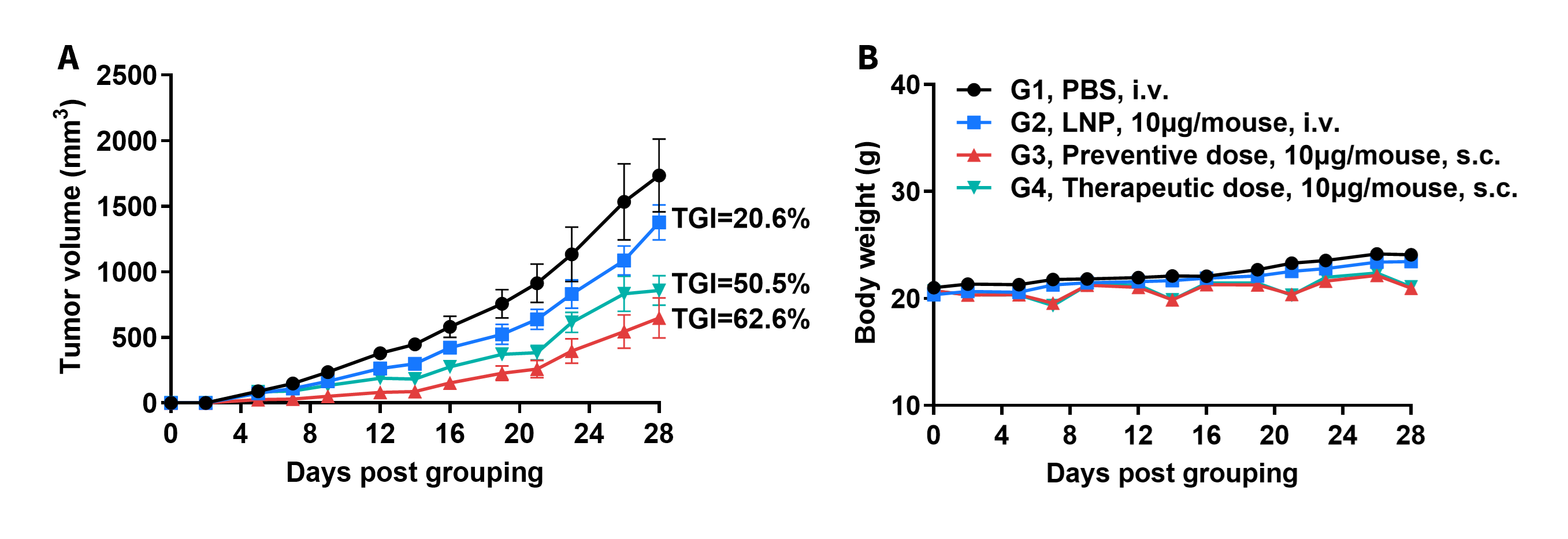
Antitumor activity of LMP2 mRNA vaccine against syngeneic tumors. (A) Tumor growth curves. (B) Body weight changes during treatment. The overage of this tumor model is 40%.

Antitumor activity of LMP2 mRNA vaccine against syngeneic tumors. Individual tumor growth curves show mouse-level responses after LMP2 mRNA vaccine treatment, supporting use of B-HLA-A2.1 mice for in vivo LNP-mRNA vaccine evaluation.

Antitumor activity of HPV16 E6/E7 LNP-mRNA vaccine against syngeneic tumors. Readouts include tumor growth curves(A), body weight changes(B), and individual tumor growth(C). B-HLA-A2.1 mice provide a preclinical model for LNP-mRNA vaccine evaluation. The overage of this tumor model is 40%.
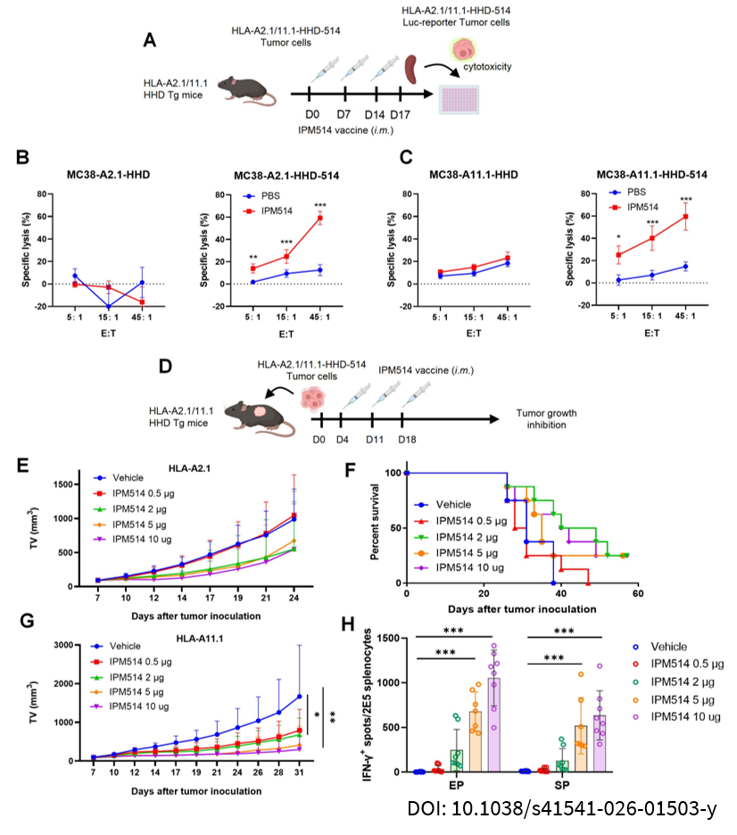
(A) Schematic of the luciferase-based ex vivo cytotoxicity assay. HLA-A2.1 or HLA-A11.1 transgenic mice received three intramuscular injections of IPM514 at weekly intervals. Spleens were harvested three days after the third immunization, and CD8+ T cells were isolated for co-incubation with tumor cells to assess ex vivo cytotoxicity. (B and C) CD8+ T cell derived from IPM514 immunized HLA-A2.1 (B) or HLA-A11.1 (C) transgenic mice exhibited specific lysis of MC38-HHD-A2.1-514 or MC38-HHDA11.1-514 cells, in contrast to MC38-HHD-A2.1 or MC38-HHD-A11.1 cells, respectively. Results are presented as means (SD) for each group (n = 5 per group). Statistical analyses were performed using two-way ANOVA with Bonferroni’s correction for multiple comparisons. (D) Treatment schematic for the heterogeneous HLA-A2.1/11.1- HHD-514 tumor model. HLA-A2.1 or HLA-A11:01 transgenic mice were grafted with 1 × 10⁶ MC38-HHD-A2.1-514 or MC38-HHD A11.1-514 tumor cells, followed by intramuscular immunization with either IPM514 or vehicle four days later. (E and F) The kinetics of tumor growth until the first mouse reached the humane endpoint are illustrated (E) and overall survival rates (F) for HLA-A2.1 mice treated with various doses IPM514 vaccines. Means (SD) are plotted for each group (n = 8 per group). (G) Kinetics of tumor growth until the vehicle group reached the humane endpoint are shown for HLA-A11.1 mice treated with varying doses of IPM514 vaccine. Means (SD) are plotted for each group (n = 8 per group).
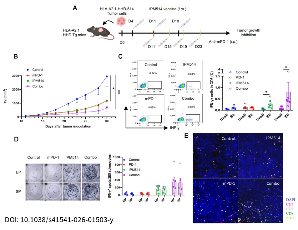
(A) Schematic of the combination therapy involving IPM514 and mPD-1 inhibitor. HLA-A2.1 transgenic mice were grafted with 1 × 10⁶ MC38-HHDA2.1-514 tumor cells and immunized intramuscularly four days later with three doses of either 2µg IPM514 or empty LNP. When the average tumor volume reached approximately 60 mm3 (D11), mice were administered isotype antibodies (Ab) or anti-mPD-1 (clone RMP1-14, BioXCell) at a dose of 1.6 mg/kg via intraperitoneal injection (i.p.) every four days for a total of four doses. (B) Kinetics of tumor growth are displayed until the control group reached the humane endpoint. Means (SD) are plotted for each group (n = 6 per group). Statistical analyses were performed using one-way ANOVA with Bonferroni’s correction for multiple comparisons at the endpoint. (C and D) Evaluation of the T-cell immune response is demonstrated through intracellular cytokine staining (C) and ELISpot assay (D). Data are shown as means (SD) for each group (n = 6 per group). Statistical analyses were performed using two-way ANOVA with Bonferroni’s correction for multiple comparisons. (E) Analysis of immune cell subsets in tumor microenvironment (TEM) using multiplex immunohistochemistry (IHC). Multiplex IHC showing the distribution of CD3⁺ cells (pink), CD4⁺ cells (gray), CD8⁺ cells (green), PD-1⁺ cells (yellow), and DAPI-stained nuclei (blue). Scale bar = 100 μm.

Q1: What are B-HLA-A2.1 mice?
B-HLA-A2.1 mice are HLA-A2.1 and human B2M gene-humanized mice on a C57BL/6N background, developed for HLA-A2.1-restricted antigen presentation, vaccine efficacy, and T-cell response studies.
Q2: Why is HLA-A2.1 important?
HLA-A2.1 is a human HLA class I molecule that presents endogenous peptide antigens to CD8+ T-cell receptors, making it important for cancer vaccine, TCR mimic antibody, T-cell engager, and TCR-T research.
Q3: How was Human B2M and HLA-A2.1 expression validated in B-HLA-A2.1 mice?
Human B2M and HLA-A2.1 expression were validated in homozygous B-HLA-A2.1 mice by flow cytometry, while mouse B2M and H-2Kb/H-2Db expression were detected in wild-type C57BL/6N mice.
Q4: Can B-HLA-A2.1 mice be used for vaccine efficacy studies?
Yes. HER2, NY-ESO-1, WT1, GP100, MAGEA3, AFP, LMP2, and HPV16 E6/E7 vaccine studies showed antigen-specific immune responses, antitumor activity, IFN-γ ELISpot responses, ICS readouts, and CTL cytotoxicity in B-HLA-A2.1 mice.
Q5: What are the main applications of B-HLA-A2.1 mice?
Applications include peptide vaccine evaluation, LNP-mRNA vaccine evaluation, tumor-peptide/HLA-A2.1 targeting, TCR mimic antibody research, T-cell engager studies, TCR-T research, and antigen-specific CD8+ T-cell response analysis.
