Basic Information

Strain Name
C57BL/6-Trbc1tm1(TRBC1)BcgenTrbc2tm1(TRBC2)Bcgen/Bcgen
Stock Number
112003
Common Name
B-hTRBC1/hTRBC2 mice
Background
C57BL/6
NCBI Gene ID
Aliases 
TCRBC1; TCRBC2

Targeting strategy

Gene targeting strategy for B-hTRBC1/hTRBC2 mice. The exon1~2, partial exon 3 and intron 1~2 of mouse Trbc1 and Trbc2 were replaced by human The exon1~2, partial exon 3 and intron 1~2 of human TRBC1 and TRBC2 in B-hTRBC1/hTRBC2 mice.

mRNA expression analysis in humanized B-hTRBC1/hTRBC2 mice

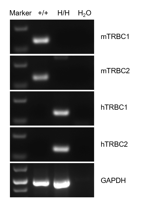
Species specific analysis of TRBC1 and TRBC2 gene expression in wild-type C57BL/6 mice and homozygous humanized B-hTRBC1/hTRBC2 mice by RT-PCR. Splenocytes were collected from wild-type C57BL/6 mice (+/+) and homozygous B-hTRBC1/hTRBC2 mice (H/H). Mouse TRBC1 and TRBC2 mRNA were detectable only in wild-type C57BL/6 mice. Human TRBC1 and TRBC2 mRNA were detectable only in homozygous B-hTRBC1/hTRBC2 mice, but not in wild-type C57BL/6 mice.

Protein expression analysis in spleen B, NK and T cells

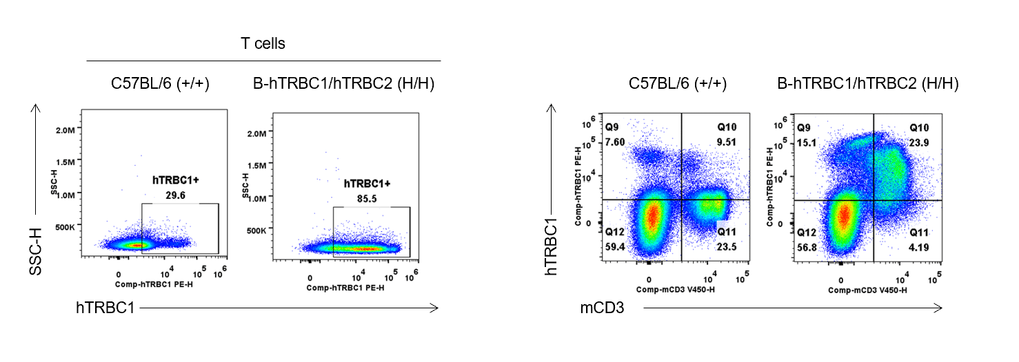
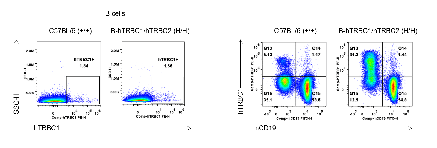
Strain specific TRBC1 expression analysis in wild-type C57BL/6 mice and homozygous humanized B-hTRBC1/hTRBC2 mice by flow cytometry. Splenocytes were collected from wild-type C57BL/6 mice (+/+) and homozygous B-hTRBC1/hTRBC2 mice (H/H), and analyzed with anti-human TRBC1 antibody (Biolegend,383502) by flow cytometry. Human TRBC1 was not detectable in wild-type C57BL/6 mice. Human TRBC1 was exclusively detectable in homozygous B-hTRBC1/hTRBC2 mice.

T cell activation analysis-CD69 and CD25

Activation of T cells was induced with human TRBC1 antibody and mouse CD3E/28 antibodies in wild-type C57BL/6 mice and homozygous B-hTRBC1/hTRBC2 mice. Splenocytes were collected from wild-type C57BL/6 mice and homozygous B-hTRBC1/hTRBC2 mice (H/H, 6-week-old, female, n=3) and purified for pan T cells using mouse Pan T Cell Isolation Kit II (Miltenyi Biotec, 130-095-130). Pan T cells were respectively stimulated with anti-mouse CD3E (BioXcell, BE0001-1), anti-mouse CD28 (BioXcell, BE0015-1) and human TRBC1 (Biolegend, 383502) antibodies for 24 hours in vivo. Activation of T cells was analyzed with anti-mouse CD69 (Biolegend, 104514) (A) and CD25 (Biolegend, 102008) (B) antibodies. T cells from WT and homozygous mice were successfully activated with anti-mCD3E antibody after 24 hours, and T cells from homozygous mice, but not from WT mice, were activated with anti-human TRBC1 antibody after 24 hours.

Activation of T cells was induced with human TRBC1 antibody and mouse CD3E/28 antibodies in wild-type C57BL/6 mice and homozygous B-hTRBC1/hTRBC2 mice. Splenocytes were collected from wild-type C57BL/6 mice and homozygous B-hTRBC1/hTRBC2 mice (H/H, 6-week-old, female, n=3) and purified for pan T cells using mouse Pan T Cell Isolation Kit II (Miltenyi Biotec, 130-095-130). Pan T cells were respectively stimulated with anti-mouse CD3E (BioXcell, BE0001-1), anti-mouse CD28 (BioXcell, BE0015-1) and human TRBC1 (Biolegend, 383502) antibodies for 24 hours in vivo. Activation of T cells was analyzed with anti-mouse CD69 (Biolegend, 104514) (A) and CD25 (Biolegend, 102008) (B) antibodies. T cells from WT and homozygous mice were successfully activated with anti-mCD3E antibody after 24 hours, and T cells from homozygous mice, but not from WT mice, were activated with anti-human TRBC1 antibody after 24 hours.

T cell activation analysis-CD69 and CD25

Activation of T cells was induced with human TRBC1 antibody and mouse CD3E/28 antibodies in wild-type C57BL/6 mice and homozygous B-hTRBC1/hTRBC2 mice. Splenocytes were collected from wild-type C57BL/6 mice and homozygous B-hTRBC1/hTRBC2 mice (H/H;H/H, 6-week-old, female, n=3) and purified for pan T cells using mouse Pan T Cell Isolation Kit II (Miltenyi Biotec, 130-095-130). Pan T cells were respectively stimulated with anti-mouse CD3E (BioXcell, BE0001-1), anti-mouse CD28 (BioXcell, BE0015-1) and human TRBC1 (Biolegend, 383502) antibodies for 24 hours in vivo. Activation of T cells was analyzed with anti-mouse CD69 (Biolegend, 104514) (A) and CD25 (Biolegend, 102008) (B) antibodies. T cells from WT and homozygous mice were successfully activated with anti-mCD3E antibody after 24 hours, and T cells from homozygous mice, but not from WT mice, were activated with anti-human TRBC1 antibody after 24 hours.

Activation of T cells was induced with human TRBC1 antibody and mouse CD3E/28 antibodies in wild-type C57BL/6 mice and homozygous B-hTRBC1/hTRBC2 mice. Splenocytes were collected from wild-type C57BL/6 mice and homozygous B-hTRBC1/hTRBC2 mice (H/H;H/H, 6-week-old, female, n=3) and purified for pan T cells using mouse Pan T Cell Isolation Kit II (Miltenyi Biotec, 130-095-130). Pan T cells were respectively stimulated with anti-mouse CD3E (BioXcell, BE0001-1), anti-mouse CD28 (BioXcell, BE0015-1) and human TRBC1 (Biolegend, 383502) antibodies for 24 hours in vivo. Activation of T cells was analyzed with anti-mouse CD69 (Biolegend, 104514) (A) and CD25 (Biolegend, 102008) (B) antibodies. T cells from WT and homozygous mice were successfully activated with anti-mCD3E antibody after 24 hours, and T cells from homozygous mice, but not from WT mice, were activated with anti-human TRBC1 antibody after 24 hours.

Activation of T cells was induced with human TRBC1 antibody and mouse CD3E/28 antibodies in wild-type C57BL/6 mice and homozygous B-hTRBC1/hTRBC2 mice. Splenocytes were collected from wild-type C57BL/6 mice and homozygous B-hTRBC1/hTRBC2 mice (H/H;H/H, 6-week-old, female, n=3) and purified for pan T cells using mouse Pan T Cell Isolation Kit II (Miltenyi Biotec, 130-095-130). Pan T cells were respectively stimulated with anti-mouse CD3E (BioXcell, BE0001-1), anti-mouse CD28 (BioXcell, BE0015-1) and human TRBC1 (Biolegend, 383502) antibodies for 24 hours in vivo. Activation of T cells was analyzed with anti-mouse CD69 (Biolegend, 104514) (A) and CD25 (Biolegend, 102008) (B) antibodies. T cells from WT and homozygous mice were successfully activated with anti-mCD3E antibody after 24 hours, and T cells from homozygous mice, but not from WT mice, were activated with anti-human TRBC1 antibody after 24 hours.

Human CD3+ T cell activation analysis-hCD69

Activation of human T cells was induced with human CD3E antibody and human TRBC1 antibodies in purified human CD3+ T cells. Human CD3+ T cells were respectively stimulated with anti-human CD3E (BioXcell, BE0001-2) and human TRBC1 (Biolegend, 383502) antibodies for 24 hours in vivo. Activation of T cells was analyzed with anti-human CD69 (Biolegend, 310912) antibody. Human T cells were successfully activated with anti-hCD3E and anti-hTRBC1 antibodies after 42 hours.

Human CD3+ T cell activation analysis-hCD25

Activation of human T cells was induced with human CD3E antibody and human TRBC1 antibodies in purified human CD3+ T cells. Human CD3+ T cells were respectively stimulated with anti-human CD3E (BioXcell, BE0001-2) and human TRBC1 (Biolegend, 383502) antibodies for 24 hours in vivo. Activation of T cells was analyzed with anti-human CD25 (Biolegend, 302610) antibody. Human T cells were successfully activated with anti-hCD3E and anti-hTRBC1 antibodies after 42 hours.

Frequency of leukocyte subpopulations in spleen

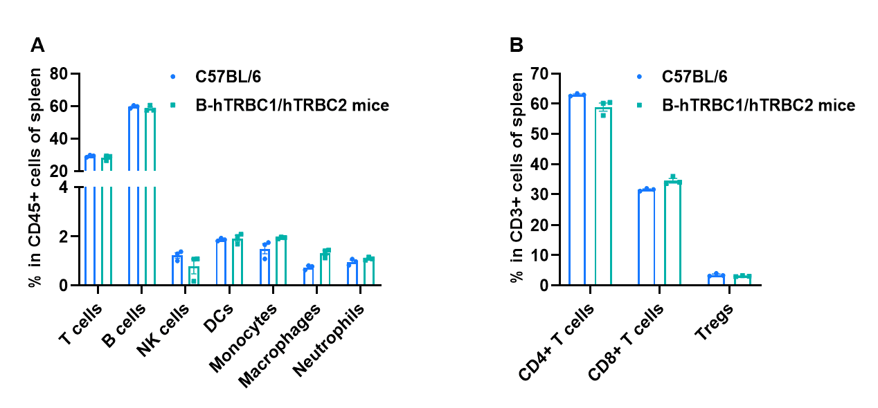
Frequency of leukocyte subpopulations in spleen by flow cytometry. Splenocytes were isolated from wild-type C57BL/6 mice and homozygous B-hTRBC1/hTRBC2 mice (n=3, 7-week-old). A. Flow cytometry analysis of the splenocytes was performed to assess the frequency of leukocyte subpopulations. B. Frequency of T cell subpopulations. Percentages of T cells, B cells, NK cells, dendritic cells, granulocytes, monocytes, macrophages, CD4+ T cells, CD8+ T cells and Tregs in B-hTRBC1/hTRBC2 mice were similar to those in C57BL/6 mice, demonstrating that humanization of TRBC1 and TRBC2 do not change the frequency or distribution of these cell types in spleen. The frequency of leukocyte subpopulations in blood and lymph node of B-hTRBC1/hTRBC2 mice were also comparable to wild-type C57BL/6 mice (Data not shown). Values are expressed as mean ± SEM. Significance was determined by two-way ANOVA test.  *P < 0.05, **P < 0.01, ***p < 0.001.

Back to top
WordPress Double Opt-in by Forge12