
Basic Information
-
Targeting strategy

-
Gene targeting strategy for B-NDG MHC I/II DKO mice plus. The murine B2m and H2-Ab1 gene were knocked out while a fused gene composed of murine B2m and Fcgrt gene was inserted after the signal peptide sequence of murine Fcgrt gene in B-NDG MHC I/II DKO mice plus.
-
Protein expression analysis
-
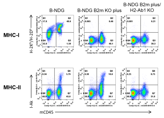
Strain specific H-2Kb/H-2Db (MHC-I) and I-Ak (MHC-II) expression analysis in B-NDG mice, B-NDG B2m KO plus mice and B-NDG MHC I/II DKO mice plus by flow cytometry. Splenocytes were collected from the three mice and analyzed by flow cytometry. Mouse H-2Kb/H-2Db was only detectable in B-NDG mice but not in B-NDG B2m KO plus mice and B-NDG MHC I/II DKO mice plus. Mouse I-Ak was only detectable in B-NDG mice and B-NDG B2m KO plus mice but not in B-NDG MHC I/II DKO mice plus.
-
Analysis of leukocyte subpopulations in spleen, blood, and bone marrow
-

Analysis of leukocyte subpopulations in spleen, blood, and bone marrow by flow cytometry. Blood, spleen, and bone marrow were collected from B-NDG mice and B-NDG MHC I/II DKO mice plus (male, 9-week-old, n=3). Leukocyte subpopulations were analyzed by flow cytometry analysis. Results showed that T cells, B cells, and NK cells were not detectable in all tissues of B-NDG mice and B-NDG MHC I/II DKO mice plus. Values are expressed as mean ± SEM. Significance was determined by a two-way ANOVA test. *P < 0.05, **P < 0.01, ***p < 0.001.
-
Significantly reduced severity of GvHD induced with human PBMC engraftment in B-NDG MHC I/II DKO mice plus
-
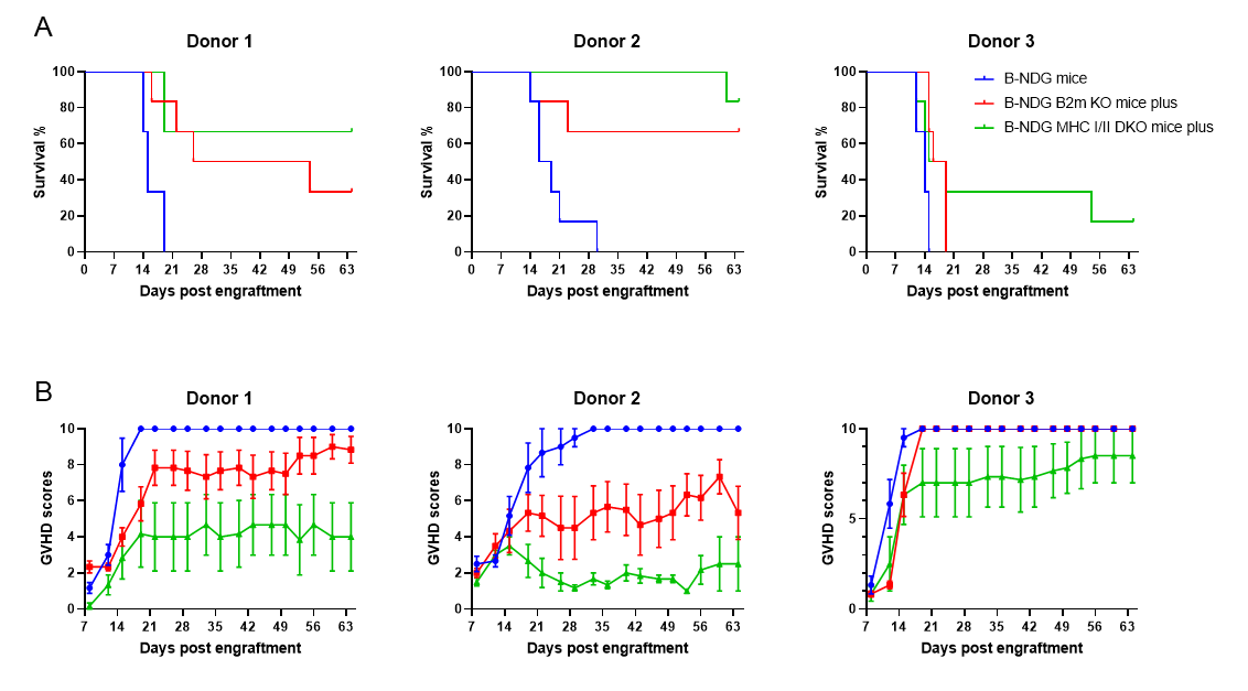
Comparison of the severity of GvHD induced with human PBMC engraftment in B-NDG mice, B-NDG B2m KO mice plus and B-NDG MHC I/II DKO mice plus.
Five weeks old of female B-NDG mice, B-NDG B2m KO mice plus and B-NDG MHC I/II DKO mice plus were respectively engrafted intravenously with human PBMCs (5×10⁶) from three healthy donors (Donor1-3) on day 0 (n=5). A. Survival rates of the mice were analyzed with Kaplan Meier survival curves. B. Body weight changes. C. Clinical signs of GvHD were scored twice a week. Results showed that MHC I/II double knocked-out in B-NDG MHC I/II DKO mice plus can significantly extend the life span and reduced the GvHD induced with human PBMC engraftment when compared that in B-NDG mice or in B-NDG B2m KO mice plus. Therefore B-NDG MHC I/II DKO mice plus are more suitable mouse model for human PBMC engraftment into the immunodeficient mice. Values were expressed as mean ± SEM.
Comparison of the peripheral blood leukocyte subpopulations in B-NDG mice, B-NDG B2m KO mice plus, and B-NDG MHC I/II DKO mice plus after human PBMC engraftment. At five weeks of age, female B-NDG mice, along with B-NDG B2m KO mice and B-NDG MHC I/II DKO mice, were intravenously engrafted with human peripheral blood mononuclear cells (PBMCs) from three healthy donors (Donors 1-3) at a dose of 5 × 10^6 cells per mouse on day 0 (n=5 per group). Blood samples were collected weekly post-engraftment for flow cytometric analysis. The levels of reconstituted leukocytes were comparable among all three mouse groups.

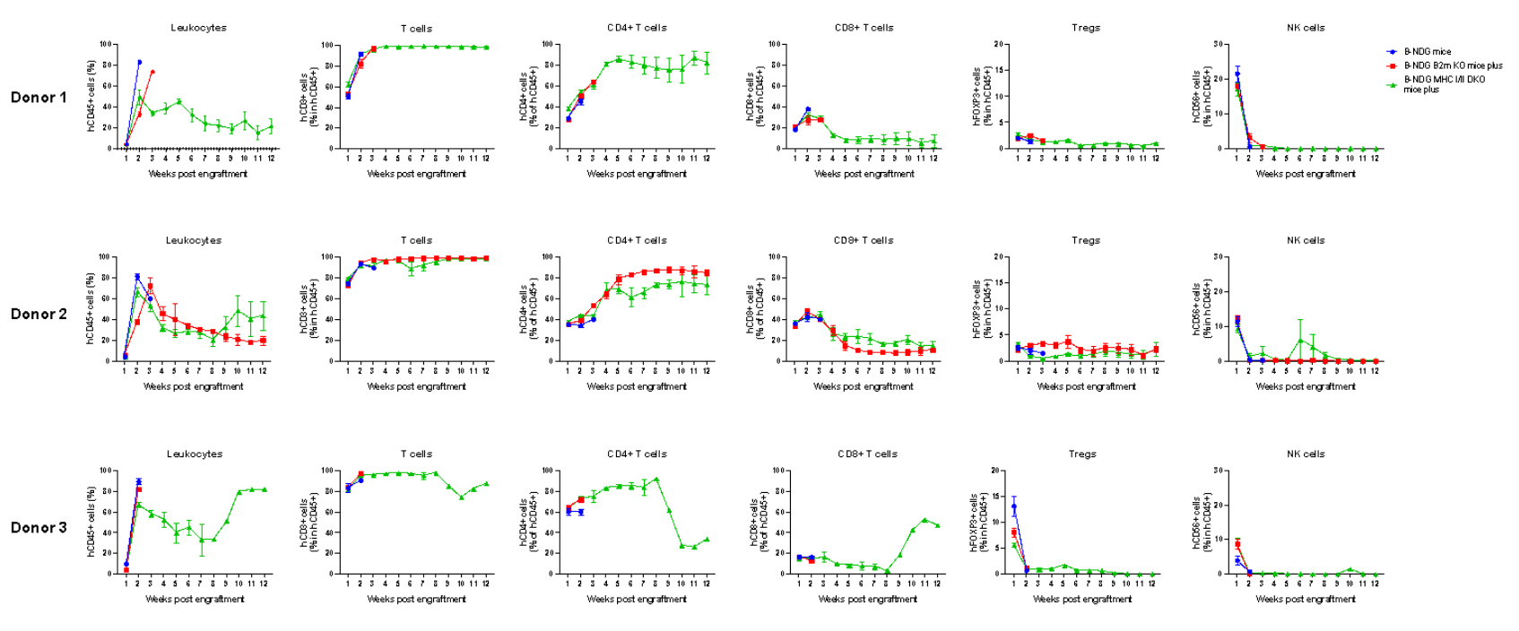
Comparison of reconstitution levels of human PBMCs among B-NDG mice, B-NDG B2m KO mice plus, and B-NDG MHC I/II DKO mice plus. Five-week-old female B-NDG mice, B-NDG B2m KO mice plus, and B-NDG MHC I/II DKO mice plus were irradiated with 1.0 Gy before being intravenously engrafted with human peripheral blood mononuclear cells (PBMCs) from three healthy donors (Donor 1-3) on day 0 (n=6 per group). Peripheral blood samples were collected weekly to assess the reconstitution level of human immune cells. The experiment concluded 90 days post-reconstitution. The findings revealed that regardless of the donor source, various subsets of human T cells could be reconstituted in B-NDG MHC I/II DKO mice plus. However, the reconstituted cell numbers in B-NDG MHC I/II DKO mice plus were consistently lower than those in B-NDG mice and B-NDG B2m KO mice plus. Notably, Donor 3 exhibited the most severe graft-versus-host disease (GvHD) and also displayed the highest counts of reconstituted human T cells and CD4+ T cells.
-
Human PBMC engraftment model for efficacy evaluation
-

B-NDG MHC I/II DKO mice have a long life span and reduced severity of GvHD when engrafted with human PBMCs. B-NDG MHC I/II DKO mice plus were engrafted intravenously with human PBMCs (1 × 10⁷) on day 0 (n=6). Survival rates of the mice were analyzed with Kaplan-Meier survival curves. Body weight was measured twice a week. Clinical signs of GvHD were scored once a week. Euthanasia was implemented when the body weight decreased by more than 20%. Meanwhile, the GvHD disease severity was scored on a scale from 0-10. Results showed that all the mice can live up to 96 days. During this period, apart from weight loss, there were no other obvious symptoms of GvHD. Values were expressed as mean ± SEM.

Human PBMCs were successfully reconstituted in B-NDG MHC I/II DKO mice plus. B-NDG MHC I/II DKO mice plus were intravenously engrafted with human peripheral blood mononuclear cells (PBMCs) at a dose of 1 × 10^7 cells on day 0 (n=6). Peripheral blood samples were collected weekly to assess the reconstitution level of human immune cells. The experiment concluded 112 days (16 weeks) post-engraftment. The analysis focused on (A) the frequency and (B) the absolute cell number of reconstituted human immune cells. The findings revealed that two weeks post-reconstitution of human PBMCs in B-NDG MHC I/II DKO mice plus, both the frequency and absolute cell number of CD45+ cells in peripheral blood began to increase, maintaining a relatively stable level of reconstitution throughout the 16-week observation period. The frequency of reconstituted human T cells surpassed 90% by the second week and continued to rise, eventually nearing 100%. Reconstituted human T cells included CD4+ T cells, CD8+ T cells, and regulatory T cells (Tregs), with a small population of dendritic cells (DCs) also detectable. Moreover, human programmed cell death protein 1 (PD-1) was widely expressed on both CD4+ T cells and CD8+ T cells, indicating robust engagement of the human immune system within the B-NDG MHC I/II DKO mice plus model. These findings underscore the utility of B-NDG MHC I/II DKO mice plus as a potent immunodeficient mouse model for the reconstitution of the human immune system using human PBMCs.
-
Summary
-
Protein expression analysis:
- Mouse H-2Kb/H-2Db was only detectable in B-NDG mice but not in B-NDG B2m KO plus mice and B-NDG MHC I/II DKO mice plus. Mouse I-Ak was only detectable in B-NDG mice and B-NDG B2m KO plus mice but not in B-NDG MHC I/II DKO mice plus.
Analysis with GvHD mouse model:
- MHC I/II double knocked-out in B-NDG MHC I/II DKO mice plus can significantly extend the life span and reduce the severity of GvHD induced with human PBMCs engraftment when compared that in B-NDG mice or B-NDG B2m KO mice plus.
- Human PBMCs from different donor sources affected reconstitution, but the influence in B-NDG MHC I/II DKO mice plus was much lower than that on B-NDG mice and B-NDG B2m KO plus mice.
- Reconstitution levels in B-NDG MHC I/II DKO mice plus, although slightly lower than the other two strains of mice, remained high.
- Regardless of the strains of mice, the higher the reconstitution level of human T cells, especially human CD4+ T cells, the more severe the GvHD.
- B-NDG MHC I/II DKO mice plus are a more suitable immunodeficient mouse model for reconstitution of the human immune system using human PBMCs.
Human PBMCs engraftment model for efficacy evaluation:
- All the mice engrafted with human PBMCs can live up to 96 days.
- Two weeks after the reconstitution of human PBMCs in B-NDG MHC I/II DKO mice plus, the frequency and absolute cell number of CD45+ cells in peripheral blood began to increase, and a relatively stable level of reconstitution was maintained until the end point at 16 week. The frequency of reconstituted human T cells exceeded 90% from two weeks and continued to rise, eventually reaching nearly 100%. Reconstituted human T cells include CD4+T cells, CD8+T cells, and Tregs. A small amount of DCs can also be detected.
-
Poster
