
Basic Information
Description
This B-luc-GFP Raji cell line expresses firefly luciferase as a marker of Raji cells. Luminescence can be observed B-luc-GFP Raji cells.
-
In vitro luciferase activity of B-luc-GFP Raji cells

-
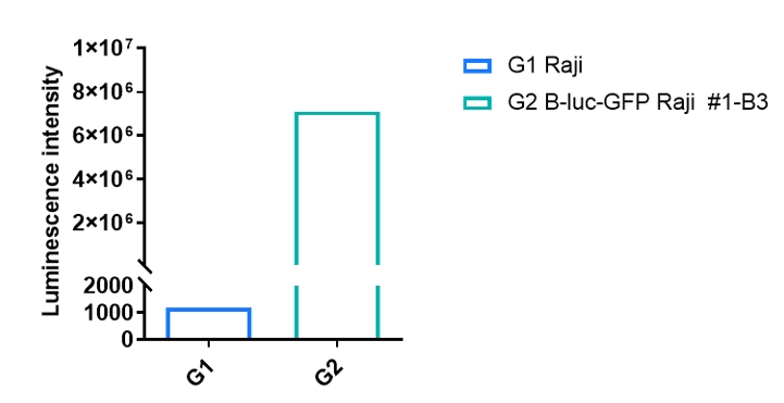
Luminescence signal intensity of B-luc-GFP Raji cells. Single cell suspensions from wild-type Raji and B-luc-GFP Raji #1-B3 (1×10⁶) cultures were measured using the Bright-GloTM luciferase Assay (Promega, Catalog No. E4030). B-luc-GFP Raji cells have a strong luminescence signal that is not present in wild-type Raji cells.
-
Quantitative imaging data & body weight changes
-
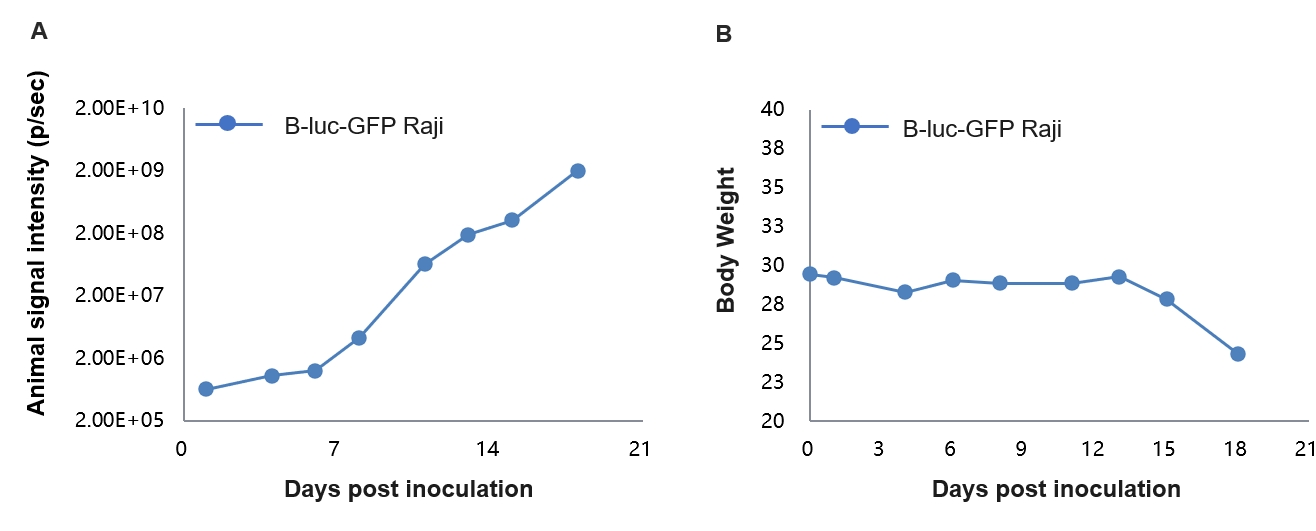
Tumor growth and in vivo imaging of B-luc-GFP Raji cells. B-luc-GFP Raji cells (1×10⁶) were injected by tail vein into B-NDG mice. Signal intensity and body weight were measured twice a week. (A) Imaging was performed on days 0, 4, 6, 8, 11, 15, and 18 , (B) Mice body weight (Mean ± SEM). B-luc-GFP Raji cells can be used for in vivo efficacy evaluation.
-
Human CD47Ab efficacy based on B-NDG mice
-
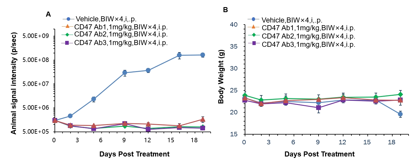
B-luc-GFP Raji cells were injected intraperitoneally into B-NDG and observed with a small animal imager. The animals were enrolled into the control group and the treatment group (n=6) when the tumor fluorescence intensity reached 1.5E6.
The results showed that different anti-human CD47 antibodies had different inhibitory effects on tumor growth at the same dose. A. Tumor fluorescence intensity ± SEM, B. Mean body weight ± SEM.
