Basic Information
Description
Background:
The immunodeficient B-NDG mice (NOD.CB17-PrkdcscidIl2rgtm1/Bcgen) was independently designed and generated by Biocytogen. B-NDG mice were generated by deleting the Il2rg gene from NOD scid mice with severe immunodeficient phenotype. This mouse model lacks mature T cells, B cells and functional NK cells. It is internationally recognized as an immunodeficient mouse model well suited for human-derived tissue or cell engraftment.
- NOD (non-obese diabetes) genetic background: Spontaneous type I diabetes; defective function of T cells, NK cells, macrophages, and dendritic cells and lack hemolytic function of complement.
- Prkdc (protein kinase, DNA activated, catalytic polypeptide) gene mutation: no mature T cells and B cells, severe combined immunodeficiency (scid) of cellular and humoral immunity.
- Il2rg gene knockout: the gamma chain of IL2 receptor (IL2Rγc, CD132) is located on the X chromosome of mouse, and is the common receptor subunit of cytokines IL2, IL4, IL7, IL9, IL15 and IL21 with important immune functions. The immune function of Il2rg knockout mice was severely reduced, especially the activity of NK cells was almost lost.
B-NDG mice: Combined NOD scid-Il2rg null background features, severe immunodeficient phenotype, absence of mature T cells, B cells, and functional NK cells, decreased function of macrophages and dendritic cells. It is very suitable for the transplantation of human hematopoietic stem cells (CD34+ HSCs) and peripheral blood mononuclear cells (PBMC) to obtain humanized mice with human immune system.
Advantages of B-NDG mice
- One of the most immunodeficient mouse models
- Longer lifespan than NOD scid mice; 1.5 years on average
- Minimal to absent rejection of human-derived cells or tissues
- More efficient for CDX and PDX model generation
Major applications
- Human-derived cell or tissue engraftment
- Tumor and tumor stem cell research
- ES and iPS cell research
- Hematopoiesis and immunology studies
- Human infectious disease studies
- Development of new humanized mouse models
Immunodeficient mice (B-NDG background)
Immunodeficient mouse models (non B-NDG background)
-
Targeting strategy

-

Gene targeting strategy for B-NDG mice. The exons 1-8 of mouse Il2rg gene that encode the full-length protein were knocked out in NOD scid mice. Homozygous mice with Il2rg null were named as B-NDG mice.
-
Phenotypic analysis

-
Analysis of T, B and NK cells in blood, spleen and bone marrow

Complete loss of T, B and NK cells in B-NDG mice. Blood, spleen and bone marrow were collected from C57BL/6, NOD scid and B-NDG mice (female, 6-week-old, n=3). Leukocyte subpopulations were analyzed by flow cytometry analysis. A. Representative FACS plots. B. Statistical analysis of FACS. Results showed that T cells, B cells and NK cells were detectable in all tissues of C57BL/6 mice. Only NK cells were detectable in blood and spleen of NOD scid mice. But none of the three cells were detectable in any tissue of B-NDG mice.

Analysis of myeloid cells in B-NDG mice. Blood, spleen and bone marrow were collected from C57BL/6, NOD scid and B-NDG mice (female, 6-week-old, n=3). Leukocyte subpopulations were analyzed by flow cytometry analysis. A. Representative FACS plots. B. Statistical analysis of FACS. Results showed that percentages of granulocytes and monocytes/macrophages of blood, spleen and bone marrow in B-NDG mice were relatively higher than that in wild-type C57BL/6 and NOD scid mice. But the percentages of DCs were lower in B-NDG mice than that in wild-type C57BL/6 and NOD scid mice.

Complete loss of antibody production in B-NDG mice. Serum were collected from BALB/c mice and B-NDG mice. 1% BSA was added as control group. Mouse antibodies were detected with ELISA method. (A) Mouse IgG and IgM detection. (B) mouse IgG subclasses detection. Results showed that mouse IgG, IgM and IgG subclasses were detectable in serum of BALB/c mice, but not detectable in serum of B-NDG mice. These results demonstrate a complete loss of antibody production in B-NDG mice.
Histological analysis of spleen

Histological analysis of mouse spleen. Spleen tissues of C57BL/6, NOD scid and B-NDG mice were respectively subjected to HE staining and immunohistochemical analysis (female, 9-week-old, n=3). (A) Splenic tissue HE staining. The results showed that the splenic structure of C57BL/6 mice was normal and the follicles were clear; The splenic structure of NOD scid mice showed leukopulp hypoplasia; But B-NDG mice showed a complete disappearance of follicular structure. (B) Immunohistochemical analysis of T cells and B cells in spleen. Mouse T cells and B cells were respectively detected with anti-mouse CD3 antibody or anti-mouse CD19 antibody. The results showed that the number and distribution of mCD3+ T cells and mCD19+ B cells in the spleen tissue of C57BL/6 mice were normal (brown); But both T cells and B cells were not detectable in NOD scid mice and B-NDG mice, indicating that there were no T cells and B cells in spleen of NOD scid mice and B-NDG mice.
Growth curve

Growth curve of B-NDG mice
Three weeks old of newborn pups were obtained at weaning (50 males and 50 females, respectively). Body weight was measured on the same day of every week and lasted for 12 weeks. The lowest and highest values of mouse body weight in the table were calculated from the mean ± SD. The growth curve conforms to the normal distribution and the probability of random error falling within ± SD is 68%.
Survival curve

Survival curve of B-NDG mice. Health status of 50 male and 50 female mice were observed. Results showed that female mice did not die before 12 months of age and the survival rate at 13 months was 86%. One male mouse died at 6 months of age and the survival rate of male mice at 13 months was 70%.
Histopathological analysis

Histopathological analysis of organs in B-NDG mice. The main organs of B-NDG mice were isolated at 32 weeks of age and analyzed with HE staining (male, n=15; female, n=16). Results showed that the follicular structure of the spleen of all mice were completely lost. 80% of the male mice had small calcifications in the testicular tissue. No obvious abnormalities were found in other organs (brain, heart, lung, liver, spleen, stomach, small intestine, colon, kidney and ovary).
-
CDX tumor models and efficacy evaluation in B-NDG mice

-
Successfully establishing a CDX lymphoma model with B-NDG mice
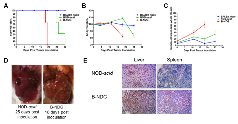
Raji lymphoma cell line can successfully establish hematologic and metastatic mouse tumor model in B-NDG mice. Raji cells (5×10⁵) were injected via tail vein of B-NDG mice, NOD scid mice and BALB/c nude mice. (A) Survival curves of the three strain of mice; (B) Body weight change; (C) The whole blood were collected from the orbital venous plexus of mice every week. Percent of human cells in the whole blood cells were assessed by qPCR; (D) B-NDG mice and NOD scid mice were euthanized and livers were respectively dissected at 18 or 25 days after cell inoculation. (E) Immunohistochemical analysis of human cells in mouse liver and spleen. Human lymphoma cells were detected with anti-human mitochondrial membrane protein antibody. These results showed that CDX mouse model with B-NDG mice had the lowest survival rate, fasted body weight loss compared to the CDX model with nude mice and NOD scid mice. The number of liver metastases in B-NDG mice was significantly higher than that of NOD scid mice. More human lymphoma cells were detected in liver and spleen of B-NDG mice compared to that in NOD scid mice. The results demonstrated that B-NDG mice was the best strain of mice to establish human-derived tumor models compared with nude mice and NOD scid mice.
Successfully establishing a CDX lymphoma model and verifying the efficacy of antibody drugs with B-NDG mice

A Raji lymphoma mouse model was established using B-NDG mice and the efficacy of antibody X was verified. B-luc-GFP Raji cells (5×10⁵) were injected via tail vein of B-NDG mice and divided into 3 groups: no dosing group, day 10 dosing group, day 3 and day 10 dosing group. Both dosing groups were given the same dose of antibody X and tumor growth was observed with imaging at different time points. (A) Imaging of mice to observe tumor growth; (B) Fluorescence intensity curve of tumor cells. The results showed that early antibody therapy (started 3 days after Raji cell transplantation and re-administered 10 days later) was significantly more effective than late antibody therapy (started 10 days after Raji cell transplantation). B-NDG mice is a powerful model for efficacy verification of anti-human antibodies. Values are expressed as mean ± SEM.
Successfully establishing a CDX lymphoma model and verifying the efficacy of anti-hCD47 antibodies with B-NDG mice
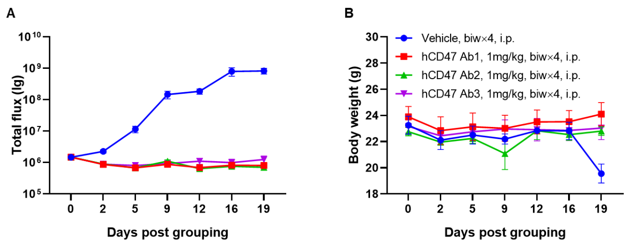
A Raji lymphoma mouse model was established using B-NDG mice and the efficacy of anti-human CD47 antibodies were verified. B-luc-GFP Raji cells (5×10⁵) were injected via tail vein of B-NDG mice. Small animal imager was used to observe tumor growth. When the fluorescence intensity of the tumor reaches about 1×10⁶ p/sec, the animals were divided into one control group and three treatment group (n=6). (A) Fluorescence intensity curve of tumor cells; (B) Body weight. The results showed that all three anti-human CD47 antibodies could significantly inhibit tumor growth. B-NDG mice is a powerful model for efficacy verification of anti-human CD47 antibodies. Values are expressed as mean ± SEM.
Successfully establishing a CDX lymphoma model and verifying the efficacy of CAR-T therapy with B-NDG mice
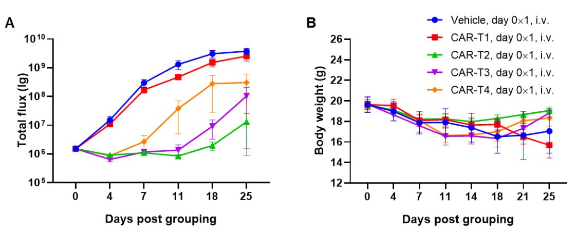
A Raji lymphoma mouse model was established using B-NDG mice and the efficacy of CAR-T therapy was verified. B-luc-GFP Raji cells (5×10⁵) were injected via tail vein of B-NDG mice. Small animal imager was used to observe tumor growth. When the fluorescence intensity of the tumor reaches about 1×10⁶ p/sec, the animals were divided into one control group and four treatment group (n=6). CAR-T cells (1×10⁷) were also injected via tail vein of B-NDG mice. (A) Fluorescence intensity curve of tumor cells; (B) Body weight. The results showed that four CAR-T cells differently inhibited tumor growth in B-NDG mice. B-NDG mice is a powerful model for efficacy verification of human CAR-T cells. Values are expressed as mean ± SEM.
Successfully establishing CDX model and verifying the efficacy of ADC drugs with B-NDG mice
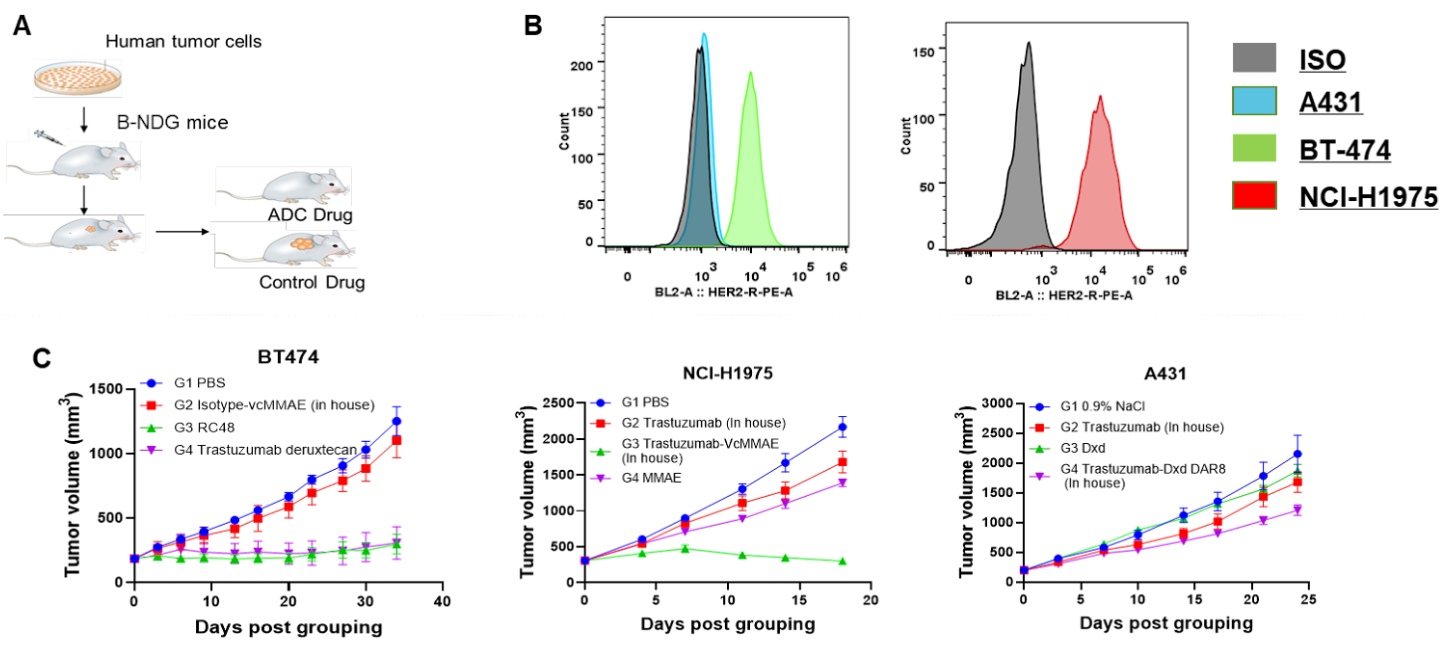
Comparing three cell lines with different Her2 expression level and establishing CDX models in B-NDG mice to verify the difference in efficacy of Her2-targeting ADC drugs. A431, BT-474 and NCI-H1975 cells were implanted subcutaneously in B-NDG mice. Mice were grouped when tumor volume reached to a suitable size, at which time they were treated intravenously with ADC drugs. The drug synthesized internally were marked with “in house”, while the commercial drugs were marked nothing after the drug name. (A) Schematic diagram of CDX model establishment and drug delivery strategy; (B) Expression level of human Her2 in in vitro cultured A431, BT-474, NCI-H1975 cell line; (C) CDX models were established for each of the three cell lines and the efficacy of the Her2-targeting ADC drugs were verified. The results showed that the expression level of human Her2 was high in BT-474 and NCI-H1975 cell line, while the expression level of Her2 in A431 cells was very low. Her2-targeting ADC drugs can significantly inhibit the tumor growth in CDX models established with high-Her2-expressing cell lines (BT-474, NCI-H1975), but had a weak inhibitory effect on low-Her2-expressing tumor (A431). B-NDG mice is a powerful model for efficacy verification of human ADC drugs. Values are expressed as averages ±SEM.
Successfully establishing CDX orthotopic model for pancreatic cancer with B-NDG mice
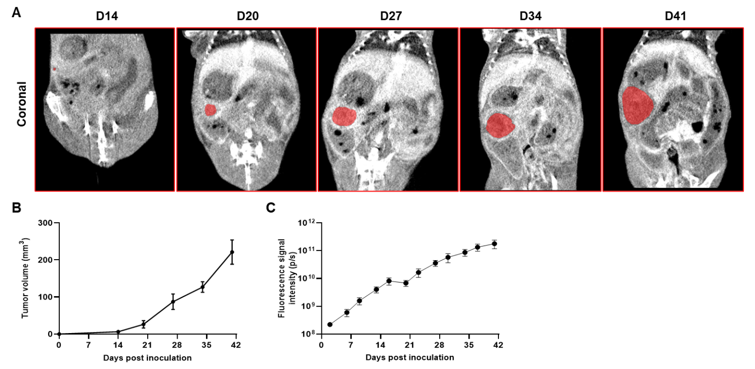
Imaging of orthotopic model for pancreatic cancer by Micro-CT. B-luc MIA Paca-2 cells were orthotopically inoculated into B-NDG mice (n=5). (A) Micro-CT images of orthotopic model in B-NDG mice were scanned from day 14 after inoculation. The images showed the coronal plane of the abdomen, with red areas representing the actual tumor. (B) Tumor growth curve was recorded by contrast-enhanced CT. (C) Dynamic changes of total fluorescence signal in the tumor site after inoculation. The results showed that human pancreatic cancer cell line (B-luc MIA Paca-2 cells) could successfully form tumors in situ pancreas of B-NDG mice.
Successfully establishing CDX orthotopic model for pancreatic cancer and verifying the efficacy of ADC drugs with B-NDG mice
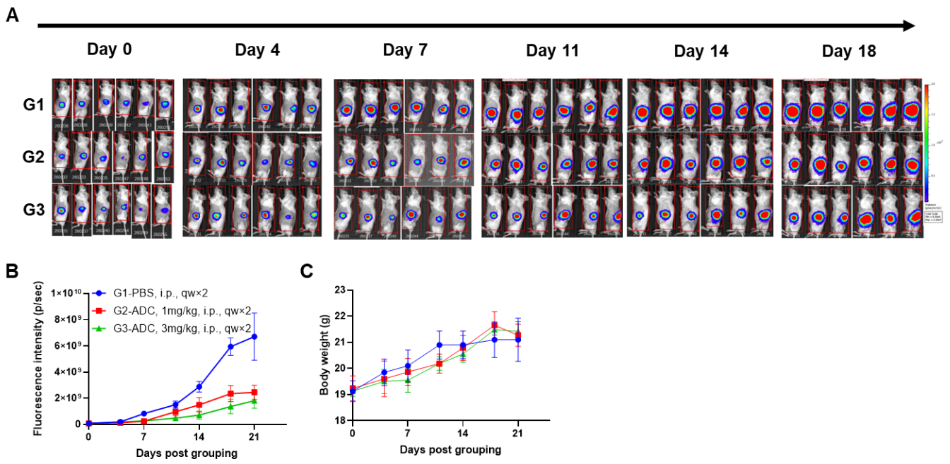
A MIA Paca-2 pancreatic orthotopic model was established using B-NDG mice and the efficacy of ADC drug was verified. B-luc MIA Paca-2 cells(1E5)were orthotopically implanted into B-NDG mice (female, 7-week-old, n=6). Mice were grouped when imaging signal value reached approximately 7E7 p/sec, at which time they were treated with the ADC drug with different doses and schedules were indicated in panel. (A) Body weight changes during treatment. As shown in panel B, this ADC drug was efficacious but mild, demonstrating that B-luc MIA Paca-2 pancreatic orthotopic model could provide a powerful preclinical model for in vivo evaluation of ADC drug. Values are expressed as mean ± SEM.
-
Human immune system reconstitution with human PBMCs in B-NDG mice and efficacy evaluation

-
Engraftment of human PBMCs in B-NDG mice to reconstitute human T cells
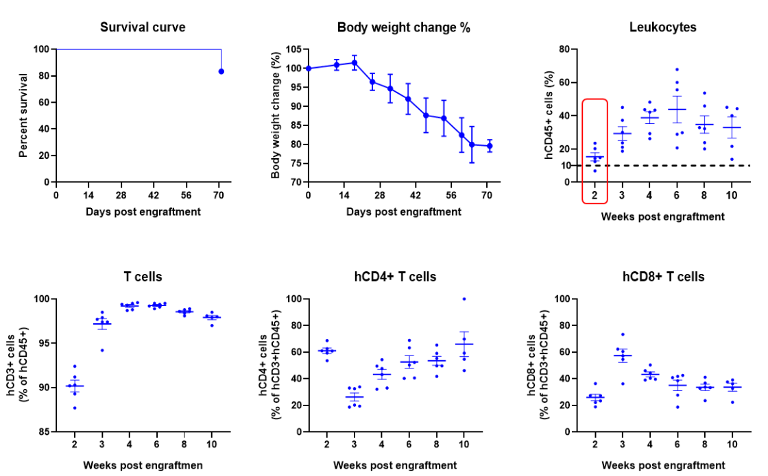
Engraftment of human PBMCs in B-NDG mice enhances reconstitution of human T cells. Human PBMCs (5E6) were intravenous engrafted into B-NDG mice (female, 6-week-old, n=6). Body weight of each mouse was recorded every week. Death data were collected every day. The peripheral blood was taken at different time points to analyze the reconstitution levels of human immune cells. B-NDG mice showed a high percentage of human CD45+ cells and T cells. B-NDG mice exhibit shortened survival and reduced body weight likely due to GvHD caused by a high Percentages of human T cell reconstitution. Values are expressed as mean ± SEM.
Successfully establishing CDX model in B-NDG mice engrafted with human PBMCs and verifying the efficacy of bispecific antibody
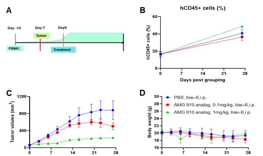
A NUGC4 human gastric cancer model was established using human PBMCs engrafted B-NDG mice and the efficacy of anti-human CD3/CLDN18.2 bispecific antibody was verified. Human PBMCs (5E6) were intravenously engrafted into B-NDG mice (female, 7-week-old, n=6). Human gastric cancer cell line NUGC4 (5E6) were inoculated subcutaneously 7days after PBMCs engraftment. AMG 910 (in house) was injected intraperitoneally 7 days after tumor inoculation. The animals were grouped into control and treatment when the tumor volume reached to 50-80 mm3 and the percentage of human blood hCD45+ cells ≥ 10%, at which time they were treated with drugs. (A) Schematic diagram of tumor model and drug delivery strategy; (B) Percentages of human CD45+ cells taken from peripheral blood on the day of administration (Day 0) and at the end point (Day 27); (C) Tumor volume; (D) Body weight. Results showed that the percentages of reconstituted human CD45+ cells continued to increase during administration. The anti-human CD3/CLDN18.2 bispecific antibody showed significant dose-dependent tumor suppression. B-NDG mice engrafted with human PBMCs can be used to establish tumor models and verify the efficacy of bispecific antibodies in vivo. Values are expressed as averages ±SEM.
Successfully establishing CDX orthotopic model for colon cancer in B-NDG mice engrafted with human PBMCs and verifying the efficacy of anti-human PD-L1 antibody
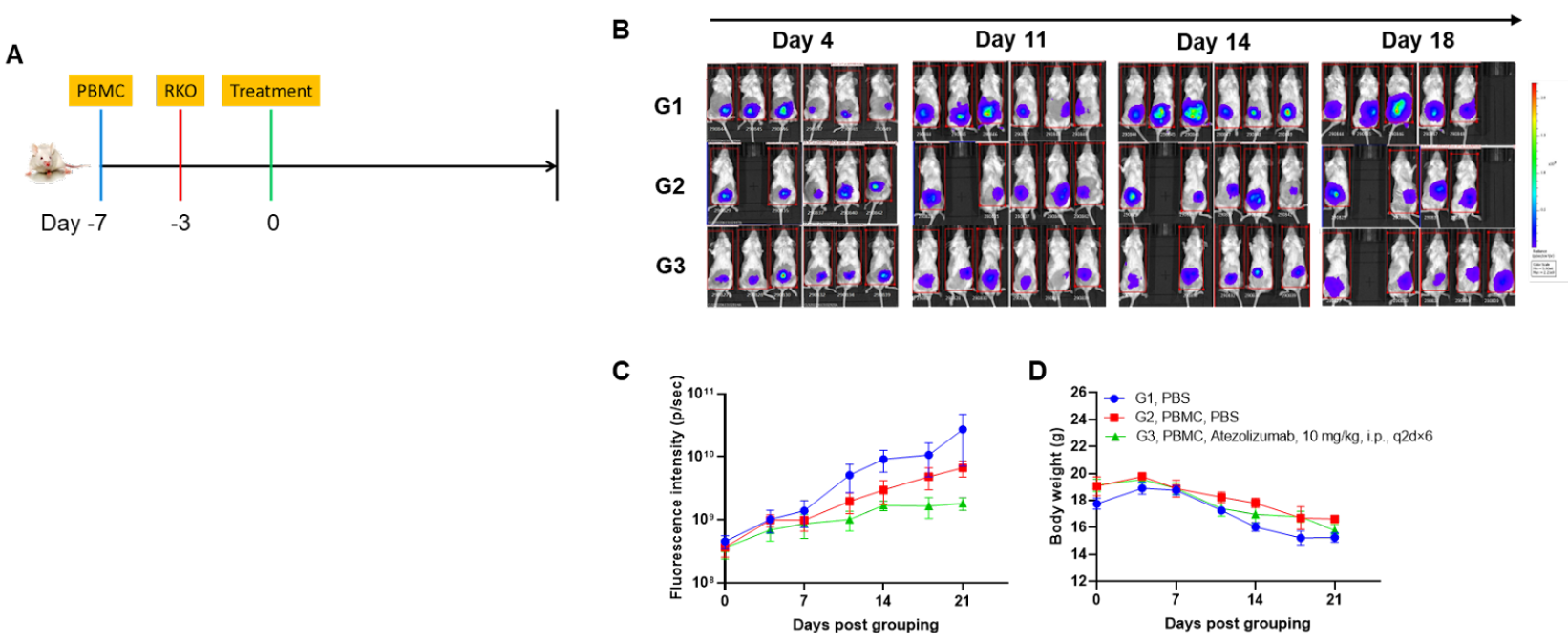
A CDX orthotopic model for human colon cancer was established using human PBMCs engrafted B-NDG mice and the efficacy of anti-human PD-L1 antibody was verified. Human PBMCs (5E6) were intravenously engrafted into B-NDG mice (female, 7-week-old, n=6). Human colonic cancer cell line B-Tg(Luc) RKO cells (1E6) were orthotopically inoculated into colonic tissue of B-NDG mice 4 days after PBMCs engraftment. Atezolizumab (in house) was injected intraperitoneally 3 days after tumor inoculation. (A) Schematic diagram of tumor model and drug delivery strategy; (B) Imaging of mice to observe tumor growth; (B) Fluorescence intensity curve of tumor cells; (D) Body weight. Results showed that anti-human PD-L1 antibody significantly inhibited tumor growth in the orthotopic colon cancer model, demonstrating that B-NDG mice engrafted with human PBMCs can be used to establish colonic orthotopic tumor model and verify the efficacy of anti-human antibodies in vivo. Values are expressed as mean ± SEM.
Engraftment of human PBMCs in B-NDG mice to establish GvHD model
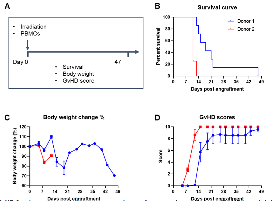
Engraftment of human PBMCs in B-NDG mice successfully constructed a graft-versus-host disease (GvHD) model. Human PBMCs (1E7) were intravenously engrafted into B-NDG mice 4 hrs after 1.0 Gy irradiation (female, 8-week-old, n=7/8). Mice were weighed twice a week and the health status of mice were recorded every day. Each mouse was scored according to the GvHD scoring standard. (A) Schematic diagram of the model-building strategy; (B) Survival curve; (C) Changes of body weight; (D) GvHD score. The results showed that the survival rate and the body weight of B-NDG mice were significantly reduced after irradiated and PBMC engraftment. GvHD scores were significant increased. But the GvHD symptoms vary between different donor sources of PBMCs. Therefor, B-NDG mice can be used to establish GvHD mouse model. Values are expressed as mean ± SEM.
Engraftment of human PBMCs in B-NDG mice to establish GvHD model and evaluate the efficacy of anti-human OX40 antibody

Engraftment of human PBMCs in B-NDG mice successfully constructed a graft-versus-host disease (GvHD) model and evaluated the efficacy of anti-human OX40 antibody. Human PBMCs (1E7) were intravenously engrafted into B-NDG mice 4 hrs after 1.0 Gy irradiation (female, 6-week-old, n=6). A anti-human OX40 antibody telazorlimab (in house) was intravenously injected into the mice of the treatment group. Mice were weighed three times a week and the health status of mice were recorded every day. Each mouse was scored according to the GvHD scoring standard. (A) Survival curve; (B) Changes of body weight; (C) GvHD score. The results showed that the GvHD model was successfully constructed. The survival rate, body weight and GvHD score of mice in the treatment group (telazorlimab) were significantly improved compared with those in the vehicle group. Therefor, B-NDG mice can be used to construct GvHD model and verify the efficacy of antibodies in vivo. Values are expressed as mean ± SEM.
-
Human immune system reconstitution with human CD34+ HSCs in B-NDG mice and efficacy evaluation

-
Engraftment of human CD34+ HSCs in B-NDG mice to reconstitute human immune system (adult mice)

Engraftment of human CD34+ HSCs in adult B-NDG mice successfully reconstituted human T and B cells, a small number of NK and myeloid cells. Human CD34+ HSCs (1.5E5) were engrafted via the tail veins of B-NDG mice (female, 6-week-old, n=5) within 4-12 hours after being irradiated with 1.6 Gy of X-ray. The reconstitution level of human immune cells in peripheral blood was analyzed by flow cytometry. (A) Survival curve of human CD34+ HSCs engrafted mice; (B) Body weight; (C) Percentages of reconstituted human immune cells. The results showed that no mice died before 22 weeks of human CD34+ HSCs engraftment. Only one mouse died after 160 days of reconstitution. All the mice continued to gain body weight. The percentage of human CD45+ cells exceeded 25% at 6 weeks of reconstitution and can still be maintained at about 25% after 24 weeks of reconstitution. The percentage of human T cells begins to increase persistently at 10 weeks of reconstitution. The percentage of human B cells reached more than 90% at 6 weeks of reconstitution and continued to decline after 8 weeks. A small percentage of human NK cells, total myeloid cells, monocytes and granulocytes can also be detected throughout the reconstitution process.
Engraftment of human CD34+ HSCs in B-NDG mice to reconstitute human immune system (neonatal mice)
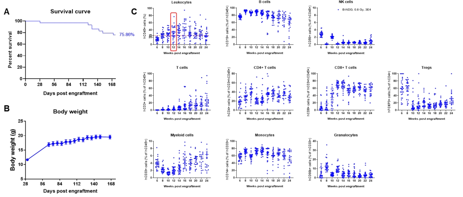
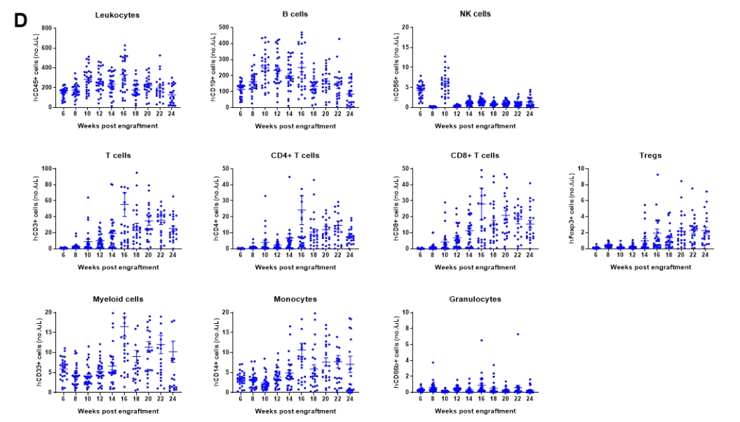
Engraftment of human CD34+ HSCs in neonatal B-NDG mice successfully reconstituted human T and B cells, a small number of NK and myeloid cells. Human CD34+ HSCs (1.5E5) were engrafted via the facial vein of B-NDG mice (male and female, 24-8 hour-old, n=28) within 4-12 hours after being irradiated with 0.8 Gy of X-ray. The reconstitution level of human immune cells in peripheral blood was analyzed by flow cytometry. (A) Survival curve of human CD34+ HSCs engrafted mice; (B) Body weight; (C) Percentages of reconstituted human immune cells; (D) Number of human immune cells. The results showed that the mice began to die from 17 weeks of reconstitution. But the lived mice continued to gain body weight. The percentage of human CD45+ cells exceeded 25% from 8 weeks of reconstitution. But there are obvious individual differences. The percentage of human T cells began to increase continuously at 10 weeks of reconstitution. Human CD4+ T cells, CD8+ T cells and Tregs could be detected at a relatively high level. The percentage of human B cells reached more than 70% at 6 weeks of reconstitution and continued to decline slowly after reaching the highest value at 8 weeks. A small percentage of human NK cells, total myeloid cells, monocytes and granulocytes can also be detected throughout the reconstitution process.
Engraftment of human CD34+ HSCs in B-NDG mice to reconstitute human immune system and evaluate the efficacy of anti-human PD-1 antibody
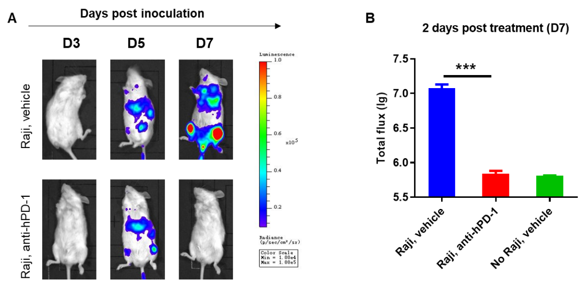
B-NDG mice reconstituted with CD34+ HSCs were used for drug efficacy evaluation. Human lymphoma cell line B-luciferase-GFP Raji cells (5E5) were inoculated via tail veins of adult B-NDG mice reconstituted with human CD34+ HSCs. Mice were treated with anti-human PD-1 antibody 5 days after tumor cell implantation. A dramatic inhibitory effect of the anti-human PD-1 antibody on tumor cell growth was observed at day 7. The results indicate that establishing a CDX tumor model in B-NDG mice with reconstituted HSCs provide a powerful preclinical model for in vivo evaluation of antibodies. Values are expressed as mean ± SEM.
-
PDX tumor models and efficacy evaluation in B-NDG mice

-
PDX models are successfully established in B-NDG mice
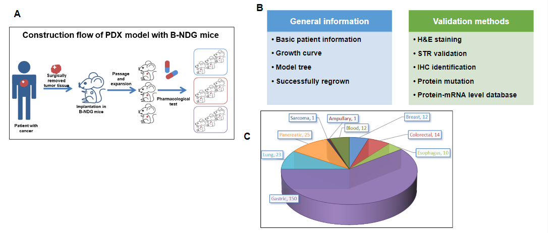
PDX (Patient-derived xenograft) models were successfully established in B-NDG mice. (A) General construction flow of the PDX model; (B) General information of tumor to be collected and the validation methods for the PDX model. (C) 248 PDX models derived from 9 types of tumors have been successfully established with B-NDG mice in Biocytogen by now.
Growth curve and STR identification for different generation of PDX tumor
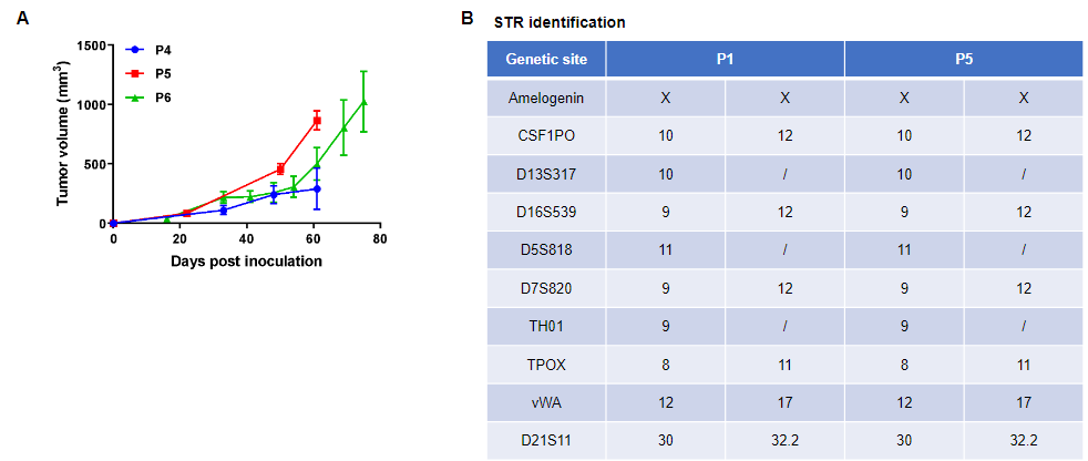
Pancreatic cancer BP0062 can successfully establish PDX model. Primary tumor sample was obtained from patient undergoing surgery for pancreatic ductal adenocarcinoma. Tumor pieces were subcutaneously inoculated into B-NDG mice. (A) Growth curve of passage 4, 5, 6 of the PDX tumor; (B) STR identification results of passage 1 and passage 5. Results showed that PDX model was successfully established in B-NDG mice implanted with pancreatic cancer BP0062. The STR of 5th generation of tumor cells is 100% matched to that of 1st generation, indicating that there was no change in the genetic background of the 5th generation of tumor tissue. Values are expressed as mean ± SEM.
Histopathological analysis of different generation of PDX tumor tissue
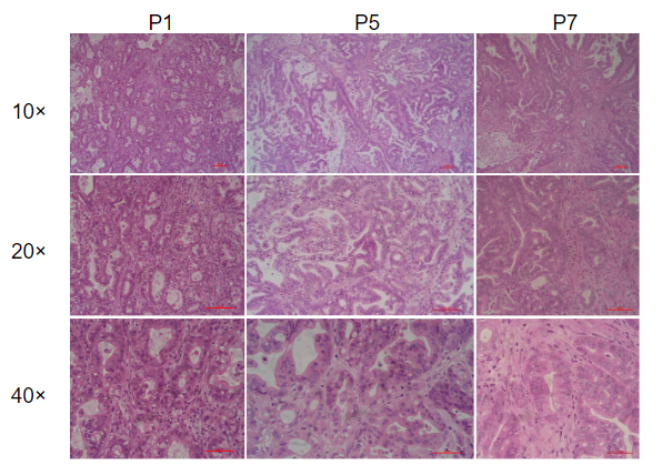
Histopathological analysis of BP0062 PDX tumor in B-NDG mice. Tumor tissue was collected from PDX model established with Pancreatic cancer BP0062 and analyzed with H&E staining. Results showed that patient-derived xenografts were found to well recapitulate the structures in original patient sample and maintain similar heterogeneity in different generations.
Expression analysis of CLDN18.2 and EGFR in PDX tumor by IHC staining
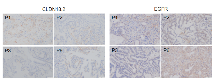
Immunohistochemical analysis of CLDN18.2 and EGFR in different generation of BP0062 PDX tumor tissue. Human CLDN18.2 and EGFR were respectively detected with anti-human CLDN18.2 antibody or anti-human EGFR antibody. The results showed that the number and distribution of CLDN18.2+ cells or EGFR+ cells in different generation of PDX tumor were obviously different (brown), indicating that the heterogeneity was maintained in different generation of PDX models.
In vivo efficacy verification of small molecule drugs for EGFR target with PDX model BP0062 in B-NDG mice
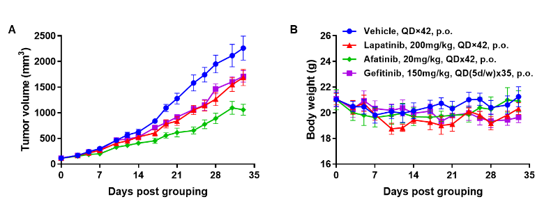
Antitumor activity of EGFR targeted drugs in PDX model BP0062 of pancreatic cancer established with B-NDG mice. (A) Molecular targeted small-molecule anti-cancer drugs slightly inhibited tumor growth of BP0062 in B-NDG mice. PDX tumor pieces of BP0062 were subcutaneously implanted into B-NDG mice (female, 6 week-old, n=6). Mice were grouped when tumor volume reached approximately 100 mm3, at which time they were treated with different targeted drugs and schedules indicated in panel (B) Body weight changes during treatment. As shown in panel A, Molecular targeted small-molecule anti-cancer drugs were efficacious, demonstrating that PDX model of BP0062 can be used to establish tumor model and provide a powerful preclinical pancreatic tumor model with EGFR positive cells. Values are expressed as mean ± SEM.
In vivo efficacy verification of ADC drugs with NSCLC PDX model BP0638 in B-NDG mice
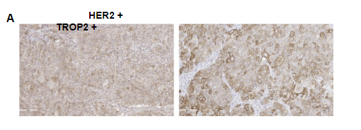
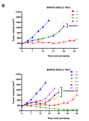
Antitumor activity of HER2-targeting and TROP2-targeting ADC drugs in NSCLC PDX model. (A) Expression analysis of HER2 and TROP2 in NSCLC (Non small Cell Lung Cancer) PDX (BP0638) tumor tissue by immunohistochemistry. The results showed both HER2 and TROP were low expression in this PDX model. (B) Efficacy verification of ADC drugs. PDX tumor pieces of BP0638 were subcutaneously implanted into B-NDG mice (n=5). Mice were grouped when tumor volume reached approximately 250-300 mm3, at which time they were treated with (HER2/TROP2-BsADC), two parental mono ADCs of the BsADC and other three commercial-derived ADC drugs (sacituzumab govitecan, disitamab vedotin, trastuzumab deruxtecan). The results showed that HER2/TROP2-BsADC had a stronger and longer inhibitory effect on tumor growth than ADCs. This inhibition was dose-dependent. Therefore, NSCLC PDX model of BP0062 provides a powerful preclinical NSCLC model for efficacy evaluation of various types of ADC drugs. Values are expressed as mean ± SEM.
In vivo efficacy verification of chemotherapy drugs with AML PDX model BP2010 in B-NDG mice
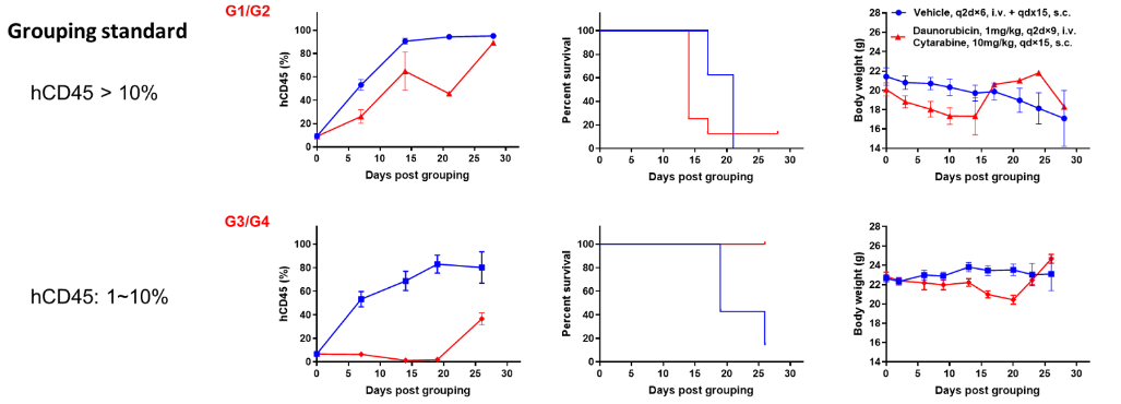
Antitumor activity of small molecule drugs in AML PDX model BP2010 established with B-NDG mice. According to the proportion of hCD45 in peripheral blood, PDX model mice were divided into 2 groups. One group of mice had a proportion of hCD45 in blood greater than 10% and the proportion of hCD45 in the blood of the other group of mice was between 1% and 10%. Each group was then divided into two sub-groups: the vehicle group and the treatment group. Mice in the treatment group were injected intravenously with daunorubicin and subcutaneously with cytarabine. The results showed that the efficacy of the group with low proportion of hCD45 (1%-10%) was better than that of the group with high proportion of hCD45 (>10%). Therefore, AML PDX model of BP2010 provides a powerful preclinical AML model for efficacy evaluation of chemotherapy drugs. Values are expressed as mean ± SEM.
In vivo efficacy verification of anti-human CD47 antibody with AML PDX model BP2010 in B-NDG mice

Antitumor activity of anti-human CD47 antibody in AML PDX model BP2010 established with B-NDG mice. (A) Human CD47 was highly expressed on the surface of tumor cells of BP2010; (B) The proportion of hCD45+ cells in the anti-human CD47 antibody treatment group was significantly reduced. PDX tumor cells of BP2010 were injected into B-NDG mice via tail vein (female, n=7). Mice were grouped when hCD45+ cells of PB reached approximately 1%, at which time they were treated with anti-human CD47 antibody Hu5F9 (in house) and schedules indicated in panel; (C) The survival rate of mice in the treatment group was significantly improved; (D) Body weight changes during treatment. The results indicated that AML PDX model of BP2010 provides a powerful preclinical AML model for efficacy evaluation of anti-human CD47 antibodies. Values are expressed as mean ± SEM.
-
Animal Breeding and Maintenance

-
1. Animal Housing and Husbandry
1.1 Health Status of Housing
Health status of housing: B-NDG mice are housed in isolators instead of IVCs in our facility. Based on our experience, the mice can live up to 2 months in SPF standard IVCs. This time frame matches the requirements of most experiments performed with B-NDG mice. To improve facility standards, strict sanitation procedures are recommended: cages and bedding need to be sterilized by autoclaving or Co60 irradiation before use, and cages need to be changed in laminar flow hoods weekly. Keeping a clean, high standard housing environment helps to improve the life span of B-NDG mice.
1.2. Animal Husbandry
Food
5CJL from Labdiet (USA) is recommended to use for breeding B-NDG mice (19.3% protein, 6.2% fat, 20 ppm Vitamin K). Co60 radiation is recommended to sterilize the food before use.
Water
B-NDG mice are housed in pathogen-free isolators in our facility. Autoclaved purified water is used.
For SPF standard facilities, we recommend following the Jackson Lab standard for water supply: acidified water (adjust pH to 2.5-3.0 using HCl), autoclaved to prevent Pseudomonas and Staphylococcus aureus infection. Autoclaved purified water can also be used with more frequent water changes. Bottle must be changed every 3 days regardless if there is still water left in the bottle.
Bedding
Shavings are the recommended bedding material for B-NDG mice. The bedding material needs to be sterile, soft, dust-free, odor-free and have high moisture absorbance. Sterilization by autoclaving or irradiation is required before use.
Bedding needs to be changed weekly in laminar flow hoods if the mice are not housed in isolators. Mice need to be transferred into new cages with fresh bedding using sterile tweezers or forceps.
Housing Environment
Enough light time and appropriate light intensity are necessary for breeding. We use a standard light cycle, which is 12-hours of light followed by 12-hours of dark.
Housing temperature is strictly 20-26 °C. The temperature difference between day and night should not be more than 4 °C.
Cages need to be made from non-toxic material and must be easy to clean and disinfect. Thorough cleaning and disinfection is required every week at least.
Parameters Range recommended Temperature 20℃-26℃ Humidity 40%-70% Ventilating rate 15 times per hr Light Cycle 12:12(standard) Light intensity 15-20 lux (in cage) Noise ≤60 db 2. Transportation
Biocytogen’s B-NDG mouse can be shipped using land and/or air. Although the courier is notified to handle the crate with care, stress response of mice during shipment is still inevitable. Although enough supply of water jelly and food will be provided in cages, increased metabolism and fecal excretion caused by the stress may result in dehydration and loss of body weight. General percentage weight loss due to shipment is ~10%. The percentage can be as high as 15% if the shipment procedure is longer and the cage is populated. Usually, most of the lost body weight is regained (although cannot reach 100%) after 5-7 days of adaptive feeding (Labdiet food is recommended).
3. Adaptive Feeding
Importance of adaptive feeding
Before performing experiments, at least 5-7 days of feeding in the receiving facility are required so that the animals can adapt to their new environment, and the stress response caused by transportation can be eliminated or alleviated.
Brief procedure description of adaptive feeding
Perform animal husbandry following 1.10.1.2. Monitor the health status of animals by observing their appearance (e.g. hair), feces and activity. Separate the animals from other animals in the facility as the sound and smell (e.g. Ammonia smelling feces) from other animals may be stimuli. Adaptive feeding is a critical prerequisite for successful experiments.
-
Anti-Tumor Efficacy in CDX & Humanized Models

-
Publications

-
- Guiding T lymphopoiesis from pluripotent stem cells by defined transcription factors
- An unexpected role for p53 in regulating cancer cell–intrinsic PD-1 by acetylation
- Exosome-derived miR-142-5p remodels lymphatic vessels and induces IDO to promote immune privilege in the tumour microenvironment
- piRNA-30473 contributes to tumorigenesis and poor prognosis by regulating m6A RNA methylation in DLBCL
- Chimeric Antigen Receptor Designed to Prevent Ubiquitination and Downregulation Showed Durable Antitumor Efficacy
- Leukemogenic Chromatin Alterations Promote AML Leukemia Stem Cells via a KDM4C-ALKBH5-AXL Signaling Axis
- Multiple Signaling Roles of CD3ε and Its Application in CAR-T Cell Therapy
- Circular RNA cESRP1 sensitises small cell lung cancer cells to chemotherapy by sponging miR-93-5p to inhibit TGF-β signalling
- Protease-activated receptor 2 stabilizes Bcl-xL and regulates EGFR–targeted therapy response in colorectal cancer
- Alpha lipoic acid promotes development of hematopoietic progenitors derived from human embryonic stem cells by antagonizing ROS signals
- Epigenetically silenced linc00261 contributes to the metastasis of hepatocellular carcinoma via inducing the deficiency of FOXA2 transcription
- Therapeutic Targeting of CDK7 Suppresses Tumor Progression in Intrahepatic Cholangiocarcinoma
- Expression levels of a gene signature in hiPSC associated with lung adenocarcinoma stem cells and its capability in eliciting specific antitumor immune-response in a humanized mice model
- Promising xenograft animal model recapitulating the features of human pancreatic cancer
- Effective antitumor activity of 5T4-specific CAR-T cells against ovarian cancer cells in vitro and xenotransplanted tumors in vivo
- NEK2 induces autophagy-mediated bortezomib resistance by stabilizing Beclin-1 in multiple myeloma
- Cancer-secreted exosomal miR-1468-5p promotes tumor immune escape via the immunosuppressive reprogramming of lymphatic vessels
- HDAC2 inhibits EMT-mediated cancer metastasis by downregulating the long noncoding RNA H19 in colorectal cancer
- Integrative multi-omics analysis of a colon cancer cell line with heterogeneous Wnt activity revealed RUNX2 as an epigenetic regulator of EMT
- Sequential treatment with aT19 cells generates memory CAR-T cells and prolongs the lifespan of Raji-B-NDG mice
- Preliminary biological evaluation of 123I-labelled anti-CD30-LDM in CD30-positive lymphomas murine models
- Glycyrrhizic acid improves cognitive levels of aging mice by regulating T/B cell proliferation
- Degradable Carbon-Silica Nanocomposite with Immunoadjuvant Property for Dual-Modality Photothermal/Photodynamic Therapy
- Long non-coding RNA SOX2OT promotes the stemness phenotype of bladder cancer cells by modulating SOX2
- Long non-coding RNA CASC9 promotes tumor growth and metastasis via modulating FZD6/Wnt/β-catenin signaling pathway in bladder cancer
- A Tumor-Targeted Replicating Oncolytic Adenovirus Ad-TD-nsIL12 as a Promising Therapeutic Agent for Human Esophageal Squamous Cell Carcinoma
- TriBAFF-CAR-T cells eliminate B-cell malignancies with BAFFR-expression and CD19 antigen loss
- Two-step protocol for regeneration of immunocompetent T cells from mouse pluripotent stem cells
- R9AP is a functional receptor for Epstein-Barr virus infection in both epithelial cells and B cells
- Licochalcone A improves the cognitive ability of mice by regulating T- and B-cell proliferation
- Downregulation of lncRNA ZNF582-AS1 due to DNA hypermethylation promotes clear cell renal cell carcinoma growth and metastasis by regulating the N(6)-methyladenosine modification of MT-RNR1
- Cutting Edge: Inhibition of Glycogen Synthase Kinase 3 Activity Induces the Generation and Enhanced Suppressive Function of Human IL-10 + FOXP3 +-Induced Regulatory T Cells
- Anti-leukemia activities of selenium nanoparticles embedded in nanotube consisted of triple-helix β-d-glucan
- Oncolytic adenovirus targeting TGF-β enhances anti-tumor responses of mesothelin-targeted chimeric antigen receptor T cell therapy against breast cancer
- A PTK7-targeted antibody-drug conjugate reduces tumor-initiating cells and induces sustained tumor regressions
- Long-Term Engraftment Promotes Differentiation of Alveolar Epithelial Cells from Human Embryonic Stem Cell Derived Lung Organoids
- LunX-CAR T Cells as a Targeted Therapy for Non-Small Cell Lung Cancer
- A Tumor-Targeted Replicating Oncolytic Adenovirus Ad-TD-nsIL12 as a Promising Therapeutic Agent for Human Esophageal Squamous Cell Carcinoma
- MUC1-Tn-targeting chimeric antigen receptor-modified Vγ9Vδ2 T cells with enhanced antigen-specific anti-tumor activity
- Natural small molecule triptonide inhibits lethal acute myeloid leukemia with FLT3-ITD mutation by targeting Hedgehog/FLT3 signaling
- Targeting epidermal growth factor-overexpressing triple-negative breast cancer by natural killer cells expressing a specific chimeric antigen receptor
- The combination of CUDC-907 and gilteritinib shows promising in vitro and in vivo antileukemic activity against FLT3-ITD AML
- BMI1 regulates multiple myeloma-associated macrophage’s pro-myeloma functions
-
References

-
1) Xinhua Xiao, Huiliang Li, Huizi Jin, Jin Jin, Miao Yu, Chunmin Ma, Yin Tong, Li Zhou, Hu Lei, Hanzhang Xu, Weidong Zhang, Wei Liu, and Yingli Wu. 2017. Identification of 11(13)-dehydroivaxillin as a potent therapeutic agent against non-Hodgkin’s lymphoma. Cell death & disease. 8(9):e3050.
2) Ito M, Hiramatsu H, Kobayashi K, Suzue K, Kawahata M, Hioki K, Ueyama Y, Koyanagi Y, Sugamura K, Tsuji K, Heike T, Nakahata T. 2002. NOD/SCID/gamma(c)(null) mouse: an excellent recipient mouse model for engraftment of human cells. Blood 100(9):3175-82. [PMID: 12384415]
3) Shultz LD, Lyons BL, Burzenski LM, Gott B, Chen X, Chaleff S, Kotb M, Gillies SD, King M, Mangada J, Greiner DL, Handgretinger R. 2005. Human lymphoid and myeloid cell development in NOD/LtSz-scid IL2R gamma null mice engrafted with mobilized human hemopoietic stem cells. J Immunol 174(10):6477-89. [PMID: 15879151]
4) McDermott SP, Eppert K, Lechman ER, Doedens M, Dick JE. 2010. Comparison of human cord blood engraftment between immunocompromised mouse strains. Blood 116(2):193-200. [PMID: 20404133]
5) Lepus CM, Gibson TF, Gerber SA, Kawikova I, Szczepanik M, Hossain J, Ablamunits V, Kirkiles-Smith N, Herold KC, Donis RO, Bothwell AL, Pober JS, Harding MJ. 2010. Comparison of human fetal liver, umbilical cord blood, and adult blood hematopoietic stem cell engraftment in NOD-scid/gammac-/-, Balb/c-Rag1-/-gammac-/-, and C.B-17-scid/bg immunodeficient mice. Blood 70(10):790-802. [PMID: 19524633]
6) Shultz LD1, Brehm MA, Bavari S, Greiner DL. 2011. Humanized mice as a preclinical tool for infectious disease and biomedical research. Ann N Y Acad Sci 1245:50-4. [PMID: 22211979]
7) Covassin L1, Jangalwe S, Jouvet N, Laning J, Burzenski L, Shultz LD, Brehm MA. 2013. Human immune system development and survival of non-obese diabetic (NOD)-scid IL2rγ (null) (NSG) mice engrafted with human thymus and autologous haematopoietic stem cells. Clin Exp Immunol 174(3):372-88. [PMID: 23869841]
8) Wege AK, Schmidt M, Ueberham E, Ponnath M, Ortmann O, Brockhoff G, Lehmann J. 2014. Co-transplantation of human hematopoietic stem cells and human breast cancer cells in NSG mice: a novel approach to generate tumor cell specific human antibodies. MAbs 6(4):968-77. [PMID: 24870377]


